Source: Laboratories of Jonas T. Kaplan and Sarah I. Gimbel—University of Southern California
Traditional brain imaging techniques using MRI are very good at visualizing the gross structures of the brain. A structural brain image made with MRI provides high contrast of the borders between gray and white matter, and information about the size and shape of brain structures. However, these images do not detail the underlying structure and integrity of white matter networks in the brain, which consist of axon bundles that interconnect local and distant brain regions.
Diffusion MRI uses pulse sequences that are sensitive to the diffusion of water molecules. By measuring the direction of diffusion, it is possible to make inferences about the structure of white matter networks in the brain. Water molecules within an axon are constrained in their movements by the cell membrane; instead of randomly moving in every direction with equal probability (isotropic movement), they are more likely to move in certain directions, in parallel with the axon (anisotropic movement; Figure 1). Therefore, measures of diffusion anisotropy are thought to reflect properties of the white matter such as fiber density, axon thickness, and degree of myelination. One common measure is fractional anisotropy (FA). FA values range from 0, which represents completely isotropic movement, to 1, reflecting maximum anisotropy.
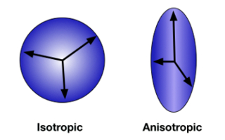
Figure 1: Diffusion anisotropy. When the direction of diffusion is unconstrained and random, movement is measured in all directions equally. This is isotropic diffusion (A). When water molecules are contained within the axon of a neuron, diffusion is anisotropic, tending to occur more frequently along the direction of the axon (B). Please click here to view a larger version of this figure.
In this experiment we will use diffusion tensor imaging (DTI) to measure white matter integrity in traumatic brain injury (TBI). TBI occurs when an external force injures the brain, such as a blow to the head or a sudden movement like the kind that might occur in a car accident. This type of brain injury from mechanical forces is associated with diffuse axonal injury-damage to white matter throughout the brain. Because it is an injury affecting white matter integrity, standard neuroimaging techniques may not reveal the damage. However, measures of diffusion are especially sensitive to these anatomical changes. Following a study by Kraus et al.1, we compare a group of healthy controls to a group of people with TBI and use diffusion imaging to measure the effect of TBI on cerebral white matter. Furthermore, we will test the relationship between white matter integrity and cognitive function using an attention task.2 This study uses a region of interest (ROI) approach focusing on three white matter tracts: the splenium of the corpus callosum, the anterior corona radiata, and the superior longitudinal fasciculus (Figure 2).
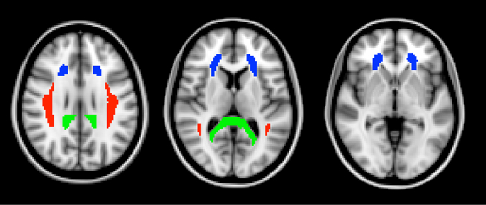
Figure 2: Regions of interest. The three ROIs, defined from the ICBM DTI-81 atlas, are shown here in horizontal slices through the brain. In green is the splenium of the corpus callosum. The splenium is the most posterior part of the corpus callosum. In blue is the anterior corona radiata. The superior longitudinal fasciculus is shown in red. Please click here to view a larger version of this figure.
Neuropsychology

