Overview
Source:Tracey A. Milligan, MD; Tamara B. Kaplan, MD; Neurology, Brigham and Women's/Massachusetts General Hospital, Boston, Massachusetts, USA
During each section of the neurological testing, the examiner uses the powers of observation to assess the patient. In some cases, cranial nerve dysfunction is readily apparent: a patient might mention a characteristic chief complaint (such as loss of smell or diplopia), or a visually evident physical sign of cranial nerve involvement, such as in facial nerve palsy. However, in many cases a patient's history doesn't directly suggest cranial nerve pathologies, as some of them (such as sixth nerve palsy) may have subtle manifestations and can only be uncovered by a careful neurological exam. Importantly, a variety of pathological conditions that are associated with alterations in mental status (such as some neurodegenerative disorders or brain lesions) can also cause cranial nerve dysfunction; therefore, any abnormal findings during a mental status exam should prompt a careful and complete neurological exam.
The cranial nerve examination is applied neuroanatomy. The cranial nerves are symmetrical; therefore, while performing the examination, the examiner should compare each side to the other. A physician should approach the examination in a systematic fashion and go through the cranial nerves in their numerical order.
| I | Olfactory | Smell |
| II | Optic | Visual acuity, afferent pupillary response |
| III | Oculomotor | Horizontal eye movements (adduction), efferent pupillary response |
| IV | Trochlear | Downward vertical eye movement, internal rotation of eye |
| V | Trigeminal | Facial sensation, jaw movement |
| VI | Abducens | Horizontal eye movement (abduction) |
| VII | Facial | Facial movement and strength, taste, dampening of loud sounds, sensation; anterior wall of external ear canal |
| VIII | Acoustic | Hearing, vestibular functioning |
| IX | Glossopharyngeal | Movement of pharynx, sensation of pharynx, posterior tongue (including taste of posterior tongue), and most of ear canal |
| X | Vagal | Movement and sensation of palate, pharynx, gag reflex, guttural sounds |
| XI | Spinal accessory | Strength of sternocleidomastoids and trapezius muscles |
| XII | Hypoglossal | Tongue protrusion and lateral movements |
Table 1. The 12 Cranial Nerves and Their Basic Functions
Cranial nerve I (the olfactory nerve) is a purely sensory nerve that conveys the sense of smell, and is not routinely tested during most examinations. Cranial nerve II (the optic nerve) is the only cranial nerve that can be directly visualized as it exits from the central nervous system. Its axons convey visual information and compose the afferent limb of the pupillary reflex. Testing of the pupillary responses also assesses the function of cranial nerve III (the oculomotor nerve), parasympathetic fibers of which form the efferent limb of the pupillary reflex. Cranial nerve exam includes assessment of the extraocular movements, which are controlled by cranial nerves III, IV and VI. Cranial nerve III innervates the superior, medial, and inferior rectus muscles, as well as the inferior oblique muscle, which together function to move eyes medially and in the vertical plane. Cranial nerve IV (the trochlear nerve_ innervates the superior oblique muscles, which moves the eye downward and outward. Cranial nerve VI (the abducens nerve) innervates the lateral rectus muscles, which abducts the eyes. The function of the medial and lateral rectus muscles is straightforward: Lateral rectus is involved in abduction, meaning lateral movement along the horizontal plane. Medial rectus adducts moving the eye medially movement along the horizontal plane. The remaining muscles each causes movement in more than one direction and some combination of elevation/depression, abduction/adduction, intorsion/extorsion.
| Muscle | Innervation | Primary action | Secondary action | Tertiary action |
| Medial rectus | CN III | Adduction | -- | -- |
| Superior rectus | CN III | Elevation | Intortion | Adduction |
| Inferior rectus | CN III | Depression | Extortion | Adduction |
| Inferior oblique | CN III | Extorsion | Elevation | Abduction |
| Superior oblique | CN IV | Intorsion | Depression | Abduction |
| Lateral rectus | CN VI | Abduction | -- | -- |
Table 2. The function of the six extraocular muscles.
This first part of the cranial nerve exam concludes by testing cranial nerve V (the trigeminal nerve). This nerve has both motor and sensory components. It controls facial sensation, masticatory movements and forms the afferent limb of the corneal reflex. There are 3 major sensory branches of the trigeminal nerve - the ophthalmic, maxillary, and mandibular (also labeled V1, V2, and V3, respectively).
Procedure
1. Cranial Nerve I (Olfactory Nerve)
The olfactory nerve exam is performed on patients acknowledging a decreased sense of smell, especially after an acceleration/deceleration head injury, as the olfactory nerves are prone to such shearing injuries.
- Have the patient close the eyes and occlude one nostril with an index finger.
- Present the patient with a recognizable smell, such as coffee granules, peppermint, cinnamon, or commercially available sniff cards. Have the patient try to identify the smell.
- Repeat the procedure for the other nostril.
2. Cranial Nerve II (Optic Nerve).
The assessment of the optic nerve assessment includes fundoscopy, visual acuity testing, visual field examination, and testing for pupillary responses.
- Directly examine the fundus using an ophthalmoscope.
- Have the patient fixate gaze across the room at a slightly upward angle.
- Use your left eye to examine the patient's left eye and your right eye to visualize the patient's right fundus.
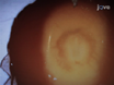
- Look for signs of optic nerve injury, such as optic nerve pallor or increased intracranial pressure with blurring of the optic disc and the loss of spontaneous venous pulsations. If venous pulsations are not observed (which can be normal in many patients), inform the patient that you are going to press lightly on the orbit of the eye, while looking for venous collapse on the optic nerve head. Papilledema is a late sign of increased intracranial pressure. Note the color of the optic nerve, which is normally a yellowish color.
- Check visual fields by direct confrontation.
- Test one eye at a time, and test the four quadrants of the visual field of each eye.
Stand facing the patient about 3 feet away and stretch your arms forward and to the sides so that your hands are just barely visible in your peripheral vision. Your fingers should be equidistant between the patient's eyes and your own. - Quickly wiggle your index finger on either the left or right in both the superior and inferior visual field quadrants, and ask the patient to look directly at your nose and identify where the movement occurs. For the patient's left eye use your right eye as a control. Then test analogous portions of the visual field on the other side.
- Next, check for loss of double simultaneous stimulation by asking the patient if one or both fingers wiggling. Then present to the patient moving stimuli in multiple visual fields simultaneously, such as moving fingers in both upper temporal fields and bilateral inferior fields. Field cuts that respect the vertical meridian are usually due to cortical lesions such as stroke. Field cuts that respect the horizontal meridian are usually related to ocular lesions such as ischemic optic neuropathy.
- Alternative ways to test visual fields:
- Ask the patient to tell you when your finger is first seen as you slowly move it in from the periphery inwards for lower and upper quadrants of vision.
- Ask the patient to count the number of fingers you hold up at various spots in the field.
- Ask the patient if any part of your face is missing or appears distorted.
- Test one eye at a time, and test the four quadrants of the visual field of each eye.
- Check visual acuity with a hand-held card:
- Have the patient alternately cover each eye and read the smallest line they can see with the card held about 14 inches away. If the patient wears corrective lenses, they should be used while checking acuity.
- Record the smallest line read correctly.
- Repeat process for other eye.
3. Cranial Nerves II and III.
Pupillary light reflex controls the diameter of the pupil in response to the light intensity. Both cranial nerves II and III are being tested when the pupillary response is checked, as the optic nerve carries the afferent fibers of the reflex, and the efferent limb is supplied by cranial nerve III (the oculomotor nerve).
- Check pupillary response, directly and consensually.
- Reduce the room illumination as much as possible.
- Shine a penlight in the direction of the patient's nose so that you can see both pupils without directing light at either of them, and check that the pupils are the same size. About 10% of normal patients will have a slight pupil asymmetry of ~1mm (aniscoria).
- Ask the patient to look across the room, and shine bright light into each pupil.
- Look for brisk constriction of the pupil as you shine the light into the patent's eye (direct response), and the corresponding constriction in the contralateral eye (consensual response).
- The swinging-flashlight test is done to look for an afferent pupillary defect, also known as a Marcus-Gunn pupil.
- To perform the test, move the flashlight between the eyes every two to three seconds. If there is no afferent or efferent abnormality, there is no change in the pupil size with the swinging light test.
If the patient has a disorder such as optic neuritis (as may be seen in multiple sclerosis), the affected eye may have a decreased response to direct illumination from a bright light. However, the efferent pathways are intact; therefore, the pupil of the diseased eye will initially briskly contract when the unaffected eye is exposed to bright light (as the consensual response is preserved). When the affected eye is then directly exposed to the bright light, the diseased optic nerve does not mount as strong a response as the consensual response had been, and the pupil paradoxically enlarges as it is being directly stimulated.
- To perform the test, move the flashlight between the eyes every two to three seconds. If there is no afferent or efferent abnormality, there is no change in the pupil size with the swinging light test.
- Finally, test the pupillary response to accommodation.
- Ask the patient to focus on your finger or the penlight itself, and move it closer to the patient's nose.
- Normally, the pupils constrict while fixating on an object being moved from far away to near the eyes.
4. Cranial Nerves III, IV, and VI.
- Ask the patient to follow your finger with the eyes while keeping the head in one position
- Using your finger, trace an imaginary letter "H" shape in front of the patient, making sure that your finger moves far enough out and up so that you're able to see all appropriate eye movements. The patient's eyes should move together throughout all planes of vision without the development of any double vision or eye muscle weakness.
- Check convergence: instruct the patient to follow your finger as you slowly moving it towards the patient's eyes. Look for restrictions of gaze, as may be seen with a sixth nerve palsy when a patient cannot fully abduct one or both eyes.
- Look for nystagmus, which are rapid rhythmic jerking movements of the eye that can be especially seen on horizontal gaze. In some instances, nystagmus may be due to the effects of medications (such as benzodiazepines or some antiepileptic medications), but it can also be associated with cerebellar dysfunction and vestibular disorders.
- The third cranial nerve also controls elevation of the eyelid. Observe for ptosis (drooping of the upper eyelids) as can be seen in lesions of the third nerve, Horner's syndrome (ptosis, miosis, and decreased ipsilateral facial sweating caused by sympathetic lesions), or muscle diseases such as myasthenia gravis.
5. Cranial Nerve V (Trigeminal Nerve).
- Evaluate sensory function by testing for pain and light touch sensation.
- Test for light touch by asking the patient if sensation is normal on each side while touching the patient in each of the 3 divisions of the trigeminal nerve separately. Touch the patient on the left and right side and compare to see if the sensation is the same on both sides.
- Test for pain sensation in all 3 sections of the trigeminal nerve with the touch of a sharp object, such as the tip of a safety pin. Ask the patient to close the eyes and to describe a sensation as sharp or dull. Again, compare the left and right side to see if the sensation is equal.
- To test for the motor function, have the patient bite down hard and palpate, the masseter muscles. Feel for contraction of the muscle and assess for symmetry between sides.
- The corneal reflex is also an objective measure of cranial nerve V function and cranial nerve VII. This reflex is usually only tested if there is suspicion of a cranial nerve lesion or in an unresponsive patient. If the patient is wearing contact lenses, the corneal reflex cannot be tested.
- To test for corneal reflexes, prepare a cotton swab by taking the end of the swab and pulling it out, leaving just a few strands of cotton projecting out so as to not injure the patient's cornea.
- After warning the patient to expect a poke in the eye, tell the patient to look to the left while you test the right eye, and look to the right while you test the left eye.
- Gently touch the patient's corneal with a wisp of cotton and observe if there is a reflexive blink. Make sure to test response on cornea, not just conjunctiva. Determine if any difference between eyes.
The examination of the cranial nerves is essentially applied neuroanatomy, and often the location of a lesion can be identified solely on the basis of physical findings. There are 12 pairs of the cranial nerves, numbered rostral to caudal, which arise directly from the brain. They are named as per their function or structure or the region of innervation. Here, we'll briefly discuss anatomy and physiology of the cranial nerves-one through six, and demonstrate how to examine these nerves in a systematic fashion
Let's start with a review of the basic neuroanatomy of the first six cranial nerves.
Cranial nerve I, or the olfactory nerve, is formed by projections of the specialized receptor neurons, located in the upper part of the nasal cavity. The olfactory nerve fibers convey the smell information to the olfactory bulb cells, which then relay the signal via the olfactory tract.
The second cranial nerve - also known as the optic nerve - is responsible for the visual information transmission from retina to the brain. In addition, this nerve constitutes the afferent limb of the pupillary light reflex. The efferent limb of this reflex is composed by the parasympathetic fibers travelling with the cranial nerve III, also known as the oculomotor nerve. The parasympathetic axons synapse at the ciliary ganglion, and the postganglionic fibers innervate the sphincter pupillae muscle. Thus, both the cranial nerves II and III are required for the pupillary constriction in response to light. This oculomotor nerve also controls the levator palpabrae superioris - a muscle that lifts the upper eyelid. Furthermore, this nerve controls four extraocular muscles - the superior, medial, and inferior recti and the inferior oblique, that function together to move the eyes medially and in the vertical plane.
Cranial nerve IV, the trochlear nerve, innervates the superior oblique muscles, which move the eye downward and outward. And cranial nerve VI, the abducens nerve, innervates the lateral rectus muscles, which are responsible for ocular abduction. Together, these muscles and nerves regulate the movement of the eyes in the six cardinal directions of gaze.
Lastly, we will discuss cranial nerve V, also known as the trigeminal nerve. This nerve has three major divisions-ophthalmic, maxillary and mandibular. The ophthalmic and maxillary branches have purely sensory function, whereas the mandibular nerve is formed by both sensory and motor fibers. The sensory fibers of all three branches relay facial sensation, and the ophthalmic branch also mediates the corneal reflex. The motor fibers of the mandibular division supply the muscles of mastication.
After this brief introduction, let's review how to assesses these nerves during a clinical encounter. As the cranial nerves are symmetrical, every test should be performed on both sides and the findings should be compared.
We will start with the examination of the cranial nerve I, the olfactory nerve. Instruct the patient to occlude one nostril with their index finger and close their eyes. Then, hold an odorant, such as coffee granules, beneath the patient's nose, and ask them to identify the smell. Repeat the test on the other side using a different odorant, like mint toothpaste.
Next, examine the cranial nerve II, the optic nerve. This part of the examination includes ophthalmoscopy, visual field examination, visual acuity assessment, and testing the pupillary responses, which are also controlled by the cranial nerve III. Start with the ophthalmoscopic examination. Ask the patient to look across the room at a slightly upward angle. As the patient is doing so, examine their right fundus with your right eye, and note any optic nerve or fundus abnormalities. Similarly, use your left eye to visualize the patient's left fundus. The technique and the potential findings on ophthalmoscopic exam are covered in detail in a separate JoVE Clinical Skills video.
Next, perform the visual field test. This term describes the entire area that can be seen during steady fixation of gaze in one direction. The visual field for each eye can be roughly divided into four quadrants by the vertical and the horizontal meridians. The upper and lower quadrants are referred to as the superior and inferior quadrants, outer two are the temporal, and inner two are the nasal quadrants. Start by evaluating the peripheral vision using the direct confrontation technique. Stand about three feet away from the patient, and ask them to fixate their gaze on your nose. Then extend your arms forward and to the sides, such that your hands are in patient's superior and inferior temporal quadrants. During this test, your hands should be barely visible in your own peripheral vision. Now ask the patient to cover their left eye and continue to fixate their gaze at your nose. Then cover your right eye and quickly wiggle your left index finger in all four quadrants of the patient's open eye, and ask them to identify where the movement occurs. Use your open eye as the control throughout this test. Repeat the same procedure on the other side. Subsequently, assess for the loss of double simultaneous stimulation. Ask the patient to keep both eyes open and let you know if they see one or both fingers moving. Present to the patient moving fingers in multiple visual fields simultaneously, such as in either upper temporal fields or bilateral inferior fields.
Next, check the visual acuity using a hand-held card. Ask the patient to wear corrective lenses or non-reading glasses, if normally used. For the test, have the patient cover one eye and read the smallest line they can with the card held about 14 inches away. Record the finding and repeat the same step for the other eye.
Next, test the pupillary responses, which can be affected by both-the optic and the oculomotor nerve dysfunction. Before this test, reduce the room illumination. Then shine a penlight in the direction of the patient's nose taking care not to illuminate the eyes directly. This is done for observing the pupils at rest, for size, shape and equality. Next ask the patient to look across the room and shine bright light directly into each eye. Look for a brisk constriction of the illuminated pupil - the direct response. Also observe the simultaneous constriction of the contralateral pupil - the consensual response. If the patient has a disorder such as optic neuritis-as may be seen in multiple sclerosis-the affected eye may have a decreased direct response, but the consensual response is preserved. Next, perform the swinging flashlight test by moving the flashlight between the pupils every two to three seconds and observing for direct and consensual response. The paradoxical dilation of the illuminated pupil seen during these tests indicates an afferent pupillary defect, also known as a Marcus-Gunn pupil. Subsequently, turn the room lights back on to observe the response to accommodation. Ask the patient to look into the distance and then focus on your thumb placed closer to their face. Repeat this a couple of times to check for the normal constriction of pupils in response to focusing on an object relatively near to the eyes.
Now, let's discuss the testing of extraocular movements, which are controlled by cranial nerves III, IV and VI. To test the eyeball movement in the six cardinal directions of gaze, ask the patient to keep their head steady, and follow your finger with their eyes as you trace an imaginary letter "H" shape. Normally, the eyes should move together throughout all planes of vision and there should not be any observed eye muscle weakness or development of any double vision. Next, instruct the patient to follow your finger as you move it slowly towards the patient's eyes. Check for convergence by noting if restriction of gaze is present. After that, move your finger in vertical, and then in horizontal directions and tell the patient to follow your finger with their eyes. Observe for nystagmus-the rapid rhythmic jerking movements of the eye. This may be normal sometimes on the horizontal gaze or as effect of certain medications, but it can also be associated with cerebellar or vestibular dysfunction. Since cranial nerve III also controls the levator palpebrae superioris muscle, ask the patient to focus on a spot and observe the position of the eyelids. Note if ptosis, which is drooping of the upper eyelids, is present. Ptosis can be associated with lesions of the third nerve, Horner's syndrome, and neuromuscular diseases, such as myasthenia gravis. This completes the cranial nerves III, IV and VI testing.
Next, assess the function of cranial nerve V, the trigeminal nerve. Lightly touch the patient's skin in each of the three areas innervated by trigeminal nerve divisions. Ask the patient if they can feel your touch and if the sensation is equal and normal on the both sides. Subsequently, test the pain sensation in each of the three divisions. For this, have the patient close their eyes and touch their skin with both the sharp tip and the rounded end of a safety pin on both sides. Ask the patient to describe a sensation as sharp or dull. Also ask them if the sensation is same on both sides. Next, place your hand on either side of the patient's jaw, and have them bite down hard, while you feel for the contraction of the masseter muscles. This tests the motor function of the trigeminal nerve. Conclude the trigeminal nerve assessment by testing the corneal reflex. Prepare a swab by pulling out most of the cotton at the end, leaving just a few strands projecting out, so as not to injure the patient's eye. Make sure that the patient doesn't wear contact lenses. Warn the patient that you are going to touch their right eye, and tell them to look to the left. Then, with a wisp of cotton, gently touch the right cornea and observe for the blink, or the corneal reflex. Similarly, test the left eye and compare between sides.
You've just watched JoVE's video on how to evaluate the first six cranial nerves in a systematic way. We went over the essential steps of the examination, which can help uncover signs of the neurologic disorders associated with this set of nerves. The "cranial nerve exam part II" will cover the testing associated with nerves VII through XII. As always, thanks for watching!
Applications and Summary
This video demonstrates a systematic approach to examining the first six cranial nerves. The central and peripheral nervous systems are an integrated system. Therefore, if the clues to a neurological problem are uncovered while taking medical history or during the mental status exam, it should make the clinician more vigilant during the rest of the examination of the nervous system to look for other abnormalities. A clinician should develop a pattern of going through each nerve in numerical order and only document those nerves that were actually examined in the final report. Patients are often being followed for diseases (such as multiple sclerosis) where findings may be changing over time. The documentation from one examination to another are important to follow and the findings should be carefully charted. It is not adequate to just look at the patient and then state "cranial nerves II-XII are intact," as is so often recorded during a typical physical examination.