Overview
Quelle: Peiman Shahbeigi-Roodposhti und Sina Shahbazmohamadi, Biomedical Engineering Department, University of Connecticut, Storrs, Connecticut
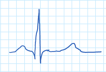
Ein Elektrokardiograph ist ein Graph, der durch elektrische Potentialveränderungen zwischen Elektroden aufgezeichnet wird, die auf den Oberkörper eines Patienten gelegt werden, um die Herzaktivität zu demonstrieren. Ein EKG-Signal verfolgt herzrhythmusstörungen und viele Herzkrankheiten, wie z. B. schlechte Durchblutung des Herzens und strukturelle Anomalien. Das DurchKontraktionen der Herzwand erzeugte Aktionspotenzial breitet elektrische Ströme aus dem Herzen im ganzen Körper aus. Die sich ausbreitenden elektrischen Ströme erzeugen an Stellen im Körper unterschiedliche Potenziale, die durch Elektroden auf der Haut wahrgenommen werden können. Die Elektroden sind biologische Messumformer aus Metallen und Salzen. In der Praxis werden 10 Elektroden an verschiedenen Punkten am Körper befestigt. Es gibt ein Standardverfahren zum Erfassen und Analysieren von EKG-Signalen. Eine typische EKG-Welle eines gesunden Individuums ist wie folgt:

Abbildung 1. EKG-Welle.
Die "P"-Welle entspricht der Vorhofkontraktion und der "QRS"-Komplex der Kontraktion der Ventrikel. Der "QRS"-Komplex ist aufgrund der relativen Dfference in der Muskelmasse der Vorhöfe und Ventrikel viel größer als die "P"-Welle, die die Entspannung der Vorhöfe verschleiert. Die Entspannung der Ventrikel ist in Form der "T"-Welle zu sehen.
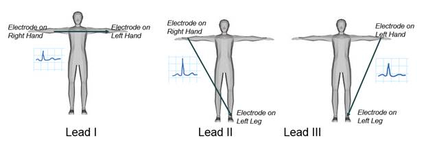
Es gibt drei Hauptleitungen, die für die Messung des elektrischen Potentials zwischen Armen und Beinen verantwortlich sind, wie in Abbildung 2 dargestellt. In dieser Demonstration wird einer der Gliedmaßen, Blei I, untersucht, und der elektrische Potentialunterschied zwischen zwei Armen wird aufgezeichnet. Wie bei allen EKG-Bleimessungen gilt die mit dem rechten Bein verbundene Elektrode als Bodenknoten. Ein EKG-Signal wird mit einem Biopotential-Verstärker erfasst und dann mit Einer Instrumentierungssoftware angezeigt, wo eine Gain-Steuerung erstellt wird, um seine Amplitude anzupassen. Schließlich wird das aufgezeichnete EKG analysiert.

Abbildung 2. EKG Gliedmaßen führt.
Principles
Der Elektrokardiograph muss nicht nur extrem schwache Signale von 0,5 mV bis 5,0 mV erkennen können, sondern auch eine DC-Komponente von bis zu 300 mV (die sich aus dem Elektroden-Hautkontakt ergibt) und eine Common-Mode-Komponente von bis zu 1,5 V, die sich aus dem zwischen den Elektroden und dem Boden. Die nutzgebrachte Bandbreite eines EKG-Signals hängt von der Anwendung ab und kann zwischen 0,5-100 Hz liegen und manchmal bis zu 1 kHz erreichen. Es ist in der Regel um 1 mV Peak-to-Peak in Gegenwart von viel größeren externen Hochfrequenz-Rauschen, 50 oder 60 Hz Interferenzen und DC-Elektroden-Offset-Potenzial. Andere Lärmquellen sind Bewegungen, die die Haut-Elektroden-Schnittstelle, Muskelkontraktionen oder elektromyographische Spitzen, Atmung (die rhythmisch oder sporadisch sein kann), elektromagnetische Interferenzen (EMI) und Geräusche von anderen elektronischen Geräten beeinflussen. das paart sich in den Eingang.
Zunächst wird ein Biopotential-Verstärker zur Verarbeitung des EKG hergestellt. Dann werden Elektroden auf den Patienten gelegt, um den potenziellen Unterschied zwischen zwei Armen zu messen. Die Hauptfunktion eines Biopotentialverstärkers besteht darin, ein schwaches elektrisches Signal biologischen Ursprungs zu nehmen und seine Amplitude zu erhöhen, so dass es weiterverarbeitet, aufgezeichnet oder angezeigt werden kann.

Abbildung 3. EKG-Verstärker.
Um biologisch nützlich zu sein, müssen alle biopotenziellen Verstärker bestimmte Grundanforderungen erfüllen:
- Sie müssen eine hohe Eingangsimpedanz haben, damit sie eine minimale Belastung des gemessenen Signals ermöglichen. Biopotente Elektroden können durch ihre Belastung beeinflusst werden, was zu einer Verzerrung des Signals führt.
- Die Eingangsschaltung eines Biopotentialverstärkers muss auch dem untersuchten Thema Schutz bieten. Der Verstärker sollte isolations- und schutzkreissicher sein, damit der Strom durch den Elektrodenkreis auf einem sicheren Niveau gehalten werden kann.
- Die Ausgangsschaltung treibt die Last an, die in der Regel ein Anzeige- oder Aufzeichnungsgerät ist. Um maximale Genauigkeit und Reichweite in der Auslesung zu erhalten, muss der Verstärker eine geringe Ausgangsimpedanz haben und in der Lage sein, die von der Last benötigte Leistung zu liefern.
- Biopotential-Verstärker müssen in dem Frequenzspektrum betrieben werden, in dem die biopotentials existieren, die sie verstärken. Aufgrund des niedrigen Pegels solcher Signale ist es wichtig, die Bandbreite des Verstärkers zu begrenzen, um optimale Signal-Rausch-Verhältnisse zu erhalten. Dies kann mit Filtern erfolgen.
Abbildung 3 ist ein Beispiel für einen EKG-Verstärker, und Abbildung 4 ist die Schaltung des EKG-Verstärkers, der während dieser Demonstration gebaut wird. Es hat drei Hauptstufen: die Schutzschaltung, den Instrumentationsverstärker und den Hochpassfilter.

Abbildung 4. Biopotential Verstärker.
Die erste Stufe ist die Patientenschutzschaltung. Eine Diode ist ein Halbleitergerät, das Strom in eine Richtung leitet. Wenn eine Diode vorwärts-voreingenommen ist, fungiert die Diode als Kurzschluss und leitet Strom. Wenn eine Diode rückwärts-voreingenommen ist, wirkt sie wie ein offener Kreislauf und leitet keinen Strom, ir 0.
Wenn sich Dioden in der vorwärtsgerichteten Konfiguration befinden, gibt es eine Spannung, die als Schwellenspannung (VT = ca. 0,7 V) bezeichnet wird, die überschritten werden muss, damit die Diode Strom leiten kann. Sobald der VT überschritten wurde, bleibt der Spannungsabfall über die Diode bei VT konstant, unabhängig davon, was Vin ist.
Wenn die Diode rückwärts-voreingenommen ist, wird die Diode wie auf offenem Stromkreis wirken und der Spannungsabfall über die Diode wird gleich Vinsein.
Abbildung 5 ist ein Beispiel für eine einfache Schutzschaltung, die auf Dioden basiert, die in dieser Demo verwendet werden. Der Widerstand wird verwendet, um den Strom zu begrenzen, der durch den Patienten fließt. Wenn ein Fehler im Instrumentationsverstärker oder dioden die Verbindung des Patienten mit einer der Stromschienen kurzschließt, würde der Strom weniger als 0,11 mA betragen. Die Leckagedioden FDH333 werden zum Schutz der Eingänge des Instrumentationsverstärkers eingesetzt. Wenn die Spannung in der Schaltung 0,8 V in der Größe überschreitet, ändern sich die Dioden in ihren aktiven Bereich oder "ON"-Zustand; der Strom fließt durch sie und schützt sowohl den Patienten als auch die elektronischen Komponenten.

Abbildung 5. Schutzschaltung.
Die zweite Stufe ist der Instrumentationsverstärker IA, der drei Operationsverstärker (Op-Amp) verwendet. An jedem Eingang ist ein Op-Amp angebracht, um den Eingangswiderstand zu erhöhen. Der dritte Op-Amp ist ein Differentialverstärker. Diese Konfiguration hat die Möglichkeit, bodenbezogene Interferenzen abzulehnen und nur den Unterschied zwischen den Eingangssignalen zu verstärken.

Abbildung 6. Instrumentierungsverstärker.
Die dritte Stufe ist der Hochpassfilter, der verwendet wird, um eine kleine Wechselstromspannung zu verstärken, die auf einer großen Gleichspannung reitet. Das EKG wird von niederfrequenten Signalen beeinflusst, die von Patientenbewegungen und Atmung kommen. Ein Hochpassfilter reduziert dieses Rauschen.
Hochpassfilter können mit RC-Schaltungen erster Ordnung realisiert werden. Abbildung 7 zeigt ein Beispiel für einen Hochpassfilter erster Ordnung und dessen Übertragungsfunktion. Die Cut-off-Frequenz wird durch die folgende Formel angegeben:
 ,
,


Abbildung 7. Hochpassfilter.
Procedure
1. Erwerb eines EKG-Signals
- Stellen Sie die Spannung der Quellen auf +5 V und -5 V ein und schließen Sie sie in Reihe an.
- Erstellen Sie die in Abbildung 4gezeigte Schaltung . Berechnen Sie die Werte der Widerstände und Kondensatoren. Für den Hochpassfilter sollte die Grenzfrequenz 0,5 Hz betragen. Der Kondensatorwert sollte aus der folgenden Tabelle ausgewählt werden (je nach Verfügbarkeit).
| Verfügbare Kondensatorwerte (F ) | ||
| 0.001 | 1 | 100 |
| 0.022 | 2.2 | 220 |
| 0.047 | 4.7 | 470 |
| 0.01 | 10 | 1000 |
| 0.1 | 47 | 2200 |

- Legen Sie Elektroden auf den rechten Arm, linken Arm und rechten Bein (dies ist Referenz) des Patienten, und verbinden Sie sie mit dem Schaltkreis.
- Verwenden Sie das Oszilloskop, um das EKG-Signal (Vo) anzuzeigen. Drücken Sie Auto Set und passen Sie die horizontalen und vertikalen Skalen nach Bedarf an. Sie sollten in der Lage sein, die R-Spitzen trotz des Rauschens im Signal zu sehen.
2. Anzeige des EKG-Signals mit Instrumention-Software
- In dieser Demo haben wir LabVIEW verwendet. Schreiben Sie ein Programm, das das EKG-Signal über eine grafische Schnittstelle zur Konfiguration von Messungen und ein Wellenformdiagramm anzeigt. Nachdem ein analoger Eingang ausgewählt wurde, konfigurieren Sie das Programm mit den folgenden Einstellungen:
- Signaleingangsbereich >> Max = 0,5; Min = -0,5
- Terminalkonfiguration >> RSE
- Erfassungsmodus >> kontinuierlich
- Lesebeispiele = 2000
- Abtastrate = 1000
- Erfassen Sie das EKG-Signal und beobachten Sie die Wellenform. Es wird ein Signal ähnlich Abbildung 1angezeigt.
- Passen Sie die Skalierung der x-Achse an, um die Zeit in Sekunden anzuzeigen.
- Bei der Instrumentierung ist es oft notwendig, das Signal des Interesses auf eine bestimmte Amplitude zu verstärken. Erstellen Sie eine Verstärkungssteuerung und legen Sie sie so fest, dass die Amplitude des EKG 2 Vp beträgt.
3. Analyse des EKG-Signals
In diesem Abschnitt wird ein EKG-Signal gefiltert und analysiert, um die Herzfrequenz zu bestimmen. Das folgende Blockdiagramm zeigt die Komponenten des Programms.
- Verwenden Sie ein Wellenformdiagramm, um das Signal anzuzeigen.
- Bewerten Sie das Spektrum des Signals mit dem Subvi amplitude und Phase Spectrum (in Der Signalverarbeitung - Spektral) und zeigen Sie seine Größe mit einem Wellenformdiagramm an. Die horizontale Achse entspricht der Frequenz. Es ist diskret, weil der Computer einen Fast Fourier Transform (FFT) Algorithmus verwendet, um das Spektrum des Signals zu berechnen. Die Frequenz geht von k = 0 bis k = (N-1)/2, wobei N die Länge der Sequenz ist, in diesem Fall 4000. Um die entsprechende analoge Frequenz zu berechnen, verwenden Sie die folgende Formel:

wobei fs die Abtasthäufigkeit ist. Beachten Sie, dass sich die meiste Energie des Signals im niederfrequenten Bereich befindet und dass es auch einen Spitzenwert hoher Intensität im mittleren Frequenzbereich gibt. Berechnen Sie die Häufigkeit dieses Peaks mit der oben angegebenen Formel. - Implementieren Sie einen Tiefpassfilter mit Butterworth of Chebyshev-Funktionen. Wählen Sie eine Grenzfrequenz von 100 Hz. Stellen Sie sicher, dass der Filter eine Dämpfung von mindestens -60 dB/Dekade im Stoppband bietet.
- Schließen Sie das Ausgangssignal des Lese-Aus-Tabellensubvi an den Eingang des Tiefpassfilters an.
- Implementieren Sie einen Stop-Band-Filter mit Butterworth- oder Chebyshev-Funktionen. Ziel ist es, die 60 Hz Interferenzen zu reduzieren, ohne die anderen Frequenzen zu verändern. Versuchen Sie Grenzfrequenzen in der Nähe von 60 Hz.
- Schließen Sie den Ausgang des Tiefpassfilters an den Eingang des Stoppbandfilters an.
- Finden Sie die Spitzen mit dem Peak-Detektor subvi (es befindet sich in Signalverarbeitung - Sig Operation). Sehen Sie sich für den Schwellenwert die Amplitude des Signals an, und wählen Sie den am besten geeigneten Wert aus.
- Extrahieren Sie die Positionen der Peaks mithilfe des Index-Array-Subvi (in Programming - Array).
- Subtrahieren Sie die untere Position von der höheren Position, multiplizieren Sie dann mit der Stichprobenperiode T = 1/fs, um das RR-Intervall zu erhalten.
- Berechnen Sie die Wechsel- und Einstelleinheiten und platzieren Sie einen Indikator, um das BPM anzuzeigen.
Elektrokardiographen zeichnen die Herzaktivität des Herzens auf und werden verwendet, um Krankheiten zu diagnostizieren, Anomalien zu erkennen und mehr über die allgemeine Herzfunktion zu erfahren. Elektrische Signale werden durch Kontraktionen in den Herzwänden erzeugt, die elektrische Ströme antreiben und unterschiedliche Potenziale im ganzen Körper erzeugen. Durch das Platzieren von Elektroden auf der Haut kann man diese elektrische Aktivität in einem EKG erkennen und aufzeichnen. EKGs sind nicht-invasiv, was sie zu einem nützlichen Werkzeug macht, um zu beurteilen, wie gut ein Patientenherz funktioniert, z. B. indem sie messen, wie gut Blut in das Organ fließt.
Dieses Video zeigt die Prinzipien von EKGs und zeigt, wie man ein typisches EKG-Signal mit einem Biopotential-Verstärker erfasst, verarbeitet und analysiert. Andere biomedizinische Anwendungen, die elektrische Signalverarbeitung nutzen, um Krankheiten zu diagnostizieren, werden ebenfalls diskutiert.
Um die Prinzipien eines EKG zu verstehen, lassen Sie uns zuerst verstehen, wie das Herz elektrische Signale erzeugt. Für ein normales, gesundes Herz, das im Ruhezustand ruht, zeigt ein EKG eine Reihe von Wellen an, die die verschiedenen Phasen eines Herzschlags widerspiegeln. Das EKG beginnt im sinoatrialen Knoten, auch bekannt als SA-Knoten, der sich im rechten Vorhof befindet und als Herzschrittmacher im Herzen fungiert. Die elektrischen Signale verursachen eine Vorhofkontraktion, die Blut in die Ventrikel zwingt. Diese Sequenz wird als P-Welle auf dem EKG aufgezeichnet. Dieses Signal geht dann von den Vorhöfen über die Ventrikel, wodurch sie sich zusammenziehen und Blut in den Rest des Körpers pumpen. Dies wird als QRS-Komplex aufgezeichnet.
Schließlich entspannen sich die Ventrikel und dies wird als T-Welle aufgezeichnet. Der Prozess beginnt dann wieder und wird für jeden Herzschlag wiederholt. Beachten Sie, dass die QRS-Welle viel größer als die P-Welle ist, dies liegt daran, dass die Ventrikel größer sind als die Vorhöfe. Das bedeutet, dass sie die Entspannung der Vorhöfe oder der T-Welle verschleiern. Andere Prozesse im Körper, wie Atmung oder Muskelkontraktionen, können die EKG-Messung stören. Wie kann Ströme aus der Schaltung verwendet, um sie zu erhalten. Oft sind die elektrischen Signale, die das EKG aufzeichnen will, recht schwach. Dazu wird ein Biopotentialverstärker eingesetzt, um deren Amplitude zu erhöhen, wodurch sie weiterverarbeitet und aufgezeichnet werden können.
Der Biopotentialverstärker, die Patientenschutzstufe, der Instrumentationsverstärker und der Hochpassfilter sind mit drei Hauptkomponenten verbunden. Wie die Hauptsache andeutet, verwendet der Patientenschutzkreis eine Kombination aus Widerständen und Dioden, um sowohl den Patienten als auch die Maschine und ausrüstung zu schützen. Die Widerstände begrenzen den Strom, der durch den Patienten fließt, wobei die Dioden den Strom in die richtige Richtung fließen lassen.
Die nächste Stufe ist der Instrumentationsverstärker, der den Unterschied zwischen den Eingängen jeder Elektrode verstärkt. Es besteht aus drei Operationsverstärkern. Zwei, um den Widerstand von jedem Eingang zu erhöhen, und der dritte, um die Differenz zwischen den Eingangssignalen zu verstärken.
Die letzte Stufe ist der Hochpassfilter, der das Rauschen reduziert und niederfrequente Signale aus der Patientenbewegung oder Atmung herausfiltert. Nun, da Sie wissen, wie ein EKG gemessen wird, sehen wir uns an, wie man einen Biopotential-Verstärker baut und die Daten verarbeitet, um ein sauberes EKG-Signal zu erhalten.
Nachdem wir die Hauptprinzipien der Elektrokardiographie überprüft haben, sehen wir uns an, wie man einen Biopotential-Verstärker baut und ein EKG-Signal erhält. Zunächst sollten Sie zunächst ein Proto-Board, einen AD-620-Instrumentierungsverstärker und alle notwendigen Schaltungskomponenten zusammentragen. Berechnen Sie dann die Werte aller Widerstände und Kondensatoren in der Schaltung mit der folgenden Gleichung.
Für den Hochpassfilter sollte die Schnittfrequenz 0,5 Hertz betragen.
Schließen Sie dann den Kondensatorwert an, um den Widerstand zu bestimmen. Als nächstes bauen Sie einen Biopotential-Verstärker nach dem mitgelieferten Diagramm. So sollte die letzte Schaltung aussehen. Befestigen Sie drei Drähte mit Alligatorclips an den Bindepfosten eines DC-Netzteils, und schalten Sie dann die Stromquelle ein. Stellen Sie die Spannung auf plus fünf Volt und minus fünf Volt ein, und schließen Sie die Drähte in Reihe an die Schaltung an.
Verwenden Sie nun ein Alkohol-Prep-Pad, um die Patienten am rechten Handgelenk, am linken Handgelenk und am rechten Knöchel zu wischen. Fügen Sie den Elektroden leitfähiges Klebegel hinzu, bevor Sie sie auf den Patienten legen. Verbinden Sie dann die Elektroden mit der Schaltung über Drähte mit Alligatorclips. Schalten Sie das Oszilloskop ein und erfassen Sie das EKG-Signal. Passen Sie die horizontalen und vertikalen Skalen nach Bedarf an. Mit diesen Einstellungen sollten Sie in der Lage sein, die R-Spitze der Wellenform zu sehen.
Schließen Sie die Schaltung an das PXI-Chassis an, öffnen Sie dann die Instrumentierungssoftware und verwenden oder schreiben Sie entweder ein Programm, das das EKG-Signal und ein Wellenformdiagramm anzeigt.
Konfigurieren Sie die Datenerfassungsschnittstelle mit den folgenden Einstellungen. Beschriften Sie den Maßstab der x-Achse, um Zeit und Sekunden anzuzeigen, und zeigen Sie dann das EKG-Signal als Wellenform an. Wenn das Signal verstärkt werden muss, erstellen Sie eine Gain-Steuerung und stellen Sie sie so ein, dass die Amplitude des EKG zwei VP beträgt.
Nun, da wir gezeigt haben, wie man ein EKG-Signal erfasst, sehen wir uns an, wie die Ergebnisse analysiert werden. Hier ist ein repräsentatives EKG-Signal. Die P-, QRS- und T-Wellen sind kaum erkennbar, da sie durch Lärm und Schwankungen verdeckt sind. Dieses Signal muss gefiltert werden. Um dieses Signal zu transformieren, wählen Sie zuerst Signalverarbeitung und dann Spektral im Menü aus. Ein Fast Fourier Transform-Algorithmus berechnet und zeichnet das Spektrum des Signals, das die Frequenz als diskrete Werte auf der horizontalen Achse anzeigt. Der größte Teil der Energie im Signal ist bei niedrigen Frequenzen.
Aber es gibt eine hohe Intensität Spitze im mittleren Frequenzbereich, die als Lärm angenommen wird. Die Frequenz wird als k auf der horizontalen Achse dargestellt und geht von Null nach N minus eins über zwei, wobei N die Länge der Sequenz ist. Für dieses Experiment entspricht N 2.000. Berechnen Sie die analoge Frequenz für jeden k-Wert mit der folgenden Gleichung, wobei f s die Abtastfrequenz ist, und bestimmen Sie die Frequenz des Hochintensitätsspitzen basierend auf dem FFT-Diagramm.
Erstellen Sie dann einen Tiefpassfilter mit einer Grenzfrequenz von 100 Hertz. Verwenden Sie entweder die Butterworth- oder Chebyshev-Funktion, um das Signal zu filtern, das mindestens 60 Dezibel pro Jahrzehnt im Stoppband dämpfen sollte. Schließen Sie das Ausgangssignal des Datensub VI an den Eingang des Tiefpassfilters an. Dieser Filter entfernt die überflüssigen Hochfrequenzwellen des EKG. Erstellen Sie nun einen Bandstop-Filter und stellen Sie die Grenzfrequenzen auf etwa 55 und 70 Hertz ein.
Um das laute Signal zu entfernen, ca. 60 Hertz. Verbinden Sie dann den Ausgang des Tiefpassfilters mit dem Eingang des Bandstop-Filters. Versuchen Sie Grenzfrequenzen, die in der Nähe von 60 Hertz liegen. Dadurch werden Interferenzen reduziert, ohne dass andere Frequenzen bewirken. Das EKG-Signal sollte nun mit deutlichen P-, QRS- und T-Komplexen klar sein.
Lassen Sie uns nun die Herzfrequenz mit dem gefilterten EKG-Signal bestimmen. Verwenden Sie zunächst den Peak-Detektor Sub VI, um die Spitzen des Signals zu finden. Wählen Sie den am besten geeigneten Wert basierend auf der Signalamplitude der R-Welle für den Schwellenwert aus. Verwenden Sie dann das Index-Array-Sub VI, um die Position der Peaks zu bestimmen.
Subtrahieren Sie die untere Spitzenposition von der höheren Position, und multiplizieren Sie diesen Wert dann mit dem Stichprobenzeitraum T, der gleich eins über f s ist. Dieser Wert ist die Zeitdauer zwischen zwei R-Wellen. Passen Sie die Einheiten an, um die Beats pro Minute zu bestimmen.
Bei dieser Demonstration betrug die gemessene Herzfrequenz etwa 60 Schläge pro Minute.
EKG und Signalverarbeitung haben wichtige Anwendungen sowohl in der Medizin als auch in der Forschung. EKGs sind nicht nur nicht invasiv, sondern auch relativ kostengünstig. Ein nützliches und zugängliches Werkzeug in Krankenhäusern. EKGs können sogar an eine komplexere und langfristigere Überwachung von Patienten angepasst werden, die wegen des akuten Koronarsyndroms behandelt werden.
Dazu werden 12 EKG-Leitungen verwendet, die bei asymptomatischen Patienten eine vorübergehende Myokard-Ischämie identifizieren können. Signalprobeundund-Verarbeitung wird auch in der Elektroenzephalographie verwendet, um elektrische Signale aus dem Gehirn zu messen. EEGs werden häufig in Verbindung mit funktioneller MRT als multimodale Bildgebungstechnik verwendet.
Die Methode nichtinvasiv erzeugt kortikale Karten der Gehirnaktivität für viele Neuroimaging-Anwendungen, wie nach visueller oder motorischer Aktivierung.
Sie haben gerade Joves Einführung in den Erwerb und die Analyse von EKG-Signalen beobachtet. Sie sollten nun verstehen, wie ein EKG-Signal erzeugt wird und wie ein Biopotential-Verstärker erstellt wird, um schwache elektrische Signale zu erkennen. Sie haben auch einige biomedizinische Anwendungen der Signalverarbeitung für die medizinische Diagnose gesehen.
Danke fürs Zuschauen.
Results
In dieser Demonstration wurden drei Elektroden an eine Person angeschlossen, und der Ausgang ging durch einen Biopotentialverstärker. Ein Beispiel-EKG-Diagramm vor der digitalen Filterung ist unten dargestellt (Abbildung 8).

Abbildung 8. EKG-Signal ohne digitale Filterung.
Nach dem Entwerfen der Filter und der Zuführung der Daten an den entwickelten Algorithmus wurden die Spitzen im Diagramm erkannt und zur Berechnung der Herzfrequenz (BPM) verwendet. Abbildung 9 zeigt die Rohdaten eines EKG-Signals (vor jeder Filterung) in Zeit- und Frequenzbereich. Abbildung 10 zeigt das Ergebnis der Filterung dieses Signals.

Abbildung 9. EKG-Signal vor dem Filtern.

Abbildung 10. Gefiltertes EKG-Signal.
Das ursprüngliche EKG-Diagramm hatte leicht sichtbare P-, QRS- und T-Komplexe, die viele Schwankungen des Rauschens aufwiesen. Das Spektrum des EKG-Signals zeigte auch eine deutliche Spitze bei 65 Hz, die als Rauschen angenommen wurde. Wenn das Signal mit einem Tiefpassfilter verarbeitet wurde, um überflüssige Hochfrequenzabschnitte zu entfernen, und dann einen Band-Stop-Filter, um die 65 Hz-Signalkomponente zu entfernen, erschien der Ausgang deutlich sauberer. Das EKG zeigt jede Komponente des Signals klar mit allen entfernten Geräuschen an.
Darüber hinaus betrug die gemessene Herzfrequenz etwa 61.8609 Schläge pro Minute.
Applications and Summary
Kontraktion des Herzmuskels während des Herzzyklus erzeugt elektrische Ströme innerhalb des Thorax. Spannungstropfen über Widerstandsgewebe werden durch Elektroden auf der Haut erkannt und von einem Elektrokardiographen aufgezeichnet. Da die Spannung schwach ist, im Bereich von 0,5 mV, und klein im Vergleich zur Größe des Rauschens, ist die Verarbeitung und Filterung des Signals notwendig. In diesem Experiment wurde ein Elektrokardiographengerät, das aus einem zweiteiligen analogen und digitalen Signalverarbeitungskreis besteht, entwickelt, um das resultierende EKG-Signal zu analysieren und die Herzfrequenz zu berechnen.
Diese Demonstration führte die Grundlagen der elektronischen Schaltung und Filterung von EKG-Signalen ein. Hier wurden praktische Signalverarbeitungstechniken eingesetzt, um ein schwaches Signal aus einem lauten Hintergrund zu extrahieren. Diese Techniken können in anderen ähnlichen Anwendungen eingesetzt werden, in denen Signalverstärkung und Rauschunterdrückung erforderlich sind.
Materialliste
| Namen | Unternehmen | Katalognummer | Kommentare |
| Ausrüstung | |||
| Stromversorgung | B&K Präzision | 1760A | |
| Multimeter | |||
| Oszilloskop | |||
| Proto-Board | |||
| 4 FDH333 Dioden | |||
| 1 AD620 | |||
| 3 47kbei Widerstand | |||
| 2 100nF Kondensatoren | |||
| 3 EKG-Elektroden | |||
| Mehrere Alligatorclips und Tektronix-Sonde. |


