Overview
מקור: פיימן שהביגי-רודפושטי וסינה שהבזמהאמדי, המחלקה להנדסה ביו-רפואית, אוניברסיטת קונטיקט, סטוררס, קונטיקט
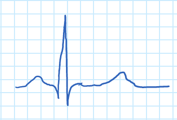
אלקטרוקרדיוגרפיה היא גרף המתועד על ידי שינויים פוטנציאליים חשמליים המתרחשים בין אלקטרודות שהונחו על פלג פלג עליון של המטופל כדי להדגים פעילות לבבית. אות אק"ג עוקב אחר קצב הלב ומחלות לב רבות, כגון זרימת דם לקויה ללב וחריגות מבניות. פוטנציאל הפעולה שנוצר על ידי התכווצויות של דופן הלב פורש זרמים חשמליים מהלב בכל הגוף. הזרמים החשמליים המתפשטים יוצרים פוטנציאלים שונים בנקודות בגוף, אשר ניתן לחוש על ידי אלקטרודות המונחות על העור. האלקטרודות הן מתמרים ביולוגיים העשויים ממתכות ומלחים. בפועל, 10 אלקטרודות מחוברות לנקודות שונות על הגוף. קיים הליך סטנדרטי לרכישה וניתוח אותות אק"ג. גל אק"ג טיפוסי של אדם בריא הוא כדלקמן:

איור 1. גל אק"ג.
גל ה-P מתאים להתכווצות האיזורים, ולמתחם ה-QRS להתכווצות החדרים. מתחם ה-QRS גדול בהרבה מגל ה-P בשל ההצטמרות היחסית במסת השריר של האטריה והחדרים, המסווה את ההרפיה של האטריה. הרפיה של החדרים ניתן לראות בצורה של גל "T".
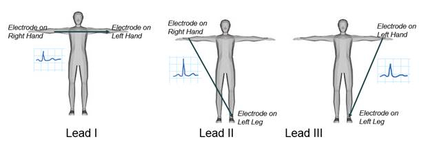
ישנם שלושה כיווני חקירה עיקריים האחראים למדידת ההבדל הפוטנציאלי החשמלי בין הידיים לרגליים, כפי שמוצג באיור 2. בהדגמה זו, אחד ממובילי הגפיים, עופרת I, ייבדק, וההבדל הפוטנציאלי החשמלי בין שתי זרועות יירשם. כמו בכל מדידות עופרת אק"ג, האלקטרודה המחוברת לרגל ימין נחשבת לצומת הקרקע. אות אק"ג יירכש באמצעות מגבר ביופוטנציאלי ולאחר מכן יוצג באמצעות תוכנת מכשור, שם תיווצר בקרת רווח כדי להתאים את המשרעת שלה. לבסוף, האק"ג המוקלט ינותח.

איור 2. עופרת גפיים א.ק.ג.
Principles
האלקטרוקרדיוגרפיה חייבת להיות מסוגלת לזהות לא רק אותות חלשים מאוד הנעים בין 0.5 mV ל 5.0 mV, אלא גם רכיב DC של עד ±300 mV (הנובע ממגע האלקטרודה-עור) ורכיב במצב משותף של עד 1.5 V, הנובע מהפוטנציאל בין האלקטרודות לקרקע. רוחב הפס השימושי של אות אק"ג תלוי ביישום ויכול לנוע בין 0.5-100 הרץ, לפעמים להגיע עד 1 kHz. זה בדרך כלל סביב 1 mV שיא לשיא בנוכחות רעש הרבה יותר גדול בתדר גבוה חיצוני, הפרעות 50 או 60 הרץ, ופוטנציאל היסט אלקטרודה DC. מקורות רעש אחרים כוללים תנועה המשפיעה על ממשק האלקטרודה-עור, התכווצויות שרירים או קוצים אלקטרומיוגרפיים, נשימה (שעשויה להיות קצבית או לא סדירה), הפרעה אלקטרומגנטית (EMI) ורעש ממכשירים אלקטרוניים אחרים המצמידים לקלט.
ראשית, מגבר ביופוטנציאלי יופק לעיבוד האק"ג. לאחר מכן, אלקטרודות יונחו על המטופל כדי למדוד את ההבדל הפוטנציאלי בין שתי זרועות. הפונקציה העיקרית של מגבר ביופוטנציאלי היא לקחת אות חשמלי חלש ממוצא ביולוגי ולהגדיל את המשרעת שלו, כך שניתן יהיה לעבד אותו עוד יותר, להקליט או להציג אותו.

איור 3. מגבר אק"ג.
כדי להיות שימושי מבחינה ביולוגית, כל המגברים הביופוטנטיים חייבים לעמוד בדרישות בסיסיות מסוימות:
- הם חייבים להיות בעליעכבת קלט גבוהה, כך שהם מספקים טעינה מינימלית של האות הנמדד. אלקטרודות ביופוטנציאליות יכולות להיות מושפעות מהעומס שלהן, מה שמוביל לעיוות האות.
- מעגל הכניסה של מגבר ביופוטנציאלי חייב גם לספק הגנה לנושא הנחקר. המגבר צריך להיות מעגלי בידוד והגנה, כך הזרם דרך מעגל האלקטרודה ניתן לשמור ברמות בטוחות.
- מעגל הפלט מניע את העומס, שהוא בדרך כלל התקן מציין או הקלטה. כדי להשיג נאמנות מרבית וטווח בקריאה, המגבר חייב להיות בעל עכבה פלט נמוכה ולהיות מסוגל לספק את הכוח הנדרש על ידי העומס.
- מגברים ביופוטנטיים חייבים לפעול בספקטרום התדרים שבו קיימים הביופוטנציאלים שהם מגבירים. בגלל הרמה הנמוכה של אותות כאלה, חשוב להגביל את רוחב הפס של המגבר כדי לקבל יחס אות לרעש אופטימלי. ניתן לעשות זאת באמצעות מסננים.
איור 3 הוא דוגמה למגבר א.ק.ג. איור 4 הוא המעגל של מגבר האק"ג שנבנה במהלך הדגמה זו. יש לו שלושה שלבים עיקריים: מעגל ההגנה, מגבר המכשור ומסנן המעבר הגבוה.

איור 4. מגבר ביופוטנציאלי.
השלב הראשון הוא מעגלי ההגנה על המטופלים. דיודה היא התקן מוליכים למחצה המוליך זרם בכיוון אחד. כאשר הדיודה מוטה קדימה, הדיודה פועלת כקצר חשמלי ומוליך חשמל. כאשר דיודה מוטה לאחור, היא פועלת כמעגל פתוח ואינה מוליכת חשמל,אני ≈ 0.
כאשר דיודות נמצאות בתצורה מוטה קדימה יש מתח המכונה מתח הסף (VT = כ 0.7 V) כי יש לחרוג על מנת הדיודה לנהל זרם. לאחר VT כבר חריגה, ירידת המתח על פני הדיודה יישאר קבוע בVT ללא קשר מה Vהוא.
כאשר הדיודה מוטה לאחור הדיודה תפעל כמו במעגל פתוח וירידת המתח על פני הדיודה תהיה שווה ל- Vב.
איור 5 הוא דוגמה למעגל הגנה פשוט המבוסס על דיודות שישמשו בהדגמה זו. הנגד משמש להגבלת הזרם הזורם דרך המטופל. אם תקלה במגבר המכשור או בדיודות מקצרת את החיבור של המטופל לאחת מפסי החשמל, הזרם יהיה פחות מ-0.11 מיליאמפר-אם-איי. דיודות הדליפה הנמוכה FDH333 משמשות להגנה על התשומות של מגבר המכשור. בכל פעם שהמתח במעגל עולה על 0.8 V בסדר גודל, הדיודות משתנות לאזור הפעיל שלהן או למצב "ON"; הזרם זורם דרכם ומגן הן על המטופל והן על הרכיבים האלקטרוניים.

איור 5. מעגל הגנה.
השלב השני הוא מגבר המכשור, IA, המשתמש בשלושה אמפרים תפעוליים (op-amp). יש מגבר אופ אחד המחובר לכל קלט כדי להגביר את התנגדות הקלט. המגבר השלישי הוא מגבר דיפרנציאלי. לתצורה זו יש את היכולת לדחות הפרעות המופנות לקרקע ורק להגביר את ההבדל בין אותות הקלט.

איור 6. מגבר מכשור.
השלב השלישי הוא מסנן המעבר הגבוה, המשמש להגברת מתח AC קטן שרוכב על גבי מתח DC גדול. האק"ג מושפע מאותות בתדר נמוך המגיעים מתנועת המטופלים ומנשימה. מסנן מעבר גבוה מפחית רעש זה.
מסנני מעבר גבוהים יכולים להתממש עם מעגלי RC מסדר ראשון. איור 7 מציג דוגמה למסנן מעבר גבוה מסדר ראשון ולפונקציית ההעברה שלו. תדירות הניתוק ניתנת על-ידי הנוסחה הבאה:
 ,
,


איור 7. מסנן מעבר גבוה.
Procedure
1. רכישת אות אק"ג
- התאם את המתח של המקורות ל+ 5 V ו- -5 V וחבר אותם בסדרה.
- בנו את המעגל המוצג באיור 4. חשב את הערכים של הנגדים והחיברים. עבור מסנן המעבר הגבוה, תדירות הניתוק צריכה להיות 0.5 הרץ. יש לבחור את ערך הקבל מהטבלה שלהלן (בכפוף לזמינות).
| ערכי קבלים זמינים (μF ) | ||
| 0.001 | 1 | 100 |
| 0.022 | 2.2 | שנות ה-12 |
| 0.047 | 4.7 | 470 |
| 0.01 | 10 | 1000 |
| 0.1 | 47 | 2200 |

- מניחים אלקטרודות על זרוע ימין, זרוע שמאל ורגל ימין (זוהי התייחסות) של המטופל, וחברו אותם למעגל.
- השתמש באוסצילוסקופ כדי להציג את אות האק"ג (Vo). הקש על הגדר אוטומטית והתאם את קנה המידה האופקי והאנכי לפי הצורך. אתה אמור להיות מסוגל לראות את פסגות R למרות הרעש באות.
2. הצגת אות האק"ג באמצעות תוכנת מכשור
- בהדגמה זו השתמשנו ב- LabVIEW. כתוב תוכנית המציגה את אות האק"ג באמצעות ממשק גרפי לתצורת מדידות וגרף צורת גל. לאחר בחירת קלט אנלוגי, קבע את תצורת התוכנית עם ההגדרות הבאות:
- טווח קלט אותות >> מקסימום = 0.5; מינימום = -0.5
- >> תצורת מסוף RSE
- מצב רכישה >> רציף
- דוגמאות לקריאה = 2000
- קצב דגימה = 1000
- לרכוש את אות האק"ג ולהתבונן צורת הגל. תראה אות דומה לאיור 1.
- כוונן את קנה המידה של ציר ה- x כדי להציג זמן בשניות.
- לעתים קרובות יש צורך מכשור כדי להגביר את האות של עניין משרעת ספציפית. צור בקרת רווח והגדר אותה כך שהמשרעת של האק"ג היא 2 סמנכ"לים.
3. ניתוח אות האק"ג
בסעיף זה, אות אק"ג יעבור סינון וניתוח כדי לקבוע את קצב הלב. דיאגרמת החסימה הבאה מציגה את רכיבי התוכנית.
- השתמש בגרף צורת גל כדי להציג את האות.
- להעריך את הספקטרום של האות באמצעות משרעת ואת ספקטרום הפאזה subvi (בעיבוד אותות → ספקטרלי) ולהציג את גודלו באמצעות גרף צורת גל. הציר האופקי תואם לתדר. הוא דיסקרטי מכיוון שהמחשב משתמש באלגוריתם של שינוי פורייה מהיר (FFT) כדי לחשב את הספקטרום של האות. התדירות עוברת מ- k = 0 ל- k = (N-1)/2, כאשר N הוא אורך הרצף, במקרה זה 4000. כדי לחשב את התדירות האנלוגית המתאימה, השתמש בנוסחה הבאה:

כאשר fs הוא תדר הדגימה. שים לב שרוב האנרגיה של האות היא בטווח התדרים הנמוכים וגם שיש שיא של עוצמה גבוהה בטווח התדרים הבינוני. חשב את התדירות של פסגה זו באמצעות הנוסחה שסופקה לעיל. - ליישם מסנן נמוך לעבור באמצעות באטרוורת ' של פונקציות Chebyshev. בחר תדירות חיתוך השווה ל- 100 הרץ. ודא שהמסנן מספק נכה של לפחות -60 dB/עשור בפס העצירה.
- חבר את אות הפלט של הקריאה מ- subvi גיליון אלקטרוני לקלט של מסנן המעבר הנמוך.
- יש ליישם מסנן סטופ-פס באמצעות פונקציות באטרוורת' או צ'בישב. המטרה היא להפחית את ההפרעה של 60 הרץ מבלי לשנות את התדרים האחרים. נסה תדרי גבול קרוב ל- 60 הרץ.
- חבר את הפלט של מסנן lowpass לקלט של מסנן פס העצירה.
- מצא את הפסגות באמצעות subvi גלאי שיא (הוא ממוקם עיבוד אותות → פעולת סיג). עבור הסף, הסתכלו על משרעת האות ובחרו את הערך המתאים ביותר.
- חלץ את מיקומי הפסגות באמצעות subvi מערך האינדקס (במערך → תכנות).
- הפחת את המיקום התחתון ממיקום גבוה יותר ולאחר מכן, הכפל בתקופת הדגימה T = 1/fs כדי להשיג את מרווח הזמן של RR.
- חשב את יחידות ההדדיות והתאם וצב מחוון כדי להציג את ה- BPM.
אלקטרוקרדיוגרפים מתעדים פעילות לבבית של הלב ומשמשים לאבחון מחלות, לזיהוי חריגות וללמוד על תפקוד הלב הכללי. אותות חשמליים מיוצרים על ידי התכווצויות בקירות הלב המניעות זרמים חשמליים ויוצרות פוטנציאלים שונים בכל הגוף. על ידי הצבת אלקטרודות על העור, ניתן לזהות ולתעד פעילות חשמלית זו בא.ק.ג. ECGs אינם פולשניים, מה שהופך אותם לכלי שימושי כדי להעריך כמה טוב הלב המטופל מתפקד, כגון על ידי מדידת כמה טוב הדם זורם לאיבר.
וידאו זה ימחיש את המנהלים של ECGs וידגים כיצד לרכוש, לעבד ולנתח אות אק"ג טיפוסי באמצעות מגבר ביופוטנציאלי. יישומים ביו-רפואיים אחרים המשתמשים בעיבוד אותות חשמליים לאבחון מחלות יידונו גם הם.
כדי להבין את העקרונות של אק"ג, בואו נבין תחילה כיצד הלב מייצר אותות חשמליים. עבור לב נורמלי ובריא, במנוחה, אק"ג מציג סדרה של גלים המשקפים את השלבים השונים של פעימת לב. האק"ג מתחיל בצומת sinoatrial, הידוע גם בשם צומת SA, אשר ממוקם באטריום הנכון ופועל כקוצב לב בלב. האותות החשמליים גורמים התכווצות של סכסוכים המכריחים דם להיכנס לחדרים. רצף זה נרשם כגל P על האק"ג. אות זה עובר מן atria על פני החדרים, גורם להם להתכווץ ולשאוב דם לשאר הגוף. זה נרשם כמתחם QRS.
לבסוף, החדרים להירגע וזה נרשם כמו גל T. לאחר מכן התהליך מתחיל שוב והוא חוזר על עצמו על כל פעימת לב. שימו לב שגל ה-QRS גדול בהרבה מגל ה-P, הסיבה לכך היא שהחדרים גדולים יותר מהאטריה. כלומר הם מסווים את ההרפיה של אטריה או גל T. תהליכים אחרים בגוף, כמו נשימה או התכווצויות שרירים, יכולים להפריע למדידת האק"ג. כמו גם זרמים מהמעגלים המשמשים להשגתם. לעתים קרובות, האותות החשמליים כי האק"ג מנסה להקליט הם חלשים למדי. לכן, מגבר ביופוטנציאלי משמש כדי להגדיל את המשרעת שלהם אשר מאפשר להם להיות מעובד נוסף ורשם.
ישנם שלושה מרכיבים עיקריים למגבר הביופוטנטי, לשלב ההגנה על המטופל, למגבר המכשור ולמסנן המעבר הגבוה. כפי שהראשית מרמזת, מעגל ההגנה על המטופל משתמש בשילוב של נגדים ודיודות כדי להגן, הן על המטופל והן על המכונה והציוד. הנגדים מגבילים את הזרם הזורם דרך המטופל, שם כמו הדיודות לשמור על הזרם זורם בכיוון הנכון.
השלב הבא הוא מגבר המכשור, אשר מגביר את ההבדל בין התשומות מכל אלקטרודה. הוא מורכב משלושה אמפרים תפעוליים. שניים כדי להגביר את ההתנגדות מכל קלט, והשלישי כדי להגביר את ההבדל בין אותות הקלט.
השלב האחרון הוא מסנן המעבר הגבוה, אשר מפחית את הרעש ומסנן אותות בתדר נמוך הנובעים מתנועת המטופל או מהנשימה. עכשיו שאתם יודעים איך נמדד אק"ג, בואו נראה איך לבנות מגבר ביופוטנציאלי ולעבד את הנתונים כדי לקבל אות אק"ג נקי.
לאחר שסקרנו את המנהלים העיקריים של אלקטרוקרדיוגרפיה, בואו נראה כיצד לבנות מגבר ביופוטנציאלי ולרכוש אות אק"ג. ראשית, אסוף תחילה לוח פרוטו, מגבר מכשור AD-620 וכל רכיבי המעגל הדרושים. לאחר מכן, חשב את הערכים של כל הנגדים והחיברים במעגל באמצעות המשוואה הבאה.
עבור מסנן המעבר הגבוה, תדירות הניתוק צריכה להיות 0.5 הרץ.
לאחר מכן, חבר את ערך הקבל כדי לקבוע את ההתנגדות. לאחר מכן, בנה מגבר ביופוטנציאלי בהתאם לדיאגרמה שסופקה. כך המעגל הסופי אמור להיראות. חבר שלושה חוטים עם קליפים תנין לעמדות הכריכה של ספק כוח DC ולאחר מכן הפעל את מקור הכוח. התאימו את המתח לפלוס חמישה וולט ומינוס חמישה וולט, וחברו את החוטים, בסדרה, למעגל.
עכשיו, להשתמש כרית הכנה לאלכוהול כדי לנגב את פרק כף היד הימנית של המטופלים, פרק כף היד השמאלית, ואת הקרסול הימני. הוסף ג'ל דבק מוליך לאלקטרודות לפני הנחתם על המטופל. לאחר מכן, חבר את האלקטרודות למעגל באמצעות חוטים עם קליפסים תנין. הפעל את האוסצילוסקופ ותשיג את אות האק"ג. התאם את הסולמות האופקיים והאנכיים לפי הצורך. עם התאמות אלה, אתה אמור להיות מסוגל לראות את פסגת R של צורת הגל.
חבר את המעגל למארז PXI, לאחר מכן פתח את תוכנת המכשור ו, או, השתמש או כתוב תוכנית שתציג את אות האק"ג וגרף טופס גל.
קבע את תצורת ממשק רכישת הנתונים עם ההגדרות הבאות. סמן את קנה המידה של ציר ה- x כדי להציג זמן ושניות ולאחר מכן הצג את אות האק"ג כצורת גל. אם יש להגביר את האות, צור שליטה על רווח והגדר אותו כך שהמשרעת של האק"ג תהיה שני סמנכ"לים.
עכשיו שהדגמנו כיצד לרכוש אות אק"ג, בואו נראה כיצד לנתח את התוצאות. הנה אות א.ק.ג. מייצג. גלי P, QRS ו- T בקושי ניתנים לגילוי מכיוון שהם מוסתרים על ידי רעש ותנודות. יש לסנן אות זה. כדי לשנות אות זה, תחילה, בחר עיבוד אותות ולאחר מכן ספקטרלי בתפריט. אלגוריתם של שינוי צורה מהיר של Fourier מחשב ומתווה את הספקטרום של האות המציג את התדירות כערכים דיסקרטיים בציר האופקי. רוב האנרגיה באות היא בתדרים נמוכים.
אבל, יש שיא בעוצמה גבוהה בטווח התדרים הבינוני, אשר מניח להיות רעש. התדירות מתווכת כ- k על הציר האופקי ומעברת מאפס ל- N מינוס אחד על פני שניים, כאשר N הוא אורך הרצף. לניסוי זה, N שווה 2,000. חשב את התדירות האנלוגית עבור כל ערך k באמצעות המשוואה הבאה, כאשר f s הוא תדר הדגימה וקבע את התדירות של שיא העוצמה הגבוהה בהתבסס על גרף FFT.
לאחר מכן, צור מסנן מעבר נמוך עם תדר ניתוק של 100 הרץ. השתמש, או, פונקציית באטרוורת 'או Chebyshev כדי לסנן את האות, אשר אמור להקליש לפחות 60 דציבלים לעשור ברצועת העצירה. חבר את אות הפלט של תת הנתונים VI לקלט של מסנן המעבר הנמוך. מסנן זה מסיר את גלי התדר הגבוהים הזרים של האק"ג. עכשיו, ליצור מסנן Bandstop ולהגדיר את תדרי הניתוק בסביבות 55 ו 70 הרץ.
כדי להסיר את האות הרועש, סביב 60 הרץ. לאחר מכן, חבר את הפלט של מסנן המעבר הנמוך לקלט של מסנן Bandstop. נסה תדרי גבול שקרובים ל-60 הרץ. פעולה זו תפחית הפרעות מבלי להשפיע על תדרים אחרים. אות האק"ג צריך להיות ברור כעת עם מתחמי P, QRS ו- T ברורים.
עכשיו, בואו לקבוע את הדופק באמצעות אות אק"ג מסונן. ראשית, השתמש בגלאי השיא תת-6 כדי למצוא את פסגות האות. בחר את הערך המתאים ביותר בהתבסס על משרעת האותות של גל R עבור הסף. לאחר מכן, השתמש בתת ה- VI של מערך האינדקסים כדי לקבוע את מיקום הפסגות.
הפחת את מיקום הפסגה התחתון מהמיקום הגבוה יותר ולאחר מכן הכפל ערך זה על-ידי תקופת הדגימה, T, השווה ל- one מעל f s. ערך זה הוא משך הזמן בין שני גלי R. התאם את היחידות כדי לקבוע את הפעימות לדקה.
בהדגמה זו, קצב הלב הנמדד היה כ-60 פעימות בדקה.
א.ק.ג. ועיבוד אותות יש יישומים חשובים, הן ברפואה והן במחקר. בנוסף להיותו לא פולשני, ECGs הם זולים יחסית. מה שהופך אותו לכלי שימושי ונגיש בבתי חולים. ECGs יכול אפילו להיות מותאם למעקב מורכב וארוך טווח יותר של חולים המטופלים לתסמונת כלילית חריפה.
בשביל זה, 12 הפניות אק"ג משמשים, אשר יכול לזהות איסכמיה שריר הלב חולף בחולים אסימפטומטיים. דגימת אותות ועיבוד משמשים גם באלקטרואנצפלוגרפיה למדידת אותות חשמליים מהמוח. EEGs משמשים בדרך כלל בשילוב עם MRI פונקציונלי כאכניקת הדמיה רב-מודאלית.
השיטה מייצרת באופן לא פולשני מפות קליפת המוח של פעילות המוח עבור יישומים רבים של דימות מוחי, כגון לאחר הפעלה חזותית או מוטורית.
הרגע צפית בהקדמה של יובה לרכישה וניתוח של אותות אק"ג. עכשיו אתה צריך להבין איך אות אק"ג מיוצר וכיצד ליצור מגבר ביופוטנציאלי כדי לזהות אותות חשמליים חלשים. ראית גם כמה יישומים ביו-רפואיים של עיבוד אותות לאבחון רפואי.
תודה שצפיתם.
Results
בהדגמה זו, שלוש אלקטרודות היו מחוברות לאדם, והתפוקה עברה דרך מגבר ביופוטנציאלי. גרף אק"ג לדוגמה לפני הסינון הדיגיטלי מוצג להלן (איור 8).

איור 8. אות אק"ג ללא סינון דיגיטלי.
לאחר עיצוב המסננים והזנת הנתונים לאלגוריתם המפותח, זוהו הפסגות בגרף ושימשו לחישוב קצב פעימות הלב (BPM). איור 9 מציג את הנתונים הגולמיים אות אק"ג (לפני כל סינון) בתחום הזמן והתדירות. איור 10 מציג את התוצאה של סינון האות.

איור 9. אות אק"ג לפני הסינון.

איור 10. אות אק"ג מסונן.
בחלקת האק"ג המקורית היו מתחמי P, QRS ו- T גלויים מעט שהציגו תנודות רבות מהרעש. הספקטרום של אות האק"ג הראה גם ספייק ברור ב 65 הרץ, אשר נחשב רעש. כאשר האות עובד באמצעות מסנן עם מעבר נמוך כדי להסיר חלקים בתדר גבוה זרים ולאחר מכן מסנן עצירת רצועה כדי להסיר את רכיב האות 65 הרץ, הפלט נראה נקי יותר באופן משמעותי. האק"ג מראה כל רכיב של האות בבירור עם כל הרעש הוסר.
בנוסף, קצב הלב הנמדד היה כ 61.8609 פעימות לדקה.
Applications and Summary
התכווצות שריר הלב במהלך מחזור הלב מייצרת זרמים חשמליים בתוך בית החזה. ירידות מתח על פני רקמה התנגדותית מזוהות על ידי אלקטרודות המונחות על העור ומוקלטות על ידי אלקטרוקרדיוגרפיה. מאז המתח חלש, בטווח של 0.5 mV, וקטן בהשוואה לעוצמת הרעש, עיבוד וסינון האות יש צורך. בניסוי זה, התקן אלקטרוקרדיוגרפיה המורכב ממעגל עיבוד אותות אנלוגי ודיגיטלי של שני חלקים תוכנן לנתח את אות האק"ג שנוצר, ולחשב את קצב פעימות הלב.
הדגמה זו הציגה את היסודות של מעגלים אלקטרוניים וסינון של אותות אק"ג. כאן, טכניקות עיבוד אותות מעשיות שימשו לחילוץ אות חלש מרקע רועש. טכניקות אלה ניתן להשתמש ביישומים דומים אחרים שבהם הגברת אותות והפחתת רעש נדרש.
רשימת חומרים
| שם | חברה | מספר קטלוג | הערות |
| ציוד | |||
| ספק כוח | דיוק B&K | 1760A | |
| ריבוי מטר | |||
| אוסצילוסקופ | |||
| לוח פרוטו | |||
| 4 דיודות FDH333 | |||
| 1 לספירה620 | |||
| 3 נגדΩ 47k | |||
| 2 קבלי 100nF | |||
| 3 אלקטרודות אק"ג | |||
| כמה קליפים תנין וגשוש Tektronix. |


