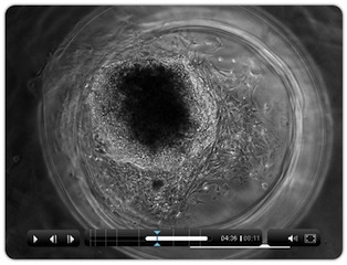
Electronic Nose Knows When a Cantaloupe is Ripe

Have you ever been disappointed by a cantaloupe from the grocery store? Too ripe? Not ripe enough? Luckily for you, researchers from the University of California, Davis might have found a way to make imperfectly ripe fruit a thing of the past.

“We are involved in a project geared towards developing rapid methods to evaluate ripeness and flavor of fruits,” said paper-author Dr. Florence Negre-Zkharov. “We evaluated an electronic nose to see it it can differentiate maturity of fruit, specifically melons. The goal is to develop a tool that can be used post-harvest to better evaluate produce, and develop better breeds.”

When fruit ripens, it develops a characteristic volatile blend, indicating its maturity. Traditionally, the gold-standard of evaluating these volatiles has been gas chromatography, but it takes up to an hour to analyze a single sample, which makes it impractical to use outside the lab. Dr. Negre-Zakharov and her team wanted to determine if the much cruder— but much faster— electronic nose was able to determine if the melon they used in the experiment were ripe. It was.

Share This Article

Twitter facebook linkedin

Eye-Tracking in Young Children with Autism

Though the prevalence of autism spectrum disorder (ASD) has been steadily climbing — from 6 in 1,000 children in 2002, to nearly 10 in 1,000 children in 2006, according to the Centers for Disease Control and Prevention —  little is known about the disorder. But, research with young kids can lead to important insights in how children with developmental abnormalities view the world. This month in JoVE, researchers demonstrate how to use eye-tracking in very young children with autism.

“Generally, individuals new to this method often struggle, as eye-tracking young children with autism involves unique challenges that are not present when tracking typically-developing older children or adult population,” said paper-author Dr. Noah Sasson.

Eye-tracking is one of the few quantifiable ways to study children with autism spectrum disorder. It requires that children look at pictures on a screen— in this study, objects and faces— and the eye-tracking technology can record where a child is looking, when.

Dr. Sasson explains that though researchers have known for a long time about the social impairment that comes with autism, they are not sure if young children with autism are actually looking at faces differently, or ignoring faces all together.

To help other scientists who are interested in answering this question, Dr. Sasson published his methodology in JoVE. According to JoVE Editor Leiam Colbert, the lack of standardization in the field is a real problem.

“There are challenges with research of this kind— both from the methodological and clinical perspectives. Less experienced researchers may or may not be aware of the difficulties inherent in eye-tracking children with autism. The need for standardization of this type is great, to prevent the publication of spurious results, or wasting scare resource funds and participant time.”

The video-article shows how Dr. Sasson sets up the lab with few distractions, so the children participating are more likely to focus on the eye-tracking screen and how cartoons and moving images with sounds are useful ways to get children to refocus their attention on the screen.

“I think visually seeing the lab, visually seeing the child and how to conduct the test is important,” said Dr. Sasson. “I think this will be a very helpful resource.”

To watch the full video-article, please click here

Share This Article

Twitter facebook linkedin

JoVE Judo in Science NOW

On Tuesday, JoVE published a fascinating article on how energy is used during complex sports, such as judo and other martial arts. Previous research on exercise science has focused on sports that can be easily recreated in the lab, such as running and cycling, but these Brazilian scientists have found a completely unique way of studying more complex sports.

Science NOW picked up the story and did an excellent job of explaining how the researchers did it:

The Science of Judo

Martial arts are exhausting, as anyone who’s traded a few punches, kicks, or throws can attest. But where exactly does the energy come from? Every form of exercise uses a different combination of the body’s metabolic systems for energy. Cyclical sports such as running and cycling are relatively easy to replicate with exercise machines in a laboratory, but that’s harder to do with more unpredictable sports such as martial arts. So a team of Brazilian researchers have taken the lab into the dojo to study the energy requirements of the Japanese art of judo.

Three different systems convert food to energy. During long periods of moderate exercise, aerobic metabolism does most of the work, using oxygen to turn sugar into energy, water, and CO2. Running a marathon or cycling for 100 miles, therefore, is almost entirely aerobic.

For shorter, more intense exertion, or when the oxygen runs out, muscles can break down sugar anaerobically, although that system is far less efficient and produces muscle-burning lactic acid as a byproduct. Lastly, for very short bursts of energy, such as a 10-second sprint, muscles can rely on another type of anaerobic system: they use up energy-storing compounds, called phosphagens, in muscular tissues.

Share This Article

Twitter facebook linkedin

Scientists Measure How Energy is Spent in Martial Arts

Two judo fighters face off, one in a white judogi (the traditional judo uniform) and one in blue. They reach for each other’s shoulders and lock arms, in what looks like an awkard dance, before the fighter in blue throws his opponent head-over-feet onto the mat.

Judo and mixed martial arts have become increasingly popular over the past few years and scientists have taken note. The two fighters were actually filmed as part of a science experiment that demonstrates how researchers can quantify exactly how the athletes are spending their energy. The video will be published in JoVE today.

Previously, researchers have only been able to study predictable sports that are easy to replicate in the laboratory, such as running. With this new method, scientists will be able to study the team and individual sports that have previously been neglected.

“Each sport has specific characteristics which confer different metabolic demands to them,” said paper-author Dr. Emerson Franchini. “One of the most important aspects of the metabolic demand is the relative contribution of the energy systems.”

Share This Article

Twitter facebook linkedin

Converting Standard InkJet Printer Into Cell Printing Machine

Researchers from Clemson University have found a way to create temporary holes in the membranes of live cells using a standard inkjet printer.

“We first had the idea for this method when we wanted to be able to visualize changes in the cytoskeleton arrangement due to applied forces on cells,” said paper-author Dr. Delphine Dean.

A fibroblast printed with the modified inkjet printer. The interior of the cell shows that the fluorescently tagged actin monomers have been incorporated.

She said other researchers have been using this method to print cells onto slide, but that they have only recently discovered that printing the cells causes the disruption in the their membranes for a few hours. Creating temporary pores allows researchers to put molecules inside of cells that wouldn’t otherwise fit, and study how the cells react.

“The authors have used an extremely innovative approach for bioprinting cells. Moreover, this approach can be used for applications other than cells printing.” said JoVE Science Editor Dr. Nandita Singh. “Matrix proteins can be printed onto substrates with this technique for cell patterning.”

The printer is modified by removing the paperfeed mechanism and adding a “stage” from which to feed the slides. The ink is replaced with a cell solution, and the cells are printed directly on to the slides.

Using this method, the researchers are able to process thousands of cells in a matter of minutes. Dr. Dean’s team used the holes to introduce fluorescent molecules that illuminate the skeleton of the cell.

“We are actually interested in the cell mechanics of compressed cells. This method allows us to push on the cells and watch the response easily,” said Dr. Dean. “We are interested in cardiovascular cells, and how they respond to mechanical force.”

Dr. Dean chose to submit her method to JoVE, the only peer reviewed, PubMed-indexed science journal to publish all of its content in both text and video format, because, she said, “until you’ve seen it done, it’s hard to understand the process.”

To see the full article, please click here.

Share This Article

Twitter facebook linkedin

Article Shows how to Open the Brain to New Types of Treatments

One of the trickiest parts of treating brain conditions is the blood brain barrier, a blockade of cells that prevent both harmful toxins and helpful pharmaceuticals from getting to the body’s control center. But, a new video shows how an MRI machine can guide the use of microbubbles and focused ultrasound to help drugs enter the brain, which may open new treatment avenues for devastating conditions like Alzheimer’s and brain cancers.

“It’s getting close to the point where this could be done safely in humans,” said paper-author Meaghan O’Reilly, “there is a push towards applications.”

The current method of disrupting the blood-brain barrier (BBB) is by using osmotic agents such as mannitol, which suck the water out of the cells that form the barrier, causing the gaps between them to get bigger. Unfortunately, this method opens large areas of the barrier, leaving the brain exposed to toxins.

The benefit of the microbubble technique is that it can be used on a very small area of the BBB. The microbubbles, made of lipids (fats) and gas, are injected into the blood stream. When focused ultrasound is applied, the bubbles expand and contract. It is thought that the force of the movement in the bubbles causes the cells that form the BBB to temporarily separate, which allows drugs to reach the brain.

“Microbubble technology has been around for years, though its applications have mostly been as contrast agents for diagnostic ultrasound,” said Editorial Director, Dr. Beth Hovey. “This newer approach, using ultrasound to help the bubbles permeablize the blood brain barrier, will hopefully allow for better treatment of diseases within the brain.”

In this method, O’Reilly and her colleagues use the MRI machine to ensure that the barrier opens, and they can also time how long it takes for it to close, which will be important for when the technique is used one patients,

O’Reilly chose to publish the technique in JoVE to help other scientists learn the method.

“The ability of focused ultrasound combined with microbubbles to disrupt the blood brain barrier has been known for over a decade. However, because the actual technique can be challenging— there are critical steps involved— the video article fills a gap in the literature that is a major hinderance to people getting into the field,” she said.

To see the full article, please click here).

Share This Article

Twitter facebook linkedin

Three Awesome Female Scientists

In honor of International Women’s Day, here is a list of three video articles from some of our super-star female authors:

Dr. Catarina Hioe: Taking Down HIV

Dr. Hioe, Professor of Pathology at New York University, is working to develop an HIV vaccine and published a piece of the puzzle in JoVE today. The article demonstrates a method of visualizing what takes place during the formation of the virological synapse, which enables cell-to-cell transfer of the virus.

Dr. Kakani Katija: Studying the Movement of Jellyfish in the Ocean

Dr. Katija is a bioengineer, and was named an Emerging Explorer by National Geographic for her work on ocean organisms. Her quote in the article sums up her work perfectly: “The creatures in our seas could be as important to ocean circulation and global climate as the winds and tides. Learning more about this animal-ocean dynamic is critical to the future.”

Dr. Laura Niklason: Engineering Lungs

Dr. Niklason is working on engineering lungs, and published a method to do just that in rats. No need to explain why that is awesome!


Share This Article

Twitter facebook linkedin

How to Culture Stem Cells in 3D

Stem cells are the body’s mechanics, repairing damaged tissues and organs. Because these cells are able to grow into any type of cell in the body, scientists believe they hold the key to groundbreaking new therapies. To help further this research, scientists from the University of Victoria demonstrate a new way to culture stem cells in 3D.

Example of a mouse induced pluripotent stem cell after three days of culture inside of a 3D fibrin scaffold. Cells have begun to migrate and differentiate inside of the fibrin scaffold.

“Cells in your body grow and divide in a 3D environment, especially when you think of stem cells, which differentiate to become all the different types of cells in your body,” said one of the authors, Dr. Stephanie Willerth. “Yet, a lot of work is still being done in a 2D environment.”

Essentially, since your body is three-dimensional, it makes sense that stem cells do their best repair work in 3D as well. By growing these cells in 3D, researchers are better able to see how these cells behave in conditions that more closely resemble those in the body.

Though Dr. Willerth’s lab specifically looks at neurons to investigate neural regeneration in the spinal cord, she chose to publish her research in JoVE because she believes her culture method is very versatile, and can be applied to other research in regenerative medicine.

“When you’re working with gels, it’s nice to have a visual representation,” she said.

To watch the video, please click here.

Share This Article

Twitter facebook linkedin