

The study of brain activity has advanced tremendously since the beginning of neurological research. A recent JoVE study from the University College London and the University of Chieti-Pescara illustrates how wearable fNIRS technology may bring that research out of the lab and into the real world.
In the last century, one of the most studied organs in human anatomy has certainly been the brain. The reason why scientists are fascinated by the brain is not only because of its complexity, but also because it is considered the biological substrate of the mind. Studying the brain, in fact, is the only way to understand how cognitive functions such as: memory, perception, attention, language, and emotions work. All these functions working together ultimately represent the identity of each of us.
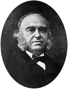 The relation between the brain and cognitive functions is a relatively new concept. In 1861, a French neurologist, Paul Broca observed that, after a brain injury, one of his patients was able to understand language but was unable to speak. Post-mortem analysis showed that the patient had a lesion in an area of his left frontal lobe – now known as Broca’s area. The discovery of the specific area responsible for language production made it possible for scientists to understand that there are specific areas of the brain responsible for the execution of specific functions.
Thanks to the development of new technologies, cognitive neuroscience together with cognitive psychology, neuropsychology, and other disciplines have tried to explain how the brain works by investigating its activity during the execution of behavioral tasks. A big jump in our understanding of the relationship between the brain and cognitive functions has been possible with the development of neuroimaging techniques. These techniques involve non-invasive tools that measure, in real time, the electrical, magnetic, or metabolic changes that occur in the brain while the subject is performing a task. For instance, if neuroimaging detects that an area is more active during a specific task rather than another one, then that area is involved in the function that is being investigated.
Numerous studies have been performed since the introduction of the neuroimaging techniques, but, although these techniques represent a powerful tool to investigate the brain activity in a non-invasive and objective way, most of these techniques (e.g., functional magnetic resonance (fMRI), magnetoencephalography (MEG), positron emission tomography (PET) require a controlled setting where the subject is required to stay still while generally performing a task on a computer monitor, which make these techniques not appropriate for everyday life situations.

The necessity of studying the brain in more ecological settings has prompted researchers to develop instruments suitable for the real world and not only for lab settings. This is the case of the wearable and wireless devices. A recent JoVE study using a fibreless, wearable functional Near Infrared Spectroscopy (fNIRS), a tool that measures changes in near-infrared light that allows researchers to monitor blood oxygenation and blood volume, was used to measure the brain activity during an ecological prospective memory task demonstrating that this system could be used to monitor brain activity during non-lab based experiments.
Researchers are still challenged by how to best study brain function in ecological settings. Although portable devices can be a powerful tool to overcome this issue, it is important to note that the use of these techniques requires a rigorous method and extra attention to the research protocol in order to avoid artifacts that could lead to misinterpretation of the results. Importantly, the visualization of these protocols also enables us to appreciate the tips and tricks associated with these techniques, thereby aiding the easy adaptation of these vital methods by researchers around the world.