زاد استخدام الخنازير في البحث في السنوات الأخيرة. ومع ذلك ، تتميز الخنازير بتشريح مجرى الهواء الصعب. من خلال توضيح كيفية إجراء التنبيب الرغامي الموجه بالمنظار ، يهدف البروتوكول الحالي إلى زيادة سلامة المختبر لتجنب معاناة الحيوانات والموت غير الضروري.
يتطلب اشتراك JoVE لعرض هذا المحتوى. تسجيل الدخول أو ابدأ نسخة تجريبية مجانية
مقالة منهجية
التنبيب الرغامي باستخدام منظار التنبيب المرن كنموذج موحد لإدارة مجرى الهواء الآمن في الخنازير
في هذه المقالة
إشعار تصحيح
Important: There has been an erratum issued for this article. View Erratum Notice
ملخص
الملخص
غالبا ما يكون التنبيب الرغامي مطلبا أساسيا للبحث الانتقالي في نماذج الخنازير للتدخلات المختلفة التي تتطلب مجرى هواء آمنا أو ضغوط تهوية عالية. التنبيب الرغامي هو مهارة صعبة ، تتطلب الحد الأدنى من عدد عمليات التنبيب الرغامي الناجحة لتحقيق معدل نجاح مرتفع في ظل الظروف المثلى ، والتي غالبا ما تكون غير قابلة للتحقيق بالنسبة للباحثين غير الخدير. بسبب تشريح مجرى الهواء الخنازير المحدد ، يمكن عادة افتراض مجرى الهواء الصعب. يمكن أن تؤدي استحالة إنشاء مجرى هوائي آمن إلى إصابة أو أحداث سلبية أو موت المختبر. باستخدام نهج تقييم مستقبلي وعشوائي ومضبوط ، فقد ثبت أن التنبيب الرغامي بمساعدة الألياف البصرية يستغرق وقتا أطول ولكن لديه معدل نجاح أعلى من التنبيب التقليدي دون التسبب في انخفاضات ذات صلة سريريا في تشبع الأكسجين. يقدم هذا النموذج نظاما موحدا للتنبيب الرغامي الموجه بالمنظار ، مما يوفر مجرى هواء آمنا ، خاصة للباحثين الذين يفتقرون إلى الخبرة في تقنية التنبيب الرغامي عن طريق تنظير الحنجرة المباشر. من المتوقع أن يقلل هذا الإجراء من معاناة الحيوانات والخسائر غير الضرورية للحيوانات.
المقدمة
غالبا ما يكون التنبيب الرغامي مطلبا أساسيا للبحث الانتقالي في نماذج الخنازير للتدخلات المختلفة التي تتطلب مجرى هواء آمنا أو ضغوط تهوية عالية (مثل التهوية أثناء الإنعاش القلبي الرئوي1 أو متلازمة الضائقة التنفسية الحادة2) أو تتطلب عدم المساس بتدفق الدم الدماغي من خلال الضغط الداخلي بواسطة أجهزة مجرى الهواء فوق المزمار3 ، والتي يتم نشرها أحيانا كبدائل في سياق مجرى الهواء الصعب المتوقع في الخنازير 4,5.
في حين أن فسيولوجيا الرئة للخنازير تظهر ميزات مماثلة لتلك الموجودة في البشر6 ، فإن تأمين مجرى الهواء يكون أحيانا أكثر صعوبةبشكل ملحوظ 7 بسبب الاختلافات المحددة في تشريح القصبة الهوائية الفموي الخنازير. خطم الخنزير له فتحة ضيقة بلسان كبير جدا ، والحنجرة متحركة للغاية ، ولسان المزمار كبير نسبيا ، مع نهاية حرة تمتد إلى الحنك الرخو. من الناحية الذيلية ، تشكل الحنجرة زاوية منفرجة مع القصبة الهوائية. الغضاريف الأريتينية كبيرة8. أضيق جزء من مجرى الهواء هو في المستوىتحت المزمار 9 ، مقارنة بتشريح مجرى الهواء للأطفال10. نظرا لأن الحنجرة في الخنازير متحركة للغاية ، فهناك خطر من أن تمر نهاية الأنبوب الرغامي عبر الحبال الصوتية ولكن لن يتم إزاحة الحنجرة إلا ذيليا بما يصل إلى عدة سنتيمترات ، والتي قد تكون مخطئة في التنبيب الصحيح 8,11. بالإضافة إلى ذلك ، يعد التنبيب المريئي خطرا شائعا عند التعامل مع إدارة مجرى الهواء الخنازير12.
لم يتم تسجيل معدلات التنبيب الرغامي الصعب أو المستحيل مع تأثير سلبي مماثل على التجربة أو الوفيات المبكرة بشكل منهجي ، ولكن تم نشر العديد من تقارير الحالات13،14. في البشر ، هناك إمكانية لاستخدام منظار التنبيب المرن في سياق التنبيب التقليدي الصعب بشكل غير متوقع15. غالبا ما تسبق عمليات التنبيب الكاذبة المختلفة هذا الإجراء. ترتبط محاولات التنبيب المتكررة هذه بالأحداث الضائرة في البشر 16,17 ، وخاصة مضاعفات مجرى الهواء18. مثل هذه الأحداث ضارة في الاختبار لأنها ، في أبسط الحالات ، تمثل متغيرا مربكا في التجربة ؛ في أسوأ الحالات ، يمكن أن تؤدي إلى خسارة غير ضرورية للحيوان.
طورت الدراسة الحالية نموذجا يعتمد على المبادئ التوجيهية لإدارة مجرى الهواء الصعب المتوقع في البشر15،19،20،21،22،23،24. في السابق ، تم وصف تقنية مماثلة لتعلم التنبيب بالألياف الضوئية في الدراسات البشرية25,26. يهدف البروتوكول المقدم في هذا التقرير إلى توفير نموذج تنبيب موحد وسهل التكيف يسمح أيضا للمتخصصين غير مجرى الهواء بإجراء تنبيب رغامي ناجح وآمن في الخنازير.
الوصول مقيد. يرجى تسجيل الدخول أو بدء فترة تجريبية لعرض هذا المحتوى.
البروتوكول
تمت الموافقة على التجارب في هذا البروتوكول من قبل لجنة رعاية الحيوان الحكومية والمؤسسية (Landesuntersuchungsamt Rheinland-Pfalz ، كوبلنز ، ألمانيا ؛ رقم الموافقة G20-1-135). أجريت التجارب وفقا لإرشادات ARRIVE. بشكل عام ، تم استخدام 10 خنازير ذكور مخدرة (Sus scrofa domestica) بمتوسط وزن 30 كجم ± 2 كجم وعمر 12-16 أسبوعا في الدراسة الحالية.
1. إعداد الحيوان
- الحفاظ على بيئة طبيعية للحيوانات لتقليل التوتر. حجب الطعام قبل 6 ساعات من التجربة المجدولة لتقليل خطر الطموح ، ولكن السماح بالوصول إلى الماء.
- قم بتخدير الخنازير بحقن مشترك من الميدازولام (0.5 مجم / كجم) والأزابيرون (2-3 مجم / كجم) (انظر جدول المواد) في عضلة الألوية أو الرقبة بإبرة (20 جم) للحقن العضلي. اترك الحيوانات دون إزعاج حتى يبدأ التخدير (15-20 دقيقة).
ملاحظة: اعتمادا على اللوائح الوطنية ، قد تخضع إدارة عوامل التخدير للتدقيق وقد تتطلب أو لا تتطلب إشراف طبيب بيطري مدرب. التشاور مع السلطات المحلية قبل التخطيط للتجارب. - نقل الحيوانات المخدرة من الاسطبلات إلى المختبر. يجب ألا يتجاوز وقت النقل وقت التخدير الكافي (هنا ، 30-60 دقيقة). تأكد من الاحتفاظ بالحرارة الكافية حتى لا يصاب الحيوان بانخفاض حرارة الجسم (أي أقل من 38 درجة مئوية) ، مثل تغطية الجسم ببطانية حسب درجة الحرارة الخارجية.
- باستخدام مستشعر (انظر جدول المواد) مثبت على الأذن أو الذيل ، راقب تشبع الأكسجين المحيطي (SpO2).
- تطهير الجلد بمطهر (كحولي) قبل إدخال قنية الوريد المحيطي (22 جم) في وريد الأذن. رش المنطقة ، امسحها مرة واحدة ، ثم رشها مرة أخرى ، واترك المطهر حتى يجف. قم بتأمين قنية الأذن باستخدام ضمادة (انظر جدول المواد).
2. التخدير والتهوية الميكانيكية
- تطبيق التسكين من خلال الحقن الوريدي بكمية 4 ميكروغرام/كغ من الفنتانيل. تحفيز التخدير بالحقن الوريدي بمقدار 3 ملغ/كغ من البروبوفول (انظر جدول المواد).
ملاحظة: بسبب تطبيق البلعة ، يتدفق الدواء بسرعة إلى المقصورة النشطة ، مما يوفر بداية سريعة للتخدير العميق. - ضع الخنزير على نقالة في وضع ضعيف وقم بإصلاحه بالضمادات. ضع مرخيات العضلات من خلال الحقن الوريدي بمقدار 0.5 مجم / كجم من الأتراكوريوم (انظر جدول المواد).
- ابدأ التهوية غير الغازية على الفور عبر قناع تهوية الكلاب (انظر جدول المواد) أو نماذج مماثلة. لضمان ملاءمة القناع بإحكام ، ضع بروز ثينار وإبهام كلتا اليدين أعلى القناع أثناء إجراء دفع الفك بالأصابع المتبقية.
ملاحظة: معلمات التهوية: FiO 2 (جزء الأكسجين الشهيقي) = 100٪ ، ذروة ضغط الشهيق = <20 سم H 2 0 ، معدلالتنفس = 18-20 نفسا / دقيقة ، PEEP (ضغط الزفير النهائي الإيجابي) = 5 سم H2 0. - الحفاظ على التخدير من خلال التسريب المستمر من 0.1-0.2 ملغ / كغ / ساعة من الفنتانيل و 8-12 ملغ / كغ / ساعة من البروبوفول. ابدأ بالغرس ب 5 مل / كجم / ساعة من محلول الإلكتروليت المتوازن (انظر جدول المواد) بشكل مستمر. الحفاظ باستمرار على عمق كاف من التخدير.
ملاحظة: المعلمات البديلة لهذا هي عدم وجود حركة ، وعدم وجود جهود تنفسية خاصة بعد التنبيب ، وعدم وجود زيادة مفاجئة في معدل ضربات القلب. إذا أمكن ، تجنب استرخاء العضلات الدائم لتمكين التفاعلات الحركية كعلامة على عدم كفاية عمق التخدير.
3. التنبيب الرغامي
- اطلب من مساعد الوقوف على الجانب الأيسر من الرأس. اجعل اليد اليسرى للمساعد تفتح فمها وتقرص اللسان للخارج واليسار بضغط. اطلب من المساعد الضغط لأسفل على الشفة العليا اليمنى بإصبع السبابة الأيمن لتوفير فتحة فم أفضل.
- إجراء تنظير الحنجرة المباشر. للقيام بذلك ، أدخل منظار الحنجرة (انظر جدول المواد) في الجانب الأيمن من الفم وادفعه للأمام أثناء دفع اللسان إلى اليسار. تقدم طرف منظار الحنجرة حتى يستقر في فاليكولا epiglottic.
ملاحظة: عادة ما يحجب لسان المزمار عن طريق الالتصاق بالحنك الرخو. - ادفع لسان المزمار جانبا بحذر باستخدام سلك توجيه أنبوبي (انظر جدول المواد) بحركة مغرفة لطيفة من تجويف الكمثري الأيمن إلى اليسار على طول الحنك الرخو.
- مرر مقبض منظار الحنجرة إلى المساعد لإصلاحه في الموضع الحالي.
- الآن ، خذ منظار التنبيب المرن الذي تم بالفعل تركيب أنبوب القصبة الهوائية عليه والمتصل بشاشة فيديو. أدخل المنظار عن طريق الفم وقم بدفعه فوق قاعدة اللسان حتى يتم تصور المزمار.
ملاحظة: لتجنب تعفير الكاميرا، يوصى بالتطبيق المسبق للعوامل المضادة للضباب (انظر جدول المواد). - دفع المنظار بين الأربطة الصوتية إلى القصبة الهوائية. تأكيد تشريح القصبة الهوائية عن طريق تحديد بصريا الحلقات الغضروفية و pars membranacea. تقدم المنظار حتى يستقر فوق كارينا. حاول ألا تلمس الغشاء المخاطي الحساس بطرف المنظار لتجنب التورم والنزيف.
- مع الحفاظ على موضع المنظار ، قم بدفع الأنبوب الرغامي حتى يصبح مرئيا في صورة الكاميرا.
ملاحظة: إذا تعذر تقدم الأنبوب الرغامي عبر المستوى المزمار ، فهناك احتمال أن يكون قد على الغضروف الأرتينيدي. في هذه الحالة ، يجب سحب الأنبوب الرغامي 1 سم وتدويره بمقدار 90 درجة قبل التقدم برفق مرة أخرى. إذا لزم الأمر ، يمكن تكرار هذه المناورة. يمكن أن تقلل الكوادر المماثلة لمنظار التنبيب المرن والأنبوب الرغامي من خطر حدوث هذه المشكلة. إذا تعذر تطوير الأنبوب الرغامي على الرغم من هذه المناورة ، فمن المحتمل أنه لا يمكن تمرير الضيق تحت المزمار - أضيق جزء من الحنجرة الخنازير. في هذه الحالة ، يجب اختيار حجم أنبوب القصبة الهوائية الأصغر. يجب أن تكون أنابيب القصبة الهوائية العادية المتاحة تجاريا بأحجام 6.5 سم أو 7.0 سم قادرة على تمرير المزمار طالما لا توجد تشوهات تشريحية. تختلف متطلبات حجم الأنبوب الرغامي حسب حجم الخنزير الصغير والسلالة. - اسحب منظار التنبيب المرن مع الحفاظ على موضع الأنبوب الرغامي.
- باستخدام حقنة 10 مل ، قم بتضخيم الكفة ب 10 مل من الهواء. تحكم في ضغط الكفة باستخدام مدير الكفة (القيمة المستهدفة: 30 cmH2O ، انظر جدول المواد).
- تأكد من الموضع الصحيح للأنبوب الرغامي والتهوية الكافية عن طريق الزفير الدوري والمنتظم لثاني أكسيد الكربون عن طريق capnography24 والتهوية على الوجهين عن طريق التسمع15.
- ابدأ التهوية الميكانيكية بعد توصيل الأنبوب بجهاز التنفس الصناعي (PEEP = 5 سم H 2 O ، معدل التنفس = متغير لتحقيق ثاني أكسيد الكربون في نهاية المد والجزر2 من <6 كيلو باسكال ، عادة 30-50 دقيقة −1 ، FiO 2 = 0.4 ، I: E (نسبة الإلهام إلى انتهاء الصلاحية) = 1:2، حجم المد والجزر = 6-8 مل / كجم).
- توسيع المراقبة (على سبيل المثال ، إنشاء قياس ضغط الدم داخل الشرايين ، وتركيب قسطرة شريانية وريدية مركزية أو رئوية27) أو الاستمرار في التدخل.
ملاحظة: اعتمادا على مسألة التجارب الإضافية ، حدد القيم الحدية للمعلمات الحيوية وخيارات التدخل وحدد المراقبة وفقا لذلك في بروتوكول الدراسة.
الوصول مقيد. يرجى تسجيل الدخول أو بدء فترة تجريبية لعرض هذا المحتوى.
النتائج
تم إجراء التنبيب الرغامي على 10 ذكور من الخنازير (تتراوح أعمارهم بين 12-16 أسبوعا ، والوزن 30 كجم ± 3 كجم) في بيئة دراسة مستقبلية وعشوائية ومضبوطة. تم تقسيم الخنازير بشكل عشوائي إلى مجموعتين: إحداهما كانت تنبيبا تقليديا بالمنظار (مجموعة CI) ، وتم تنبيب المجموعة الأخرى بمساعدة منظار التنبيب المرن كما هو موضح في البروتوكول (مجموعة FIE). تم تنفيذ مهمة المجموعة عن طريق سحب مظاريف مختومة. تم تعيين المحقق بشكل عشوائي على أساس يومي.
تم إجراء الدراسة من قبل متدربين في التخدير يتمتعون بخبرة تز...
الوصول مقيد. يرجى تسجيل الدخول أو بدء فترة تجريبية لعرض هذا المحتوى.
المناقشة
في الدراسات السابقة ، وصفت مجموعتنا البحثية بالفعل تفاصيل محددة تتعلق بالفوائد الانتقالية لنموذج الخنازير2،27،32،33. بشكل عام ، يجب أن يكون تقليل مستوى الإجهاد للحيوان والألم غير الضروري جزءا لا يتجزأ من أي بروتوكول دراسة وهو أمر بالغ الأهمية لتوليد بيانات قابلة للتكرار بشكل موثوق. لذلك ، فإن التنبيب الموجه بالمنظار للخنزير مع مجرى الهواء الصعب المتوقع ليس خيارا.
تجد...
الوصول مقيد. يرجى تسجيل الدخول أو بدء فترة تجريبية لعرض هذا المحتوى.
الإفصاحات
تم توفير منظار التنبيب المرن وملحقاته دون قيد أو شرط من قبل الشركة المصنعة لأغراض البحث فقط. ولا يعلن أصحاب البلاغ عن أي تضارب آخر في المصالح المالية أو غيرها.
شكر وتقدير
يود المؤلفون أن يشكروا داغمار ديرفونسكيس على دعمها الفني الممتاز.
الوصول مقيد. يرجى تسجيل الدخول أو بدء فترة تجريبية لعرض هذا المحتوى.
المواد
| الاسم | الشركة | رقم فهرسي | التعليقات |
|---|---|---|---|
| Ambu aScope Regular | Ambu GmbH ، Medizinprodukte ، Bad Nauheim ، ألمانيا | الألياف البصرية القابل للتصرف القطر الخارجي 5 مم | |
| Ambu aView Monitor | Ambu GmbH ، Medizinprodukté ، Bad Nauheim ، ألمانيا | مراقب | |
| Atracurium Hikma 50 مجم / 5 مل | Hikma Pharma GmbH ، Martinsried | atracurium | |
| Azaperone (Stresnil) 40 مجم / مل | Lilly Deutschland GmbH ، باد هومبورغ ، ألمانيا | azaperone | |
| BD Discardit II Spritze 2 ، 5 ، 10 ، 20 مل | Becton Dickinson S.A. Carretera ، Mequinenza Fraga ، حقنة إسبانيا | ||
| BD Luer Connecta | Becton Dickinson Infusion Therapy ، AB Helsingborg ، Schweden | 3-way-stopcock | |
| BD Microlance 3 20 G | Becton Dickinson S.A. Carretera ، Mequinenza Fraga ، إسبانيا | قنية | |
| Curafix iv. كلاسيكيات | لومان & أمبير Rauscher International GmbH & شركة KG ، Rengsdorf ، ألمانيا | صلصة الاحتفاظ بالقنية | |
| Engstr & ouml ؛ م Carestation | GE Heatcare ، جهاز التنفس الصناعي ماديسون الولايات المتحدة الأمريكية | ||
| Fentanyl-Janssen 0.05 مجم / مل | Janssen-Cilag GmbH ، Neuss | fentanyl | |
| F ü ؛ hrungsstab ، Durchmesser 4.3 | R & uuml. sch | أنبوب القصبة الهوائية مقدمة | |
| IBM SPSS Statistics for Windows، الإصدار 20 | IBM SPSS Statistics for Windows، الإصدار 20.0. أرمونك ، نيويورك: شركة IBM Corp.) | البرمجيات الإحصائية | |
| Incetomat-line 150 cm | Fresenius, Kabi Deutschland, GmbH | perfusor line | |
| Intrafix Primeline | B. Braun Melsungen AG, Melsungen, Germany | Infusion line | |
| JOZA Einmal Nitril Untersuchungshandschuhe | JOZA, Mü nchen ، ألمانيا | قفازات يمكن التخلص منها | |
| منظار الحنجرة ، 45.48.50 ، KL 2000 | مقبض منظار الحنجرة | Medicon | |
| Littmann Classic III سماعة الطبيب | 3M Deutschland GmbH ، Neuss ، ألمانيا | سماعة الطبيب | |
| Luer Lock | B.Braun Melsungen AG ، ألمانيا | ||
| Mended Vlieskompresse | MimpH GmbH ، Neuenkirchen ، ألمانيا | ضغط الصوف لإصلاح اللسان | |
| Masimo LNCS Adtx SpO2 < / sub>sensor | Masimo Corporation Irvine ، Ca 92618 مشبك | للذيل | |
| Masimo LNCS TC-I SpO2< / sub> مستشعر مشبك الأذن | Masimo Corporation Irvine ، Ca 92618 الولايات المتحدة الأمريكية | مشبك التشبع للأذن | |
| Masimo Radical 7 | Masimo Corporation Irvine ، Ca 92618 الولايات المتحدة الأمريكية | تشبع الأكسجين المحيطي | |
| ميدازولام 15 مجم / 3 مل | Hameln Pharma GmbH ، هاملن ، ألمانيا | ميدازولام | |
| Midmark قناع بلاستيك صغير مع الحجاب الحاجز FRSCM-0005 | Midmark Corp. ، دايتون ، أوهايو ، الولايات المتحدة الأمريكية | قناع تهوية | |
| Octeniderm farblos | Sch ü. lke & Mayr GmbH ، Nordenstedt ، ألمانيا | مطهر كحولي | |
| حقنة Perfusor الأصلية 50 مل | B.Braun Melsungen AG ، ألمانيا | حقنة perfusor | |
| FM Braun | B.Braun Melsungen AG ، ألمانيا | مضخة حقنة | |
| بروبوفول 2٪ 20 مجم / مل (50 مل قوارير) | Fresenius ، Kabi Deutschland ، GmbH | بروبوفول | |
| RÜ SCH Fü hrungsstab fü ص القصبة الهوائية (معرف 5.6 مم) | Teleflex Medical Sdn. دينار بحريني ، ماليزيا | PVC المغلفة أنبوب توجيه | |
| الأسلاك R & uuml ؛ schelit سوبر سلامة واضحة >معرف 6 / 6.5 / 7.0 مم | Teleflex Medical Sdn. دينار بحريني ، أنبوب | القصبة الهوائية | |
| غير القابل للصدأ Macintosh Gr & ouml ؛ ß e 4 | Welch Allyn69604 | لمنظار الحنجرة | |
| Sterofundin | B.Braun Melsungen AG ، Melsungen ، ألمانيا | محلول إلكتروليت متوازن | |
| زجاجة Ultrastop Antibeschlagmittel مع قطارة 25 مل | Sigmapharm Arzneimittel GmbH ، فيينا ، النمسا | عامل مضاد للضباب | |
| Vasofix Safety 22 G-16 G | B.Braun Melsungen AG ، القسطرة | ||
| VBM الكفة مانومتر | VBM Medizintechnik GmbH ، Sulz a.N. ، ألمانيا | مقياس ضغط الكفة | |
| Zelette | Lohmann & amp Rauscher International GmbH & شركة KG ، Rengsdorf ، ألمانيا | مسحة الأنسجة |
المراجع
- Kleinman, M. E., Oh, W., Stonestreet, B. S. Comparison of intravenous and endotracheal epinephrine during cardiopulmonary resuscitation in newborn piglets. Critical Care Medicine. 27 (12), 2748-2754 (1999).
- Rissel, R., et al. Bronchoalveolar lavage and oleic acid-injection in pigs as a double-hit model for acute respiratory distress syndrome (ARDS). Journal of Visualized Experiments. (159), e61358(2020).
- Segal, N., et al. Impairment of carotid artery blood flow by supraglottic airway use in a swine model of cardiac arrest. Resuscitation. 83 (8), 1025-1030 (2012).
- Goldmann, K., Kalinowski, M., Kraft, S. Airway management under general anaesthesia in pigs using the LMA-ProSeal: A pilot study. Veterinary Anaesthesia and Analgesia. 32 (5), 308-313 (2005).
- Wemyss-Holden, S. A., Porter, K. J., Baxter, P., Rudkin, G. E., Maddern, G. J. The laryngeal mask airway in experimental pig anaesthesia. Lab Animal. 33 (1), 30-34 (1999).
- Kobayashi, E., Hishikawa, S., Teratani, T., Lefor, A. T. The pig as a model for translational research: overview of porcine animal models at Jichi Medical University. Transplantation Research. 1 (1), 8(2012).
- Judge, E. P., et al. Anatomy and bronchoscopy of the porcine lung. A model for translational respiratory medicine. American Journal of Respiratory Cell and Molecular Biology. 51 (3), 334-343 (2014).
- Dondelinger, R. F., et al. Relevant radiological anatomy of the pig as a training model in interventional radiology. European Radiology. 8 (7), 1254-1273 (1998).
- Nickel, R., Schummer, A., Seiferle, E. Lehrbuch der Anatomie der Haustiere, Band I: Bewegungsapparat. , Parey. Singhofen, Germany. (2003).
- Wani, T. M., Rafiq, M., Akhter, N., AlGhamdi, F. S., Tobias, J. D. Upper airway in infants-A computed tomography-based analysis. Paediatric Anaesthesia. 27 (5), 501-505 (2017).
- Chum, H., Pacharinsak, C. Endotracheal intubation in swine. Lab Animal. 41 (11), 309-311 (2012).
- Ettrup, K. S., et al. Basic surgical techniques in the Göttingen minipig: Intubation, bladder catheterization, femoral vessel catheterization, and transcardial perfusion. Journal of Visualized Experiments. (52), e2652(2011).
- Steinbacher, R., von Ritgen, S., Moens, Y. P. S. Laryngeal perforation during a standard intubation procedure in a pig. Laboratory Animals. 46 (3), 261-263 (2012).
- Iliff-Sizemore, S. A., Chrisp, C. E., Rush, H. G. Peritracheolaryngeal abscess: An iatrogenic complication of endotracheal intubation in swine. Laboratory Animal Science. 39 (5), 455-458 (1989).
- Piepho, T., et al. S1 guidelines on airway management. Der Anaesthesist. 64 (11), 859-873 (2015).
- Mort, T. C. Emergency tracheal intubation: Complications associated with repeated laryngoscopic attempts. Anesthesia & Analgesia. 99 (2), 607-613 (2004).
- Hasegawa, K., et al. Association between repeated intubation attempts and adverse events in emergency departments: An analysis of a multicenter prospective observational study. Annals of Emergency Medicine. 60 (6), 749-754 (2012).
- Martin, L. D., Mhyre, J. M., Shanks, A. M., Tremper, K. K., Kheterpal, S. 3,423 emergency tracheal intubations at a university hospital: airway outcomes and complications. Anesthesiology. 114 (1), 42-48 (2011).
- Ahmad, I., et al. Difficult Airway Society guidelines for awake tracheal intubation (ATI) in adults. Anaesthesia. 75 (4), 509-528 (2020).
- Frerk, C., et al. Difficult Airway Society 2015 guidelines for management of unanticipated difficult intubation in adults. British Journal of Anaesthesia. 115 (6), 827-848 (2015).
- Cook, T. M., et al. Consensus guidelines for managing the airway in patients with COVID-19: Guidelines from the Difficult Airway Society, the Association of Anaesthetists the Intensive Care Society, the Faculty of Intensive Care Medicine and the Royal College of Anaesthetists. Anaesthesia. 75 (6), 785-799 (2020).
- Kornas, R. L., Owyang, C. G., Sakles, J. C., Foley, L. J., Mosier, J. M. Evaluation and management of the physiologically difficult airway: Consensus recommendations from Society for Airway Management. Anesthesia & Analgesia. 132 (2), 395-405 (2021).
- Higgs, A., et al. Guidelines for the management of tracheal intubation in critically ill adults. British Journal of Anaesthesia. 120 (2), 323-352 (2018).
- Apfelbaum, J. L., et al. American Society of Anesthesiologists Practice Guidelines for Management of the Difficult Airway. Anesthesiology. 136 (1), 31-81 (2022).
- Doyle, D. J. GlideScope-assisted fiberoptic intubation: A new airway teaching method. Anesthesiology. 101 (5), 1252(2004).
- Lenhardt, R., et al. Is video laryngoscope-assisted flexible tracheoscope intubation feasible for patients with predicted difficult airway? A prospective, randomized clinical trial. Anesthesia & Analgesia. 118 (6), 1259-1265 (2014).
- Ruemmler, R., Ziebart, A., Garcia-Bardon, A., Kamuf, J., Hartmann, E. K. Standardized model of ventricular fibrillation and advanced cardiac life support in swine. Journal of Visualized Experiments. (155), e60707(2020).
- Dodge, Y. Kolmogorov-Smirnov Test. The Concise Encyclopedia of Statistics. , Springer. New York, NY. 283-287 (2008).
- Ross, A., Willson, V. L. Independent Samples T-test. Basic and Advanced Statistical Tests: Writing Results Sections and Creating Tables and Figures. , SensePublishers. Rotterdam, The Netherlands. 13-16 (2017).
- Mann, H. B., Whitney, D. R. On a test of whether one of two random variables is stochastically larger than the other. The Annals of Mathematical Statistics. 18 (1), 50-60 (1947).
- Spearman, C. The proof and measurement of association between two things. American Journal of Psychology. 100 (3-4), 441-471 (1987).
- Ziebart, A., et al. Standardized hemorrhagic shock induction guided by cerebral oximetry and extended hemodynamic monitoring in pigs. Journal of Visualized Experiments. (147), e59332(2019).
- Kamuf, J., et al. Oleic acid-injection in pigs as a model for acute respiratory distress syndrome. Journal of Visualized Experiments. (140), e57783(2018).
- Kurita, T., Kawashima, S., Morita, K., Nakajima, Y. Assessment of the benefits of head-up preoxygenation using near-infrared spectroscopy with pulse oximetry in a swine model. Journal of Clinical Monitoring and Computing. 35 (1), 155-163 (2021).
- Ruemmler, R., Ziebart, A., Ott, T., Dirvonskis, D., Hartmann, E. K. Flexible fibreoptic intubation in swine - Improvement for resident training and animal safety alike. BMC Anesthesiology. 20 (1), 206(2020).
- Cook, J. A., Ramsay, C. R., Fayers, P. Using the literature to quantify the learning curve: A case study. International Journal of Technology Assessment in Health Care. 23 (2), 255-260 (2007).
- Buis, M. L., Maissan, I. M., Hoeks, S. E., Klimek, M., Stolker, R. J. Defining the learning curve for endotracheal intubation using direct laryngoscopy: A systematic review. Resuscitation. 99, 63-71 (2016).
- Knapp, S., et al. The assessment of four different methods to verify tracheal tube placement in the critical care setting. Anesthesia & Analgesia. 88 (4), 766-770 (1999).
- Schmidt, R. F. Physiologie des Menschen. 31, Springer. New York, NY. (2010).
- Eberlein, C. M., Luther, I. S., Carpenter, T. A., Ramirez, L. D. First-pass success intubations using video laryngoscopy versus direct laryngoscopy: A retrospective prehospital ambulance service study. Air Medical Journal. 38 (5), 356-358 (2019).
- Lohse, J., Noppens, R. Awake video laryngoscopy - An alternative to awake fiberoptic intubation. Anasthesiologie, Intensivmedizin, Notfallmedizin, Schmerztherapie. 51 (11-12), 656-663 (2016).
- Johnson, C., Roberts, J. T. Clinical competence in the performance of fiberoptic laryngoscopy and endotracheal intubation: A study of resident instruction. Journal of Clinical Anesthesia. 1 (5), 344-349 (1989).
- Geovanini, G. R., Pinna, F. R., Prado, F. A., Tamaki, W. T., Marques, E. Standardization of anesthesia in swine for experimental cardiovascular surgeries. Revista Brasileira de Anestesiologia. 58 (4), 363-370 (2008).
الوصول مقيد. يرجى تسجيل الدخول أو بدء فترة تجريبية لعرض هذا المحتوى.
إعادة الطباعة والأذونات
تصحيح
Formal Correction: Erratum: Endotracheal Intubation Using a Flexible Intubation Endoscope As a Standardized Model for Safe Airway Management in Swine
Posted by JoVE Editors on 4/03/2023. Citeable Link.
An erratum was issued for: Endotracheal Intubation Using a Flexible Intubation Endoscope As a Standardized Model for Safe Airway Management in Swine. The Protocol, Representative Results, and Discussion sections were updated.
In the Protocol, step 1.5 was updated from:
Disinfect the skin with a disinfectant (alcoholic) before inserting a peripheral vein cannula (22 G) into an ear vein. Spray the area, wipe once, then spray again, and allow the disinfectant to dry.
to:
Disinfect the skin with a disinfectant (alcoholic) before inserting a peripheral vein cannula (22 G) into an ear vein. Spray the area, wipe once, then spray again, and allow the disinfectant to dry. Secure the ear cannula with a band-aid (See Table of Materials).
In the Protocol, step 3.7 was updated from:
While maintaining the position of the endoscope, advance the endotracheal tube until it becomes visible in the camera image.
NOTE: If the endotracheal tube cannot be advanced through the glottic plane, there is a possibility that it has become caught on the arytenoid cartilage. In this case, the endotracheal tube must be withdrawn 1 cm and rotated by 90° before gently advancing again. If necessary, this maneuver can be repeated. Similar calibers of flexible intubation endoscope and endotracheal tube can minimize the risk of this issue occurring. If the endotracheal tube cannot be advanced despite this maneuver, it is likely that the subglottic narrowness-the narrowest part of the porcine larynx-cannot be passed. In this case, a smaller endotracheal tube size needs to be selected. Regular commercially available endotracheal tubes in sizes 6.5 cm or 7.0 cm ID should be able to pass the glottis as long as no anatomic abnormalities are present.
to:
While maintaining the position of the endoscope, advance the endotracheal tube until it becomes visible in the camera image.
NOTE: If the endotracheal tube cannot be advanced through the glottic plane, there is a possibility that it has become caught on the arytenoid cartilage. In this case, the endotracheal tube must be withdrawn 1 cm and rotated by 90° before gently advancing again. If necessary, this maneuver can be repeated. Similar calibers of flexible intubation endoscope and endotracheal tube can minimize the risk of this issue occurring. If the endotracheal tube cannot be advanced despite this maneuver, it is likely that the subglottic narrowness-the narrowest part of the porcine larynx-cannot be passed. In this case, a smaller endotracheal tube size needs to be selected. Regular commercially available endotracheal tubes in sizes 6.5 cm or 7.0 cm ID should be able to pass the glottis as long as no anatomic abnormalities are present. Endotracheal tube size requirements vary depending on the piglet size and breed.
In the Representative Results, the sixth paragraph was updated from:
Statistical analyses were performed using commercially available software (see Table of Materials). Normal distribution was examined using the Kolmogorov-Smirnoff test28. If a normal distribution was determined, group differences were analyzed using t-tests of independent samples29 or the Mann-Whitney U test30 for the non-parametric version. Data are presented as mean (± standard deviation). Correlations of ordinal-scale data were examined using Spearman's correlation coefficient31. A significance level of p < 0.05 was assumed.
to:
Statistical analyses were performed using commercially available software (see Table of Materials). Normal distribution was examined using the Kolmogorov-Smirnoff test28. If a normal distribution was determined, group differences were analyzed using t-tests of independent samples29 or the Mann-Whitney U test30 for the non-parametric version. Data are presented as mean (± standard deviation). Correlations of ordinal-scale data were examined using Spearman's correlation coefficient31. A significance level of p < 0.05 was assumed. All tests were performed with exploratory intention; therefore p-values are descriptive. Nevertheless, p < 0.05 was accepted as indicative of statistical significance.
In the Representative Results, the legend for figure 1 was updated from:
Figure 1: Number of intubation attempts in group comparison. For the group that was intubated using a flexible intubation endoscope, every intubation attempt was successful; in the group that was conventionally intubated, it took an average of 1.4 attempts before the endotracheal tube could be placed correctly. Error bars show the standard deviation. Please click here to view a larger version of this figure.
to:
Figure 1: Number of intubation attempts in group comparison. For the group that was intubated using a flexible intubation endoscope, every intubation attempt was successful; in the group that was conventionally intubated, it took an average of 1.4 attempts before the endotracheal tube could be placed correctly. Error bars show the standard deviation. n = 5 (for each group). Please click here to view a larger version of this figure.
In the Representative Results, figure 2 was updated from:
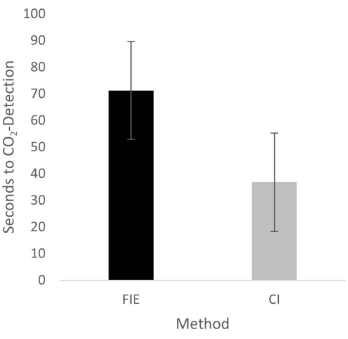
Figure 2: Time until CO2 detection in group comparison. For the group that was intubated using a flexible intubation endoscope, it took significantly longer until end-tidal CO2 could be detected, depicted as mean and standard deviation. Please click here to view a larger version of this figure.
to:
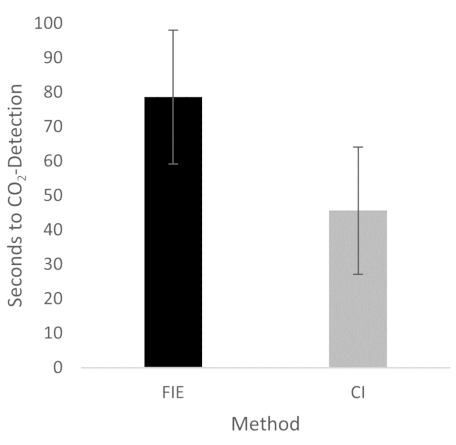
Figure 2: Time until CO2 detection in group comparison. For the group that was intubated using a flexible intubation endoscope, it took significantly longer until end-tidal CO2 could be detected, depicted as mean and standard deviation. n = 5 (for each group). Please click here to view a larger version of this figure.
In the Discussion, the fifth paragraph was updated from:
The increased duration had no clinical significance in this cohort. At no time was the termination criterion-a saturation of less than 93%-reached. This is shown in the results because a procedure change was unnecessary at any time. Prior adequate mask ventilation is a critical step to allow sufficient time for fiberoptic endotracheal tube placement to avoid rapid desaturation34. These results are consistent with previous studies comparing conventional intubation and endoscopically assisted intubations with inexperienced providers35.
to:
The increased duration had no clinical significance in this cohort. At no time was the termination criterion-a saturation of less than 93%-reached. This is shown in the results because a procedure change was unnecessary at any time. Prior adequate mask ventilation is a critical step to allow sufficient time for fiberoptic endotracheal tube placement to avoid rapid desaturation34. These results are consistent with previous studies comparing conventional intubation and endoscopically assisted intubations with inexperienced providers35. We attribute the prolonged duration of fiberoptic intubation to the fact that one must first reorient again after insertion, whereas with conventional intubation, one retains a view of the glottis. It is also important to avoid contact with the mucosa with the flexible intubation endoscope during advancement. This requires occasional corrective maneuvers. Last but not least, after successful placement, retraction of the relatively long endoscope is required, which increases the time to CO2 detection slightly.
