تعتمد وظيفة العدسة البصرية في تركيز الضوء على الشبكية على شفافية الأنسجة، والميكانيكا الحيوية، ومعامل الانكسار. تسلط هذه المراجعة الضوء على البنية ثلاثية الأبعاد للعدسة وتناقش الآليات المعقدة التي تحافظ على توازن الأنسجة.
يتطلب اشتراك JoVE لعرض هذا المحتوى. تسجيل الدخول أو ابدأ نسخة تجريبية مجانية
مقالة مراجعة
العدسة العينية للثدييات في التركيز: التطور، التشريح، الفسيولوجيا، الشفافية، الميكانيكا الحيوية، والتحديات المتعلقة بالعمر
في هذه المقالة
ملخص
الملخص
العدسة هي عضو شفاف وبيضاوي في الحجرة الأمامية للعين يركز الضوء على الشبكية. يتكون هذا النسيج ثلاثي الأبعاد من خلايا ظهارية وخلايا ليافية، وهو منظم بشكل كبير لضمان مسار ضوء واضح ويستخدم استراتيجيات متعددة لتحديد والحفاظ على معامل انكسار عالي وشفافية. في البشر، تخضع العدسة لتغيرات في الشكل لتركيز الضوء من مسافات مختلفة في عملية تعرف بالتكييف، وتعتمد هذه الخصائص المرنة والميكانيكية الحيوية للعدسة على آليات الأنسجة والخلايا والجزيئيات. إعتام عدسة العين، التي تعرف بأنها أي عتامة في العدسة الشفافة عادة، لا تزال السبب الرئيسي للعمى في العالم. تم ربط قصر النظر الشيخيخي، بسبب التغيرات المرتبطة بالعمر في صلابة العدسات، بتدهور تكييلي والحاجة إلى نظارات القراءة. ستستكشف هذه المراجعة تطور عدسات الثدييات ونموها مدى الحياة، والبلورات والبروتينات الهيكلية الخلوية، والتواصل بين الخلايا والدورة الدموية الدقيقة، وميكانيكا العدسة الحيوية، والتحديات والأساليب الجديدة لدراسة هذا النسيج الفريد.
المقدمة
العدسة العينية هي، للوهلة الأولى، نسيج بسيط نسبيا يتكون من نوعين من الخلايا، الخلايا الطهارية والخلايا الليفية. تتكون الكتلة الأكبر للعدسة من خلايا ليافية، وهناك طبقة أحادية من الخلايا الطهارية تغطي نصف الكرة الأمامي1 (الشكل 1A). يتم تغليف الأنسجة بالكامل بغشاء قاعدي رقيق يسمى كبسولة العدسة، وهو مصنوع أساسا من الكولاجين الوريدي واللامينين، كما وصف في مراجعة سابقة2. وبسبب نقص إمداد الدم والأعصاب، تعلق العدسة في الحجرة الأمامية للعين بواسطة ألياف زونية مرنة (المعروفة أيضا بالزونولز) تربط كبسولة العدسة بالجسم الهدبي1. الخلايا الظهارية الأمامية أو المركزية للعدسة مكعبة ومرتبة في نمط حصى 3,4,5 (الشكل 1A، الخلايا الزرقاء الفاتحة، الأعلى). هذه الخلايا هادئة ولا تتكاثر6. أمام خط الاستواء العدسي مباشرة، تنقسم الخلايا الظهارية في المنطقة الإنتابية (الشكل 1A، الخلايا البرتقالية، أعلى اليمين)، وتهاجر هذه الخلايا الاستوائية نحو خط الاستواء حيث تصبح مرتبة بشكل كبير إلى صفوف زالية ذات شكل خلوي سداسي 7,8,9,10 (الشكل 1A، خلايا خضراء، أعلى اليمين). تثبت خلايا الصف الخلالي أطرافها القمية عند نقطة الارتكاز، أو الموديولوس، وتبدأ في الإطالة والتمايز إلى خلايا ألياف ثانوية جديدة (الشكل 1A، أسفل اليسار). تهاجر أطراف الألياف الثانوية القمم نحو القطب الأمامي عن طريق التفاعل مع الجانب القمي لخلايا الظهارة، مكونة وصلة غير معتادة بين القمةوالقمة 3 (الشكل 1A، الخط الأحمر). تمتد أطراف القاعدة الخلفية للألياف الجديدة نحو القطب الخلفي عن طريق التحرك على طول كبسولة العدسة (الشكل 1A، أسفل اليسار). بمجرد وصول أطراف الخلايا الليفية الممتدة إلى القطب الأمامي أو الخلفي، تنفصل الخلايا عن الخلايا الظهارية الأمامية أو الكبسولة الخلفية وتلتقي بأطراف الخلايا الممتدة من الجانب المقابل للنسيج لتشكل الغرز 11,12,13 (الشكل 1A، أعلى اليسار). خلايا ألياف العدسة هي شكل سداسي ممدود في المقطع العرضي مع جانبين طويلين/عريضين، و4 جوانب قصيرة، و6 وصلات ثلاثية الخلايا (الشكل 1A، أسفل اليمين)14. المسافة بين الخلايا بين الخلايا الليفية المجاورة تقلل من تشتت الضوء14. الإضافة المستمرة لخلايا الألياف القشرية في المحيط، التي تتراكب على الأجيال السابقة من الألياف، تسهل نمو العدسة مدى الحياة. مركز العدسة يسمى النواة، وهذه المنطقة من العدسة لديها صلابة متزايدة مقارنة بالمحيط أو القشرة، كما هو موضح في مراجعة سابقةرقم 16.
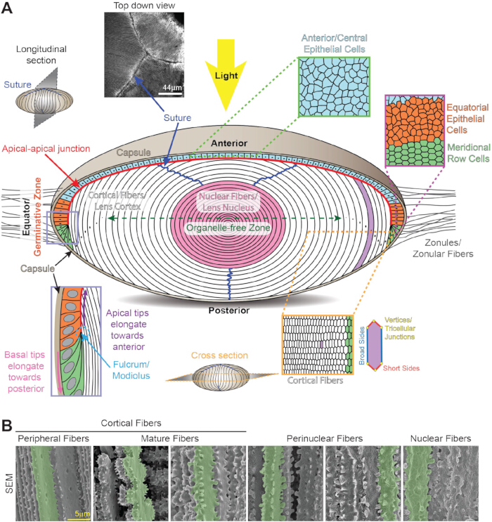
الشكل 1: تشريح العدسة وبناؤها. (أ) تتكون العدسة من طبقة أحادية من الخلايا الظهارية (أزرق فاتح، برتقالي، وأخضر فاتح) تغطي نصف الكرة الأمامي وكتلة كبيرة من ألياف العدسة (بيضاء ووردية). يغلف النسيج غشاء كولاجيني رقيق يعرف بكبسولة العدسة (تان تان). العدسة معلقة في الحجرة الأمامية للعين بواسطة ألياف زونولية (زونولات) تدخل في الكبسولة حول خط استواء العدسة. الخلايا الطهارية الأمامية/المركزية (زرقاء فاتحة، في الأعلى) ذات شكل مكعب ومرصوفة بالمقطع العرضي. تتحول خلايا الظهارة الاستوائية (البرتقالية) في المنطقة الإنزاتية من خلايا عشوائية (برتقالية) إلى صفوف منتظمة من الخلايا السداسية (خضراء). تمتد خلايا الصف الخلالي لتصبح طبقات جديدة من الخلايا الليفية الثانوية القشرية (بيضاء) عند محيط العدسة. تثبت أطراف الألياف الثانوية الجديدة في نقطة الارتكاز/النقطة الزمنية (صندوق البيريوينكل، أسفل اليسار). تهاجر أطراف الألياف الثانوية الممدودة نحو القطب الأمامي على الجانب القمي لخلايا العدسة الظهارية وتشكل وصلة غير معتادة بين القمة والقمة (خط أحمر). تنتقل أطراف الألياف الطويلة القاعدية نحو القطب الخلفي على طول الكبسولة الخلفية. عندما تكون الألياف الثانوية ممدودة بالكامل، تنفصل الأطراف عن الظهارة الأمامية أو الكبسولة الخلفية لتشكيل خياطة العدسة (خطوط زرقاء داكنة والصورة العلوية اليسرى). خلايا ألياف العدسة هي سداسيات ممتدة في المقطع العرضي (أسفل اليمين) مع جانبين طويلين/عريضين، وأربعة جوانب قصيرة، و6 وصلات ثلاثية الخلايا. خلال نضج الخلايا الليافية، تزال جميع العضيات الخلوية لتشكيل المنطقة الخالية من العضيات (سهم أخضر داكن ذو رأسين) وتقليل تشتت الضوء. أقدم خلايا ألياف العدسة تضغط في مركز النسيج لتشكيل نواة العدسة (وردية). الرسوم المتحركة لا ترسم بمقياس. (ب) تظهر صور المجهر الإلكتروني الماسح لخلايا الألياف على أعماق مختلفة في العدسة تغير شكل الخلايا أثناء التمايز والنضج. الخلايا المفردة مميزة باللون الأخضر للتأكيد. تحتوي الألياف الطرفية القشرية على تداخل أصابع كروية ومقبس على الجوانب الطويلة وبروزات صغيرة على الجوانب القصيرة. خلال نضج الخلايا الليافية، تزين الجوانب القصيرة للخلايا الناضجة مجاديف كبيرة مزينة بتروزات صغيرة وبروزات كبيرة. الألياف حول النواة تحتوي على تداخل لسان وأخاديد على طول الغشاء بأكمله وبروزات كبيرة على الجوانب القصيرة. الألياف النووية لها نتوءات كبيرة على الجوانب القصيرة وشكل الغشاء الكروي. شريط الميزة، 5 ميكرومتر. تم التعديل من 16,328. يرجى الضغط هنا لعرض نسخة أكبر من هذا الشكل.
تكمن تعقيد العدسة في ترتيب الخلايا ثلاثي الأبعاد (ثلاثي الأبعاد) المرتب للغاية، ونضج الخلايا الليفية المتخصص الذي يحلل ويزيل جميع العضيات الخلوية، والآليات الجزيئية والخلوية لتأسيس والحفاظ على الشفافية ومعامل الانكسار العالي للنسيج. نقص العضيات الخلوية في معظم خلايا ألياف العدسة يزيد من صعوبة الحفاظ على صحة وطول عمر كل خلية عدسة تم إنشاؤها طوال حياة الكائن الحي. تعد العدسة نسجا ممتازا لدراسة التغيرات المرتبطة بالشيخوخة والعمر، لأن الخلايا المركزية تتكون خلال التطور الجنيني المبكر، وتستمر العدسة في توليد خلايا جديدة على أطراف النسيج طوال الحياة، مما يسمح بدراسة الخلايا القديمة زمنيا مقابل الخلايا التي تتكون أثناء الشيخوخة. تسمح وظيفة التركيز الدقيق للعدسة بتكوين صورة واضحة على الشبكية من أجسام على مسافات مختلفة وتعتمد على قدرة العدسة على تغيير الشكل أو التكيف مع الصورة. العدسة هي نسيج غير متصل وغير عضلي حيث تتطلب خصائص بيوميكانيكية للوظيفة الطبيعية.
السبب الرئيسي لضعف البصر هو سن17. تؤدي التغيرات المرتبطة بالعمر إلى مرضين رئيسيين في العدسة، إعتام عدسة العين وقصر النظر18. تظل المياه البيضاء أو أي تعتيم في العدسة السبب الرئيسي للعمى في العالم19، وتسببه عوامل وراثية وبيئية، بما في ذلك الأشعة فوق البنفسجية، والتدخين، والسكري، والإجهاد التأكسدي، التي تؤدي إلى تجمع البروتينات و/أو عيوب الخلايا، كما هو موضح في المراجعات السابقة 18,20,21,22,23,24,25. قصر النظر المشيخيخ، وهو انخفاض مرتبط بالعمر في التكييف، ينتج عن انخفاض قدرة العدسة على تغيير شكلها عند التركيز على الأجسام القريبة، وبالتالي الحاجة إلى نظارات القراءة 26,27,28. تقدر منظمة الصحة العالمية أن هناك ما لا يقل عن مليار شخص حول العالم يعانون من ضعف بصري غير معالج، منهم 65.2 مليون شخص يعانون من إعتام عدسة عدسة عدسة و826 مليون شخص يعانون من قصر النظر الشيخيخي(17). مع تقدم السكان في العمر، هناك حاجة ملحة لفهم فسيولوجيا وبيولوجيا العدسة بشكل أفضل.
تغطي هذه المراجعة بنية الأنسجة، وترتيب الخلايا، والآليات الجزيئية المهمة لوظيفة وتوازن العدسات الثديية، مع بعض المعلومات عن عدسات من فقاريات أخرى. الهدف هو تقديم نظرة عامة عامة على علم الأحياء وعلم وظائف الأعضاء العدسية مع مراجعات إضافية وتفصيلية. تشمل المواضيع تطور العدسات الجنينية، ونمو العدسات بعد الولادة، وترتيب الخلايا والأنسجة ثلاثية الأبعاد، والخصائص البيوميكانيكية للأنسجة، والدورة الدموية الدقيقة. تناقش أدناه البروتينات الرئيسية للعدسة، وهي البلورات، والشبكات الهيكلية الخلية، والتواصل بين الخلايا والخلية. ستختتم المراجعة بالتحديات الحالية وطرق جديدة لدراسة هذا النسيج الفريد.
الوصول مقيد. يرجى تسجيل الدخول أو بدء فترة تجريبية لعرض هذا المحتوى.
مراجعة ومنظور
تطور العدسة الجنينية ونمو العدسة بعد الولادة
في الفقاريات، تتطور العدسة من خلايا الأديم الخارجي السطحي، وتبدأ العدسة المستقبلية بتكثيف الأديم الخارجي السطحي فوق الحويصلة البصرية 6,29,30 (الشكل 2A). بالتزامن مع تكوين كوب العدسة، يدخل الديكود الخارجي السميك أو لوحة العدسة إلى حفرة العدسة 6,29,30 (الشكل 2B). تربط خيوط الأكتين (F-actin) حفرة العدسة الخلفية بالسطح الأمامي لكوب البصري المتطور لتنسيق حركة وتغيرات شكل هذه الأنسجة النامية 31,32,33,34، ويتطلب الأمر تضييق قمم خلايا العدسة الظهارية لدخول حفرة العدسة 35,36,37,38,39,40(الشكل 2C). تستمر فتحة حفرة العدسة في الضيق حتى يغلق الثقب، حيث تخضع خلايا ساق العدسة لعملية الاستماتة 41,42,43. يتم فصل حويصلة العدسة عن الأديم الخارجي المستقبلي للقرنية لتشكيل حويصلة العدسة، وهي كرة مجوفة من خلايا الظهارة 6,29,30 (الشكل 2D). تمتد الخلايا الطهارية الخلفية في حويصلة العدسة نحو الظهارة الأمامية وتتمايز إلى خلايا ليفية أولية لملء التجويف وتكوين العدسة الجنينية (الشكل 2E). يتم الحفاظ على طبقة أحادية من خلايا الظهارة التي تغطي نصف الكرة الأمامي بينما لا توجد خلايا ظهارية خلفية في العدسة الجنينية الكاملة التكوين (الشكل 2F). تشكل الخلايا الليفية الأولية الجزء الأوسط من نواة العدسة وتحيط بها أجيال جديدة من ألياف العدسة الثانوية التي تضاف من محيط العدسة أو القشرة طوال الحياة 6,29,30. بينما تتكون معظم عدسات الفقاريات عن طريق الفتح، في سمك الزرد، يتزداد سمك اللوحة ثم يتقشر لتشكيل العدسة44.
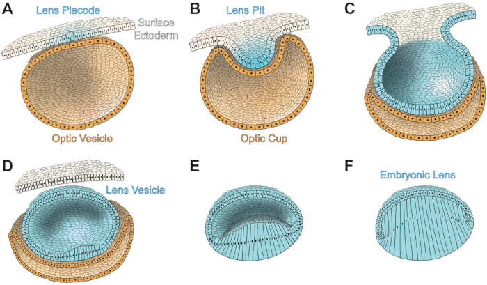
الشكل 2: تطور العدسة الجنينية في عيون الثدييات. خلايا الأديم الخارجية على السطح بيضاء، خلايا العدسة زرقاء فاتحة، وخلايا الكوب البصري/الحويصلات بلون الرمل. (أ) تتطور لوحة العدسة الجنينية (زرقاء فاتحة) من سطح الأديم الخارجي. الجزء الخلفي من الأديم الخارجي للسطح ولوحة العدسة هو الحويصلة البصرية. (ب) يشكل تداخل لوحة العدسة حفرة العدسة، وتشكل الحويصلة البصرية كوب العدسة. سيتطور كوب البصري الأمامي إلى القزحية والجسم الهدب، بينما يشكل كوب البصري الخلفي الظهارة ذات صبغة الشبكية وشبكية الشبكية. (ج) تضيق فتحة لوحة العدسة حتى تغلق الفتحة. (د) تنفصل حويصلة العدسة عن الأديم الخارجي السطحي وهي كرة مجوفة مبطنة بخلايا ظهارية العدسة. (ه) تبدأ خلايا العدسة الظهارية الخلفية في الحويصلة العدسية في الاستمرارية والتمايز إلى ألياف أولية للعدسة. (و) تملأ ألياف العدسة الأولية تجويف حويصلة العدسة لتشكيل العدسة الجنينية. الرسوم المتحركة لا ترسم بمقياس. مستوحى من 411. يرجى الضغط هنا لعرض نسخة أكبر من هذا الشكل.
خلال التطور الجننيني، تحيط العدسة بشبكة تشبه السلة من الأوعية الدموية 6,45,46. تنشأ هذه الأوعية من الشريان الهيالويد في الشبكية وتبدأ في تكوين شبكة في الجزء الخلفي من حفرة العدسة وفي النهاية تخلق شبكة منظمة من التونيكا الوعوية العدسية لتغذية العدسة الجنينية حتى تتطور الحجرة الأمامية وإنتاج الأخلاط المائي بالكامل 6,45. تتراجع هذه الأوعية وتختفي لضمان مسار ضوء واضح قبل الولادة أو قبل فتح العين، حسب النوع 6,45,46,47.
Pax6 ضروري لتطور العيون المبكر 48,49,50,51,52 وهو ضروري وكاف لتطوير العدسات 53,54,55,56. تنظم الفسفرة والتداخل في Pax6 وظيفة هذا العامل أثناء تطور الدماغ والجهاز العصبي المركزيوالعين 57,58,59,60. تتقاطع مسارات الإشارة المهمة الأخرى المطلوبة لتطور العين والعدسة الجنينية مع إشارات Pax6، بما في ذلك المعززات [Meis، البروتينات الشكلية العظمية (Bmp)، وعامل نمو الأرومات الليفية (FGF)] والبروتينات اللاحقة [Foxe3 وحمض الريتينويك (RA)]، كما وصفت في مراجعة سابقة61. يرتبط Meis1 و Meis2 بمنطقة المعزز من جين Pax6 وهما مطلوبان لتحفيز العدسة في الفقاريات62. تظهر الدراسات الجينية أن Bmp7 ضروري لتطوير لوحة العدسة من خلال تنظيم تعبير Pax663,64. Foxe3 هو بعد إشارات Pax6 ومطلوب لتطوير العدسة65,66، خاصة للفصل النظيف بين ساق العدسة والجلد الخارجي عند تكوين حويصلة العدسة. فقدان فوكس 3 يؤدي إلى عيوب خلقية في العين تعرف باسم شذوذ بيترز67. تتعاون إشارات مستقبلات FGF (FGFR) مع Bmp7 لتنظيم تعبير Pax6، مما يؤثر بدوره على مستويات Foxe3 في العدسةالنامية 68. يؤدي تعبير شكل مثبط من FGFR إلى تأخير في تكوين لوحة العدسة، وفصل غير كامل لساق العدسة، ووجود حويصلة عدسة صغيرةرقم 68. يؤدي اضطراب إشارات الروماتويديوم إلى عيوب تكوين العين69، وعندما يتعطل Pax6، ينخفض التعبير عن الحمض الروماتويدي، مما يشير إلى وجود تداخل بين مسارين للإشارة70.
بعد أن تتكون العدسة الجنينية بالكامل، يعتقد أن نمو العدسة المستمر من خلال تكاثر وتمايز خلايا الظهارة الاستوائية إلى ألياف عدسة ثانوية مدفوع بإشارات عامل النمو، خاصة من خلال FGF في عدسات الثدييات، كما هو موضح في مراجعةسابقة 71. تتغذى الألياف الصلبة على الألياف الزجاجية أكثر وفرة في الجسم الزجاجي مقارنة بالخلاط المائي، مما يخلق تدرجا لإشارات النمو والتمايز المتزايدة بالقرب من خط الاستواء العدسي72,73. تعبر العدسة عن 3 من 4 FGFRs، FGFR1، FGFR2، وFGFR3، مع زيادة في التعبير عند وخلف خط الاستواء74,75. يؤدي التعبير الزائد عن الوجبات الزائدة إلى استطالة غير طبيعية وتمايز الخلايا الظهارية الأمامية الهادئة عادة إلى خلايا شبيهة بالألياف 76,77,78. بالإضافة إلى إشارات FGF، أشارت اختبارات المثبطات إلى أن مسار Bmp يلعب دورا في استطالة خلايا الألياف79,80، وأن مستقبل بيتا محول غير طبيعي (TGFβ) يعطل نضج خلايا الأليافالطبيعي 81.
شفافية العدسة ومعامل الانكسار
تخضع خلايا ألياف العدسة لعملية متخصصة لإزالة جميع العضيات الخلوية أثناء النضج للقضاء على الأجسام المبعثرة للضوء في مسار الضوء ولتكوين المنطقة الخالية من العضيات (OFZ)82 (الشكل 1A). يحدث فقدان جميع العضيات الخلوية المنسق بسرعة عندما تصل الخلايا الليفية إلى مرحلة محددة من برمجة النضج 83,84,85,86,87,88. يؤدي التحلل غير الطبيعي للعضويات إلى إعتام عدسة العين، وقد حددت عدة نماذج لأسماك الزرد والفئران (KO) جينات لتحلل الكروماتين والحمض النووي 89,90,91,92,93، والكيناز المعتمد على السيكلين94,95، وعامل الصدمة الحرارية4 91,96,97,98، وPLATT فوسفوليباز 99 وهي مهمة لتشكيل OFZ. يمكن أن تؤثر أيضا على التعبير غير الطبيعي أو الطفرات في بروتينات غشاء الخلية والهيكل الخلوي 100,101,102,103,104، والبلورات 105,106,107، والجينات المسرحية 108,109 على إزالة العضيات وشفافية العدسة. ومع ذلك، لا تزال الإشارة الدقيقة التي تحفز تحلل العضيات في خلايا الألياف غير واضحة. كشفت الأعمال الحديثة أهمية البروتينات المرتبطة بالحمض النووي الريبي في تطور العدسات وتكون الساد، كما هو موضح في مراجعةسابقة 110. يؤدي اضطراب البروتينات المرتبطة بالحمض النووي الريبي، بما في ذلك Tdrd7111، كابرين2112، Rbm24113، وCelf1 114,115,116 إلى تطور غير طبيعي للعدسات، وتدهور العضيات، والمياه البيضية.
مقارنة بمعظم أنواع الخلايا في الجسم، تحتوي خلايا ألياف العدسة الثديية على تركيز بروتين مرتفع جدا (200–450 ملغ/مل)117,118,119,120. تشكل البروتينات 30٪–35٪ من إجمالي وزن العدسة، وبالتالي فإن العدسة تحتوي على نسبة ماء منخفضة نسبيا، حوالي 65٪–70٪121,122. تسعون بالمئة من بروتينات العدسة من حيث الكتلة هي بلورات123، وهناك ثلاث عائلات من بروتينات العدسات البلورية في عدسات الثدييات: α، β، و γ، كما هو موضح في المراجعات السابقة124,125. عادة ما يزيد المحتوى العالي من البروتين من خطر وجود تجمعات أكبر من نصف طول موجة الضوء، مما يؤدي إلى تشتت الضوء126، لكن البروتينات البلورية منظمة في سيتوبلازم الخلية الليفية بترتيب قصير المدى لتعزيز معامل انكسار عالي وشفافية117,127. يتم تخفيف تجميع البروتينات بواسطة وظائف شبيهة بالشابرونات للبلورات α، والتي تناقش أدناه. يعمل معامل الانكسار التدرج (GRIN) عبر العدسة على تعزيز تركيز الضوء، كما هو موضح في المراجعات السابقة128,129، وأقصى معامل انكسار في مركز العدسة مرتبط بضغط خلايا الألياف النووية 130,131,132. في عدسات الأسماك، تشير البيانات إلى أن معامل الانكسار العالي في النواة هو نتيجة نقل البروتين إلى مركز النسيج133. يفترض أن التوزيع الموحد لبروتينات البلورات عبر الخلايا وبين أغلفة الألياف يمكن تسهيله بواسطة مسار نقل جزيئي كبير عبر اندماجات الأغشية على أغشية الخلاياالليفية 134,135. ومع ذلك، لم تلاحظ عمليات اندماج الأغشية بوضوح في عدسات الفئران 136,137,138، كما أن دراسات المقاومة في عدسات من عدة أنواع لا تكشف عن اندماجات الأغشية 139,140,141,142. تشير بيانات نماذج الفئران التي تعطلت في التواصل بين الفجوات إلى أن التواصل بين الخلية وإطالة الخلايا الليفية تساهم أيضا في مسار النقل الجزيئي الكبير143.
البلورات بوزن جزيئي ~20–30 كيلو دالتون وتلعب دورا هيكليا وانكسارا في العدسة. تتكون البلورات ألفا من وحدات فرعية αA وαB، تشترك في 55٪–60٪ تشابه 144,145,146، وبينما يعبر عن αB بشكل واسع في معظم الخلايا، يعبر عن αA-crystallin بشكل رئيسي في العدسة وقد تم اكتشافه بمستويات منخفضة في أنسجة أخرى، بما في ذلك الطحال والزعتر 147,148. أظهرت الدراسات الحديثة أيضا أن الخلايا الظهارية المصبوغة في الشبكية وخلايا العقدة الشبكية يمكنها التعبير عن αA-crystallin أثناء الإجهاد أو المرض، على الأرجح لتوفير تأثير حماية عصبية 149,150,151. عادة ما ترتبط البلورات ألفا بوحدات غير متجانسة كبيرة تتكون من 30–35 وحدة مختلطة من αA و αB123. في ظهارة عدسة الفأر والبقر والإنسان، يكتشف فقط αB في الخلايا الأمامية، بينما يوجد αA وαB بنسبة 1:3 في الخلايا الاستوائية؛ تنقلب هذه النسبة إلى 3:1 أثناء تمايز وتمد الخلايا الليفية الثانوية 123,152,153. تنتمي البلورات ألفا إلى عائلة بروتينات الصدمة الحرارية الصغيرة ولها وظائف تشبه الشابرون154، مما يسمح لهذه البروتينات بالارتباط واحتجاز البروتينات غير الطبيعية أو غير المطوية، مما يمنع تجمع البروتينات أكثر. تم استكشاف الببتيدات الصغيرة α البلورات لمنع الأمراض الناتجة عن تجمع البروتينات، بما في ذلك المياه البيضاء (المياه البيضاء)، كما هو موضح في مراجعةسابقة رقم 155. ومع ذلك، فإن زيادة أو انخفاض النشاط الشبيه بالشابرونات للبلورات α في نماذج الفئران يظهر أن كلا السيناريوهين يؤدي إلى زيادة شدة الماء الأبيض ويشير إلى أن النشاط المناسب الشبيه بالشابرون للبلورات α ضروري لشفافية العدسة156. تنتمي البلورات بيتا و γ إلى عائلة بروتينات β/γ بلورات خاصة بها، وهي خاصة بالخلايا الأليافية؛ في الثدييات، هناك 7 أشكال متساوية من البلورات β توجد كديمرات أو رباعيات أو أوليغومرات من الدرجة الأعلى، وهناك 7 أشكال متساوية من البلورات γ هي أحاديات، كما هو موضح في مراجعةسابقة رقم 125. بالإضافة إلى الوظائف الهيكلية، ترتبط β و γ بلورات بأيونات الكالسيوم وتنظم توفر واحتياطيات أيونات الكالسيوم في ألياف العدسات 157,158,159,160,161.
بروتينات العدسة الهيكلية والغشائية
تحتوي خلايا العدسة الظهارية والألياف على شبكات من خيوط الشتاء الوسيطة F، الكيراتين أو الفيمنتين، والخيوط الوسيطة المتخصصة ذات الخرز، والأنابيب الدقيقة، كما هو موضح في مراجعات سابقة. 162، 163، 164، 165، 166. بالإضافة إلى الوظائف في تطوير العين والعدسات 31,32,33,34، فإن شبكات F-actin المتنوعة في خلايا ظهارية العدسة والألياف مهمة لشفافية العدسة167,168,169,170,171,172. أظهرت الدراسات الحديثة أن تغير شبكة F-actin يؤثر على ميكانيكا العدسةالحيوية 173، 174، 175، 176. الخيوط الوسيطة المكونة من الفيمنتين أو الكيراتين موجودة فقط في المراحل المبكرة من تمايز خلايا ألياف العدسة أو أثناء التطور الجنيني على التوالي 177,178,179,180,181; على النقيض من ذلك، جميع ألياف العدسة تحتوي على خيوط وسيطة مركبة مكونة من هتيرودايمرات من الفيلينسين (المعروف أيضا باسم CP95/CP115 ومشفرة بواسطة Bsfp1) وCP49 (المعروف أيضا باسم الفاكينين والمشفرة بواسطة Bsfp2)182,183,184. يرجع المظهر المرصع لهذه الهيترويديمرات إلى أوليجومرات مكونة من CP49 والفيلينسين تزين نواة الخيط185,186، وارتباط α-بلورات على هذه الخيوط الوسيطةالمتخصصة 187,188,189,190,191. فقدان إما فيلينسين أو CP49 يؤدي إلى فقدان كامل لهذه الشبكة 192,193,194,195. يسبب اضطراب خيوط الفيمنتين الوسيطة أو الخيوط الوسيطة المزينة بالخرز الساد في نماذج البشر والحيوانات192، 193، 196، 197، 198، 199، 200، 201، 202، 203، 204، مما يوضح أهمية هذه الشبكات الهيكلية الخلوية لشفافية العدسات. تشير الأعمال الحديثة إلى أن الأنابيب الدقيقة مهمة لتمديد خلايا ألياف العدسة205,206 وتكوين المنطقة الخالية من العضيات177.
الوصلات اللاصقة التي تتفاعل مع هيكل خلوي الأكتين مهمة لتكوين العدسة207 وتمايز الخلايا الأليافية. يتم التعبير عن E-كاديرين وN-كاديرين في خلايا العدسة الظهارية 208,209,210,211، ويتم إيقاف E-كاديرين في بداية تمايز الخلايا الليفية 212. يعبر عن N-كاديرين بشكل كبير في خلايا ألياف العدسة ويثري عند نقاط منتصف القدم السداسية القاعدية لخلايا الألياف الممتدة 8,209. فقدان N-كاديرين بعد تكوين خلايا الألياف الأولية يسبب اضطرابا في استطالة خلايا الألياف الثانوية بسبب الهجرة غير الطبيعية لأطراف القمة للألياف المتمايزة على طول السطح القمي لخلايا الظهارة الأمامية213. يسبب اضطراب الإشارات ثنائية الاتجاه من نوع Eph-Ephin في العدسة عيوبا في وصلات الالتصاقات 4,7,214,215,216.
تشمل البروتينات الرئيسية الأخرى لأغشية العدسة الأكوابورينات؛ الكونكسينز؛ MP20، كما وصف في مراجعة سابقة217؛ جاليكتين 3؛ البروتين بين الألياف المرتبط بالجالكتين (GRIFIN)؛ الإنتغرينز، كما وصف في مراجعة سابقة218؛ وأفرز بروتينا حمضيا وغنيا بالسيستين (SPARC). يتم مناقشة الأكوابورينات والكونكسين في القسم التالي. MP20 (المعروف أيضا باسم MP17 أو Lim2) هو بروتين غشائي خاص بالعدسة له وظائف التصاق الخليةبالخلايا 85,219 وموجود بكثرة في خلايا الألياف220,221؛ تؤدي الطفرات في MP20 إلى إعتام عدسةعدسة العين 222,223,224,225,226,227,228,229,230. أظهرت الأعمال الحديثة تفاعلات بين MP20 والجاليكتين 3، وهو جزيء التصاق خلوي مهم آخر 231,232,233. GRIFIN، وهو شكل خاص بالعدسة للجاليكتين، يتركز في أغشية الخلايا الليفية ويتفاعل مع Pax6، البلورات، والكاديرينات 234,235,236,237. الإنتيرينز هي روابط مهمة بين المصفوفة خارج الخلية (ECM) والهيكل الخلوي، وهي ضرورية لتطور العدسة الطبيعي. تعمل نظائر الإنتجرين المعبر عنها في العدسة كمستقبلات لمكونات الكبسولات مثل اللامينين، الكولاجين، أو الفيبرونكتين 8,238,239,240,241,242,243,244,245. يمكن لكل مكون من المصفوفات التفاعل مع عدة إنتغرينات، مما يؤدي إلى إشارات متنوعة من خلال التعبير الزماني والمكاني للإنترينات أثناء تطور العدسةونموها. هناك اهتمام خاص بفهم التغيرات في TGFβ، والإنتيرينات، ومكونات ECM خلال الانتقال من الظهارة إلى الميزانسميا في خلايا العدسة الظهارية المتبقية بعد جراحة الساد، مما يؤدي إلى تعتيم الكبسولة الخلفية، كما هو موضح في مراجعةسابقة 218. SPARC، وهو جزيء التصاق وتكاثر الخلية، متمركز في ظهارة العدسة ولا يكون موجودا في الألياف التمايزية246. فقدان SPARC يؤدي إلى إعتام عدسة العين 246,247,248، وأظهرت الدراسات الحديثة أن SPARC قد يكون له وظائف شبيهة بالشابرون 249، ويؤثر على الإنتجرينات وبروتينات ECM250,251,252، ويؤثر على مسار الدورة الدقيقة253.
المسار الدقيق الدورة الدموية للنسيج الأوعائي العلوي
بسبب نقص إمداد الدم، تولد العدسة الكاملة التكوين مسارا دوريا دقيقا خاصا بها لتوصيل المغذيات وإزالة الفضلات، كما هو موضح في المراجعات السابقة 254,255,256,257. يدخل تيار أيوني العدسة عند القطبين الأمامي والخلفي ويخرج عند خط الاستواء258. يعتمد التيار الداخلي على حركة الصوديوم إلى الفراغات خارج الخلية بين خلايا العدسة عند كل قطب عبر تدرج تركيز ناتج عن النقل النشط، ويتم تسهيل التيار الخارج بواسطة قنوات الوصلة الفجوة على أغشية الخلايا الليفية المركزة بالقرب من خط استواء العدسة للسماح لأيونات الصوديوم بالخروج عند خط الاستواء259. تحتوي خلايا الألياف الظهارية والألياف الطرفية الاستوائية على تعبير عالي عن مضخات Na+/K+ ATPase التي تتبادل بنشاط 3 أيونات صوديوم خارج الخلايا مقابل أيونين بوتاسيوم من البيئة خارج العدس 260,261,262. تحافظ العدسة على تركيز بوتاسيوم مرتفع (135 ملم مول) وتركيز منخفض من الصوديوم (16 ملم) لتوفير الظروف التي تسمح للصوديوم بالعودة إلى العدسة عند الأقطاب، على تدرج التركيز259,263. بالإضافة إلى إنزيمات Na+/K+ ATPase، يساعد ناقل الأيونات المشارك Na-K-2Cl 1 (NKCC1) أيضا في تنظيم تركيزات أيونات العدسة من خلال التنشيط عبر قنوات حساسة للميكانيكيا، كما هو موضح في مراجعة سابقة264.
تتكون وصلات الفجوات في العدسة من ثلاثة موصلات: كونكسين 43 (Cx43 أو α1)، Cx46 (أو α3)، وCx50 (أو α8)265,266. يعبر عن Cx43 بشكل رئيسي في خلايا العدسة الظهارية267، وبينما يبدأ تكوين الخلايا الليفية أمر طبيعي في عدسات Cx43 KO، فإن الواجهة بين الخلايا الظهارية والألياف توسعت في المساحة خارج الخلية والفراغات داخل الخلية، مما يشير إلى وظيفة ل Cx43 في التوازن الأسموزي268. يتركز Cx46 في خلايا ألياف العدسة، ويوجد Cx50 في كل من الخلايا الطهارية والألياف269. يعبر عن Cx23 أيضا في خلايا الألياف في عدسات الفأر، لكن ليس في العدسات البشرية 270,271,272,273. طفرة سائدة في Cx23 تسبب عيوبا في خلايا الألياف في فئران270، لكن عدسات Cx23 KO لا تعاني من عيوب ملحوظة274. تشكل ست وحدات فرعية من الكونكسين اتصالا أو نصف قناة، واتصال في خلية واحدة يرتقي مع موصل على الخلية المجاورة ليشكل وصلة فجوة، مما يخلق مسام أنغستروم ~10 بين الخلايا المجاورة، مما يسمح بتبادل الجزيئات الصغيرة والأيونات والماء275,276. تسمح قنوات الكونيكسين بمرور المواد بين السيتوبلازم داخل الخلية والبيئة خارج الخلية، كما هو موضح في المراجعات السابقة277,278، وتعد القنوات النصفية مهمة للدوران الدقيق279، والنقل الحساس للمغذيات ومضادات الأكسدة للميكانيكي280,281,282,283، والاستجابة التأكسدية284,285,286,287,288. تتجمع التقاطعات الفجوة لتشكل لويحات كبيرة على غشاء الخلية الليفية؛ في العدسة، يمكن أن تكون اللويحات بحجم ميكرون في القطر وتكون مصطفة على الجوانب الطويلة لخلايا الألياف المجاورة289. فقدان Cx46 في عدسات الفئران يؤدي إلى إعتام عدسة نووية كثيفة بسبب انسداد مسار خروج العدسة في الدورة الدقيقة290,291. يتطلب تجميع و/أو استقرار لويحات Cx46 الكبيرة على غشاء الخلية الليفية شبكة F-أكتين والخيوط الوسيطة المزدوجة289. يؤدي التهاب العين البصري Cx50 إلى الصغراف، والمياه البيضاء النووية الخفيفة، والعدسات الصغيرة بسبب تكاثر خلايا الظهارة غير الطبيعي292,293. يؤثر فقدان Cx50 على بوابات بقية لوح وصلات الفجوة في خلايا ألياف العدسة139. استبدال Cx50 ب Cx46 القابل للطرق المعبر عنه تحت الموضع الداخلي Cx50 (4 نسخ من Cx46) ينقذ المياه البيضاء بسبب فقدان Cx50 ويزيد من اقتران الفجوة بين الألياف، لكن هذه العدسات الشفافة التي تظل صغيرة142,294. لذا، لا يمكن ل Cx46 استبدال دالة نمو العدسة في Cx50. كما تم استخدام استراتيجية التوصيل لإنقاذ المياه البيضاء النووية الناتجة عن بروتينات γB-بلورات متحولة، مما يشير إلى أن زيادة الترابط الفجوي يمكن أن تكون استراتيجية لتحسين توازن العدسة والوقاية من الساد295.
يصاحب التدفق الأيوني الناتج عن أيونات الصوديوم تدفق الماء إلى خلايا العدسات عبر قنوات الأكوابورين259. تتكون قنوات الأكوابورين من Aqp1 في الخلايا الظهارية، بينما في الخلايا الليفية، تشكل قنوات المياه Aqp0 (المعروفة أيضا باسم MIP) وAqp5، حسب نضج الخلايا، كما هو موضح في المراجعات السابقة 264,296,297. حركة الماء عبر قنوات الربط الفجوة تولد تدرج ضغط هيدروستاتيكي عبر العدسة298,299، ويحافظ هذا التدرج على ضغط الأسموزي للعدسة عبر قنوات الفانيلويد الميكانيكية الحساسة لمستقبلات المستقبلات العابرة (TRPV1) وTRPV4 التي تزيد وتنخفض الضغط الهيدروستاتيكيللعدسة 299,300,301,302 على التوالي . انخفاض التوتر الزوني على العدسة يقلل الضغط الهيدروستاتيكي، مما ينشط TRPV1 ثم NKCC1 لزيادة تركيز الأيونات في العدسة ويدفع تدفق الماء إلى العدسة لاستعادة الضغط الهيدروستاتيكي الطبيعي300,301. وعلى العكس، فإن زيادة التوتر الزوني وحجم العدسة تنشط TRPV4 ثم Na+/K+ ATPase لتقليل تركيز أيونات العدسة وبالتالي تقليل الضغط الهيدروستاتيكي 300,301,302.
يتم نقل العناصر الغذائية الأساسية والأحماض الأمينية إلى العدسة عبر مسار الدورة الدقيقة303، وهذا النقل مهم بشكل خاص لألياف OFZ. توجد ناقلات الجلوكوز الميسرة (GLUT) في خلايا العدسة الظهارية والألياف؛ يعبر عن GLUT1 في كلا نوعي الخلايا، وGLUT3 يعبر عنه في الخلايا الطهارية 304,305,306,307. تصاب فئران GLUT1 KO بإعتام عدسة عدسة، مما يشير إلى أن امتصاص الجلوكوز ضروري لشفافية العدسة308. الجلوتاثيون (GSH)، وهو مضاد للأكسدة المهم لشفافية العدسات309,310، يصنع في العدسة ويمكن نقله إلى العدسة من الهيمور المائي311,312,313,314,315. السيستين والجلوتامات والجلايسين هي أحماض أمينية أساسية لتخليق GSH، ويتجمع هذه الأحماض الأمينية في القشرة والمناطق النووية من العدسة314,315. يمكن أن يؤدي انخفاض تخليق GSH المرتبط بالعمر أو زيادة استخدام GSH إلى انخفاض توفر GSH في العدسة، خاصة في النواة 316,317,318,319,320,321. تستخدم بيروكسيدازات الجلوتاثيون (GPX) GSH كإنزيم مساعد لتعادلية الهيدروبروكسيد، وتظهر الدراسات الحديثة أن الفيروبتوزيس الناتج عن بيروكسيدات الدهون السامة يمكن أن يؤدي إلى إعتام عدسة العين نتيجة اضطراب GPX4322,323.
هندسة العدسات ثلاثية الأبعاد والميكانيكا الحيوية
ينبع نمط ألياف العدسة إلى صفوف منظمة من الخلايا السداسية الشكل من نمط الخلايا الظهارية الاستوائية إلى صفوف خط الطول (الشكل 1A). الآليات التي تدفع تنظيم الخلايا الطهارية الاستوائية المكدسة عشوائيا والمرصوفة بالأحجار إلى خلايا صفية سداسية الشكل تدور حول هيكل خلوي الأكتين. هناك حاجة لإرسال إشارات مستقبل كيناز التيروزين EphA2 لدفع تراكم F-actin عند رؤوس خلايا الصفوف السداسية لتعزيز تنظيم الخلية 7,324. الميوسين IIA، وهو بروتين انقباض يرتبط بفيتامين F-actin، مهم أيضا لتعزيز شكل الخلايا السداسي 8,325,326. لطالما كان يعتقد أن هذا التعبئة المرتبة عالية لخلايا الألياف ضروري لشفافية العدسة، لكن سوء التعبئة بسبب فقدان EphA2 7,324، أو الطفرات في الميوسين IIA325,326، أو الخلايا المشوهة التي تتكون خلال سنالتقدم تشير إلى أن تعبئة الخلايا السداسية ليست ضرورية للشفافية.
الخلايا الليفية التي تمتد من الأقطاب الأمامية إلى الخلفية يمكن أن تصل إلى عدة مليمترات في الطول حسب نوع الحيوان وحجم العدسة327. عادة، تكون خلايا الألياف فقط 4–7 ميكرومتر في المقطع العرضي327، وهذه الخلايا الطويلة والنحيفة تثبت معا بواسطة تداخل ديناميكي بين الخلايا الخلوية تشكل سحابا ثلاثي الأبعاد بين الخلايا الليفية المجاورة، كما هو موضح في مراجعةسابقة 328. تداخل خلايا ألياف العدسة له أنماط معقدة تتغير مع تمايز الخلايا ونضوجها، فمن 1) ألياف جديدة تشكلت في الأطراف مع نتوءات صغيرة على الجوانب القصيرة وبروزات كروية ومجوفة على الجوانب الطويلة، 2) خلايا ليفية ناضجة تفقد العضيات وتحتوي على مجاذيف كبيرة مع نتوءات صغيرة متشابكة على الجوانب القصيرة. 3) ألياف حول النواة ذات نتوءات متشابكة كبيرة على الجوانب القصيرة وتداخل الأخدود واللسان على طول الغشاء بأكمله، و4) ألياف نووية ذات نتوءات كبيرة على الجوانب القصيرة وشكل الغشاء الكروي على طول الغشاء بأكمله137,328,329,330,331,332,333,334,335, 336,337,338,339 (الشكل 1B). قدمت أبحاث حديثة دليلا مباشرا على أن التداخل بين خلايا ألياف العدسة التي تعتمد على شبكات F-actin مهم لصلابة الأنسجة174، مما يدعم فرضية طال انتظارها. فقدان ARVCF، وهو مكون من وصلات الالتصاقات، يؤدي إلى تداخل خلايا الألياف غير الطبيعية، واتساع المساحة خارج الخلية، وظهور مبكر لساد العين القشري340. ترتبط عيوب في صلابة وحجم نواة العدسة بتغيرات في تداخل اللسان والأخاديد وتأخر الانتقال إلى شكل الغشاء الكروي131.
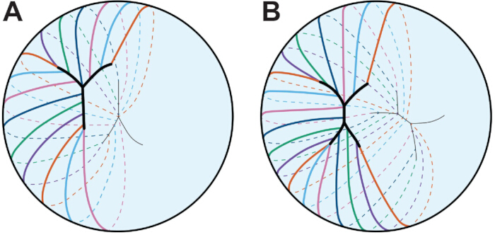
الشكل 3: نمط خياطة العدسات. يمثل كل خط ملون خلية ألياف واحدة. (أ) تشكل الخلايا الليفية في عدسة الإنسان الجنينية خياطة Y. الدرز على شكل Y معكوس عند القطبين الأمامي والخلفي. (ب) مع استمرار تطور العدسة البشرية ونموها، ستصبح الغرز أكثر تفرعا. يصور هنا غرز رباعي الفروع لبساطة. نمط الغرز متزاحج بين القطبين. الرسوم المتحركة لا ترسم بمقياس. مستوحى من 45. يرجى الضغط هنا لعرض نسخة أكبر من هذا الشكل.
يختلف الشكل العام للعدسة حسب النوع، ويفترض أن انحناء خلايا الألياف ونوع الغرز لتحديد شكل العدسة341,342. تتكون الغرز من التقاء أطراف خلايا الألياف الممتدة عند القطبين الأمامي والخلفي، وتنعكس الخياطة بين القطبين 11,12,13,343 (الشكل 3). في عدسات الفئران، تكون الخياطة على شكل حرف Y، وفي عدسات الرئيسيات والبشر، تبدأ الخياطة على شكل Y وتصبح أكثر تفرعا مععمر 11، 12، 45. ومن المثير للاهتمام أن خياطة Y في عدسات الفأر تحد من مرونة أو مرونة العدسة بعد إزالة الحمل الميكانيكي، كما أن الغرز المتفرعة في عدسات KO التي تتسبب في اضطراب إشارات إيفرين Eph ترتبط بزيادة المرونة344. التكوين الطبيعي لغرزة العدسة يتطلب اتصالا بفاصل الفجوة345، والمياه العينية الأمامية المرتبطة بالعمر في عدسات الفئران هي نتيجة لتكوين الغرز غير الكامل130.
يمكن أن تتأثر الخصائص الميكانيكية الحيوية للعدسة بمجموعة متنوعة من العوامل الجزيئية والخلوية والأنسجة، كما هو موضح في مراجعة سابقة16. يعتقد أن زيادة تيبس العدسة المرتبطة بالعمر هي أحد الأسباب الرئيسية لمرض قصر النظر346. عند النظر إلى الأجسام البعيدة، يؤدي استرخاء عضلات الهدب إلى شد الألياف الزونونية المتصلة بالكبسولة، مما يؤدي إلى تسطح العدسة1. عند التركيز على شيء قريب، تنقبض العضلة الهدبية مما يسمح للزونول بالاسترخاء، وتدور العدسة للأعلى لتركيز الضوء القادم من الجسمالقريب 1. تتطلب هذه العملية المطابقة خصائص بيوميكانيكية للعدسة، وانقباض عضلات الهدب، وتوتر زوني. مع التقدم في العمر، أظهرت الدراسات تغيرا طفيفا أو معدوما في انقباض عضلات الهداب والمرونة الزونية والتوتر347,348,349,350. أشارت عدة دراسات إلى أن زيادة حجم العدسة وحجم النواة وصلابتها تساهم في صلابة العدسة بالكامل مع عمر 26,351,352,353,354. ومع ذلك، تشير الدراسات حول صلابة العدسة مع التقدم في العمر في الفئران إلى أن هناك ارتباطا ضئيلا بين حجم العدسة وحجم النواة مع صلابة العدسةالكاملة 130,132، مما يشير إلى أن عوامل أخرى تؤثر على تصلب العدسة المرتبطة بالعمر. أظهرت الأعمال الحديثة أن التغيرات في شبكات الهيكل الخلوي تؤثر على صلابة العدسة؛ فقدان تروبوميوزين 3.5، وهو بروتين مثبت لأكتين F، يؤثر على تركيب وترتيب شبكات F-actin في غشاء خلايا الألياف ويقلل من صلابة العدسة173. وبالمثل، تؤدي الطفرات العفوية في عدة سلالات فئران برية متزاوجة إلى ضربة قاضية تلقائية لCP49 192,355 وعدسات أكثر ليونة356,357. أغشية ألياف العدسة تحتوي على نسبة كوليسترول مرتفعة بشكل غير معتاد 358,359,360,361,362,363، مما يمكنه تنظيم مرونة الغشاء364. يعتقد أن التغيرات في محتوى الكوليسترول في الغشاء وارتباط α البلورات بغشية الألياف مع التقدم في العمر تزيد من صلابة العدسة365,366,367,368,369,370,371,372.
تحديات وطرق جديدة لدراسة العدسة
يخلق الهيكل ثلاثي الأبعاد للعدسة تحديات لفهم ترتيب الخلية والخلية ولإنشاء نماذج مخبرية مناسبة تعيد تلخيص بنية الأنسجة. بينما تم توضيح رؤى قيمة حول الإشارات وتحديد مواقع البروتينات الطفرة باستخدام خلايا الزراعة الخالية أو الأولية، فإن خلايا العدسة الظهارية المعزولة لا تمتلك الشكل المكعب المميز أو تمتلك نفس التعبير الجيني أو الهياكل الخلوية الهيكلية مثل خلايا العدسة الظهاريةالأصلية 373. العدس في المختبر، كما هو موضح في مراجعة سابقة،374 ونماذجعدسة 375 تخلق خلايا شبيهة بالألياف وكرات من الخلايا تشبه العدسة الصغيرة على التوالي. تخلق هذه النماذج بنية شفافة تشبه العدسة يمكن أن تكون مفيدة لأغراض فحص الأدوية السريع، لكن لا يعكس أي من النموذجين تنظيم الخلايا الليفية الطبيعية أو البنية ثلاثية الأبعاد للعدسة. تم تطوير العديد من البروتوكولات لتحديد مواقع البروتينات في مقاطع نسيج العدسة والتلوين المناعي، ولا تزال هذه الطريقة الطريقة الأكثر كفاءة لفهم ما إذا كانت البروتينات تعبر عنها في أقسام خلايا محددة. ومع ذلك، قد يكون من الصعب تقسيم الأنسجة في علم الأنسجة والتلوين المناعي، خاصة في عينات من الثدييات البالغة الأكبر سنا، بسبب عدم التثبيت الكامل للعدسة137؛ ودراسة البنية ثلاثية الأبعاد في المقاطع ثنائية الأبعاد تزيل التفاصيل المهمة لفهم شكل الخلايا، والتفاعلات بين الخلايا، وتحديد مواقع البروتينات. بينما يظل المجهر الإلكتروني (EM) المعيار الذهبي لدراسة شكل الخلايا الليافية، فإن تحديد مواقع البروتينات في عينات الكهرومغناطيسية لا يزال تحديا 376,377,378. أعمال حديثة مع تركيبات مسطحة لطبقة الخلايا الظهاريةأحادية الطبقة 4,7,324,373، وتلوين خلية ليف واحدة 85,173,174,379، وتصوير كامل العدسة 4,7,324,344,380,381 كشفت عن معلومات مهمة حول التصاقات الخلايا الخلية، وميكانيكا العدسة الحيوية، وتغير شبكات الهيكل الخلوي في ألياف العدسة.
لخلايا العدسة الظهارية والألياف وظائف مختلفة، مثل النسخ والبروتيومات، وكانت الدراسات السابقة لمقارنة أقسام الخلايا المختلفة صعبة لأن هناك طبقة واحدة فقط من الخلايا الظهارية تغطي نصف الكرة الأمامي، مما يحد من كمية الحمض النووي الريبي أو البروتين الذي يمكن جمعه دون تجميع العديد من العينات. سمحت التطورات في تقنيات استخراج الحمض النووي الريبي، وPCR في الوقت الحقيقي، واختبارات الرحلان الكهربائي الشعري الآلي بتحليل الجينات والبروتينات لعينات خلايا العدسة الظهارية المجمعة من زوج واحد من عيون الفئران 130,132,373,382 أو المناطق المشوهة دقيقة من ظهارة العدسة، والألياف القشرية، والألياف النووية383. تسمح تقنية النسخ الأحادية الخلية بتحليل معمق لخلايا العدسة من أجنة سمك الزرد وأجنة اليرقات والفراخ384,385. تفتح هذه الطرق الجديدة الباب للكشف عن الآليات الجزيئية التي تحافظ على كل جزء من خلية العدسة خلال الشيخوخة الطبيعية والأمراض المرضية.
مع تقدم المجال، من المهم أن نتذكر أن الخلفية الوراثية للفئران يمكن أن تلعب أدوارا مهمة في علم أمراض العدسات386,387 والميكانيكاالحيوية 355,356,357، وبالتالي فإن ضوابط الأبناء في التجارب ضرورية لتحليل البيانات. توفر نماذج فئران المتحولين وKO نظاما لدراسة إعتام عدسة العين المرتبطة بالعمر وزيادة في صلابة العدسات 130,356,357,388,389,390,391,392,393,394,395,396,397، ونظرا لقصر عمر الفئران نسبيا، يجب إجراء دراسات الشيخوخة على الحيوانات القديمة بدلا من استنتاج بيانات من الحيوانات الصغيرة.
الوصول مقيد. يرجى تسجيل الدخول أو بدء فترة تجريبية لعرض هذا المحتوى.
الاستنتاجات
لقد حسنت التطورات التكنولوجية في علم النسخ، والبروتيوميات، والتصوير، والاختبارات البيوميكانيكية فهمنا لبيولوجيا العدسة، وعلم وظائف الأعراض، وعلم الأمراض. معرفتنا المتزايدة حول الفروقات بين مجموعات العدسة الظهارية وخلايا الألياف ستكون مهمة لفهم التغيرات أثناء التقدم في العمر وآليات استمرار البروتين والخلايا. زاد الاهتمام بتطوير تدخلات دوائية لمنع أو تأخير إعتام عدسة العين و/أو قصر النظر الشيخوخي، وإمكانية تجديد العدسات الطبيعية بعد جراحة المياه البيضاء. يتطلب توصيل الأدوية إلى العدسة لمنع...
الوصول مقيد. يرجى تسجيل الدخول أو بدء فترة تجريبية لعرض هذا المحتوى.
الإفصاحات
المؤلفون ليس لديهم ما يكشفون عنه.
شكر وتقدير
تم تمويل هذا العمل من خلال منحة R01 EY032056 (إلى CC) من المعهد الوطني للعيون. يشكر المؤلفان إيزايا ج. إينيس وغريفين م. فلاورز على تعليقاتهم المفيدة.
الوصول مقيد. يرجى تسجيل الدخول أو بدء فترة تجريبية لعرض هذا المحتوى.
المراجع
- Freddo, T. F., Chaum, E. Anatomy of the Eye and Orbit: The Clinical Essentials, 1e. , Lippincott Williams & Wilkins, a Wolters Kluwer business. (2018).
- Danysh, B. P., Duncan, M. K. The lens capsule. Exp Eye Res. 88 (2), 151-164 (2009).
- Zampighi, G. A., Eskandari, S., Kreman, M. Epithelial organization of the mammalian lens. Exp Eye Res. 71 (4), 415-435 (2000).
- Cheng, C., Gong, X. Diverse roles of Eph/ephrin signaling in the mouse lens. PLoS One. 6 (11), e28147(2011).
- Parreno, J., Cheng, C., Nowak, R. B., Fowler, V. M. The effects of mechanical strain on mouse eye lens capsule and cellular microstructure. Mol Biol Cell. 29 (16), 1963-1974 (2018).
- Lovicu, F. J., Robinson, M. L. Development of the Ocular Lens. , Cambridge University Press. (2011).
- Cheng, C., Ansari, M. M., Cooper, J. A., Gong, X. EphA2 and Src regulate equatorial cell morphogenesis during lens development. Development. 140 (20), 4237-4245 (2013).
- Bassnett, S., Missey, H., Vucemilo, I. Molecular architecture of the lens fiber cell basal membrane complex. J Cell Sci. 112 (Pt 13), 2155-2165 (1999).
- Bassnett, S., Sikic, H. The lens growth process. Prog Retin Eye Res. 60, 181-200 (2017).
- Sikic, H., Shi, Y., Lubura, S., Bassnett, S. A full lifespan model of vertebrate lens growth. R Soc Open Sci. 4 (1), 160695(2017).
- Kuszak, J. R. The development of lens sutures. Progress in Retinal and Eye Research. 14 (2), 567-591 (1995).
- Kuszak, J. R., Mazurkiewicz, M., Zoltoski, R. Computer modeling of secondary fiber development and growth: I. Nonprimate lenses. Mol Vis. 12, 251-270 (2006).
- Kuszak, J. R., Zoltoski, R. K., Tiedemann, C. E. Development of lens sutures. Int J Dev Biol. 48 (8-9), 889-902 (2004).
- Kuszak, J. R. The ultrastructure of epithelial and fiber cells in the crystalline lens. Int Rev Cytol. 163, 305-350 (1995).
- Kuszak, J. R., Macsai, M. S., Rae, J. L. Stereo scanning electron microscopy of the crystalline lens. Scan Electron Microsc. (Pt 3), 1415-1426 (1983).
- Cheng, C. Tissue, cellular, and molecular level determinants for eye lens stiffness and elasticity. Front Ophthalmol (Lausanne). 4, 1456474(2024).
- World Health Organization. World report on vision. , (2019).
- Michael, R., Bron, A. J. The ageing lens and cataract: a model of normal and pathological ageing. Philos Trans R Soc Lond B Biol Sci. 366 (1568), 1278-1292 (2011).
- Asbell, P. A., et al. Age-related cataract. Lancet. 365 (9459), 599-609 (2005).
- Hejtmancik, J. F. Oxidative Stress in Genetic Cataract Formation. Antioxidants (Basel). 13 (11), (2024).
- Shiels, A., Hejtmancik, J. F. Biology of Inherited Cataracts and Opportunities for Treatment. Annu Rev Vis Sci. 5, 123-149 (2019).
- Truscott, R. J. W., Friedrich, M. G. Molecular Processes Implicated in Human Age-related Nuclear Cataract. Invest Ophthalmol Vis Sci. 60 (15), 5007-5021 (2019).
- Fan, X., Monnier, V. M. Protein posttranslational modification (PTM) by glycation: Role in lens aging and age-related cataractogenesis. Exp Eye Res. 210, 108705(2021).
- Budnar, P., Tangirala, R., Bakthisaran, R., Rao, C. M. Protein Aggregation and Cataract: Role of Age-related Modifications and Mutations in alpha Crystallins. Biochemistry (Mosc). 87 (3), 225-241 (2022).
- Shiels, A., Hejtmancik, J. F. Inherited cataracts: Genetic mechanisms and pathways new and old. Exp Eye Res. 209, 108662(2021).
- Heys, K. R., Cram, S. L., Truscott, R. J. Massive increase in the stiffness of the human lens nucleus with age: the basis for presbyopia? Mol Vis. 10, 956-963 (2004).
- Glasser, A., Campbell, M. C. Biometric, optical and physical changes in the isolated human crystalline lens with age in relation to presbyopia. Vision Res. 39 (11), 1991-2015 (1999).
- Weeber, H. A., et al. Dynamic mechanical properties of human lenses. Exp Eye Res. 80 (3), 425-434 (2005).
- Piatigorsky, J. Lens differentiation in vertebrates. A review of cellular and molecular features. Differentiation. 19 (3), 134-153 (1981).
- Gunhaga, L. The lens: a classical model of embryonic induction providing new insights into cell determination in early development. Philos Trans R Soc Lond B Biol Sci. 366 (1568), 1193-1203 (2011).
- Chauhan, B. K., et al. Cdc42 and IRSp53 dependent contractile filopodia tether presumptive lens and retina to coordinate epithelial invagination. Development. 136 (21), 3657-3667 (2009).
- McAvoy, J. W. Cytoplasmic processes interconnect lens placode and optic vesicle during eye morphogenesis. Exp Eye Res. 31 (5), 527-534 (1980).
- Mann, I. The development of the human eye. , 3d, Grune & Stratton. (1964).
- Eiraku, M., et al. Self organizing optic cup morphogenesis in three dimensional culture. Nature. 472 (7341), 51-56 (2011).
- Lang, R. A., Herman, K., Reynolds, A. B., Hildebrand, J. D., Plageman, T. F. Jr p120 catenin dependent junctional recruitment of Shroom3 is required for apical constriction during lens pit morphogenesis. Development. 141 (16), 3177-3187 (2014).
- Plageman, T. F. Jr, et al. A Trio RhoA Shroom3 pathway is required for apical constriction and epithelial invagination. Development. 138 (23), 5177-5188 (2011).
- Borges, R. M., Lamers, M. L., Forti, F. L., Santos, M. F., Yan, C. Y. Rho signaling pathway and apical constriction in the early lens placode. Genesis. 49 (5), 368-379 (2011).
- Quintin, S., Gally, C., Labouesse, M. Epithelial morphogenesis in embryos: asymmetries, motors and brakes. Trends Genet. 24 (5), 221-230 (2008).
- Muccioli, M., Qaisi, D., Herman, K., Plageman, T. F. Jr Lens placode planar cell polarity is dependent on Cdc42 mediated junctional contraction inhibition. Dev Biol. 412 (1), 32-43 (2016).
- Houssin, N. S., Martin, J. B., Coppola, V., Yoon, S. O., Plageman, T. F. Jr Formation and contraction of multicellular actomyosin cables facilitate lens placode invagination. Dev Biol. 462 (1), 36-49 (2020).
- Glucksmann, A. Cell deaths in normal vertebrate ontogeny. Biol Rev Camb Philos Soc. 26 (1), 59-86 (1951).
- Ozeki, H., Ogura, Y., Hirabayashi, Y., Shimada, S. Suppression of lens stalk cell apoptosis by hyaluronic acid leads to faulty separation of the lens vesicle. Exp Eye Res. 72 (1), 63-70 (2001).
- Yan, Q., Liu, J. P., Li, D. W. Apoptosis in lens development and pathology. Differentiation. 74 (5), 195-211 (2006).
- Easter, S. S. Jr, Nicola, G. N. The development of vision in the zebrafish (Danio rerio). Dev Biol. 180 (2), 646-663 (1996).
- Mann, I. The development of the human eye. , The University Press. (1928).
- Darche, M., et al. Three dimensional characterization of developing and adult ocular vasculature in mice using in toto clearing. Communications Biology. 5 (1), 1135(2022).
- Wang, Z., Liu, C. H., Huang, S., Chen, J. Assessment and Characterization of Hyaloid Vessels in Mice. J Vis Exp. (147), e59222(2019).
- Hill, R. E., et al. Mouse small eye results from mutations in a paired like homeobox containing gene. Nature. 354 (6354), 522-525 (1991).
- Jordan, T., et al. The human PAX6 gene is mutated in two patients with aniridia. Nat Genet. 1 (5), 328-332 (1992).
- Hogan, B. L., et al. Small eyes (Sey): a homozygous lethal mutation on chromosome 2 which affects the differentiation of both lens and nasal placodes in the mouse. J Embryol Exp Morphol. 97, 95-110 (1986).
- Hanson, I. M., et al. Mutations at the PAX6 locus are found in heterogeneous anterior segment malformations including Peters' anomaly. Nat Genet. 6 (2), 168-173 (1994).
- Grindley, J. C., Davidson, D. R., Hill, R. E. The role of Pax 6 in eye and nasal development. Development. 121 (5), 1433-1442 (1995).
- Fujiwara, M., Uchida, T., Osumi Yamashita, N., Eto, K. Uchida rat (rSey): a new mutant rat with craniofacial abnormalities resembling those of the mouse Sey mutant. Differentiation. 57 (1), 31-38 (1994).
- Collinson, J. M., Hill, R. E., West, J. D. Different roles for Pax6 in the optic vesicle and facial epithelium mediate early morphogenesis of the murine eye. Development. 127 (5), 945-956 (2000).
- Quinn, J. C., West, J. D., Hill, R. E. Multiple functions for Pax6 in mouse eye and nasal development. Genes Dev. 10 (4), 435-446 (1996).
- Ashery Padan, R., Marquardt, T., Zhou, X., Gruss, P. Pax6 activity in the lens primordium is required for lens formation and for correct placement of a single retina in the eye. Genes Dev. 14 (21), 2701-2711 (2000).
- Kim, E. A., et al. Phosphorylation and transactivation of Pax6 by homeodomain interacting protein kinase 2. J Biol Chem. 281 (11), 7489-7497 (2006).
- Mikkola, I., Bruun, J. A., Bjorkoy, G., Holm, T., Johansen, T. Phosphorylation of the transactivation domain of Pax6 by extracellular signal regulated kinase and p38 mitogen activated protein kinase. J Biol Chem. 274 (21), 15115-15126 (1999).
- Yan, Q., et al. Sumoylation activates the transcriptional activity of Pax 6, an important transcription factor for eye and brain development. Proc Natl Acad Sci U S A. 107 (49), 21034-21039 (2010).
- Yan, Q., et al. Protein phosphatase 1 modulates the function of Pax 6, a transcription factor controlling brain and eye development. J Biol Chem. 282 (19), 13954-13965 (2007).
- Cvekl, A., Zhang, X. Signaling and Gene Regulatory Networks in Mammalian Lens Development. Trends Genet. 33 (10), 677-702 (2017).
- Zhang, X., Friedman, A., Heaney, S., Purcell, P., Maas, R. L. Meis homeoproteins directly regulate Pax6 during vertebrate lens morphogenesis. Genes Dev. 16 (16), 2097-2107 (2002).
- Dudley, A. T., Lyons, K. M., Robertson, E. J. A requirement for bone morphogenetic protein 7 during development of the mammalian kidney and eye. Genes Dev. 9 (22), 2795-2807 (1995).
- Wawersik, S., et al. BMP7 acts in murine lens placode development. Dev Biol. 207 (1), 176-188 (1999).
- Blixt, A., et al. A forkhead gene, FoxE3, is essential for lens epithelial proliferation and closure of the lens vesicle. Genes Dev. 14 (2), 245-254 (2000).
- Brownell, I., Dirksen, M., Jamrich, M. Forkhead Foxe3 maps to the dysgenetic lens locus and is critical in lens development and differentiation. Genesis. 27 (2), 81-93 (2000).
- Dimanlig, P. V., Faber, S. C., Auerbach, W., Makarenkova, H. P., Lang, R. A. The upstream ectoderm enhancer in Pax6 has an important role in lens induction. Development. 128 (22), 4415-4424 (2001).
- Faber, S. C., et al. Fgf receptor signaling plays a role in lens induction. Development. 128 (22), 4425-4438 (2001).
- Bavik, C., Ward, S. J., Chambon, P. Developmental abnormalities in cultured mouse embryos deprived of retinoic by inhibition of yolk sac retinol binding protein synthesis. Proc Natl Acad Sci U S A. 93 (7), 3110-3114 (1996).
- Enwright, J. F. 3rd, Grainger, R. M. Altered retinoid signaling in the heads of small eye mouse embryos. Dev Biol. 221 (1), 10-22 (2000).
- Lovicu, F. J., McAvoy, J. W. Growth factor regulation of lens development. Dev Biol. 280 (1), 1-14 (2005).
- Schulz, M. W., Chamberlain, C. G., de Iongh, R. U., McAvoy, J. W. Acidic and basic FGF in ocular media and lens: implications for lens polarity and growth patterns. Development. 118 (1), 117-126 (1993).
- Lovicu, F. J., Chamberlain, C. G., McAvoy, J. W. Differential effects of aqueous and vitreous on fiber differentiation and extracellular matrix accumulation in lens epithelial explants. Invest Ophthalmol Vis Sci. 36 (7), 1459-1469 (1995).
- de Iongh, R. U., Lovicu, F. J., Chamberlain, C. G., McAvoy, J. W. Differential expression of fibroblast growth factor receptors during rat lens morphogenesis and growth. Invest Ophthalmol Vis Sci. 38 (9), 1688-1699 (1997).
- de Iongh, R. U., Lovicu, F. J., Hanneken, A., Baird, A., McAvoy, J. W. FGF receptor 1 (flg) expression is correlated with fibre differentiation during rat lens morphogenesis and growth. Dev Dyn. 206 (4), 412-426 (1996).
- Lovicu, F. J., Overbeek, P. A. Overlapping effects of different members of the FGF family on lens fiber differentiation in transgenic mice. Development. 125 (17), 3365-3377 (1998).
- Robinson, M. L., et al. Extracellular FGF 1 acts as a lens differentiation factor in transgenic mice. Development. 121 (2), 505-514 (1995).
- Robinson, M. L., et al. Disregulation of ocular morphogenesis by lens specific expression of FGF 3/int 2 in transgenic mice. Dev Biol. 198 (1), 13-31 (1998).
- Faber, S. C., Robinson, M. L., Makarenkova, H. P., Lang, R. A. Bmp signaling is required for development of primary lens fiber cells. Development. 129 (15), 3727-3737 (2002).
- Belecky Adams, T., et al. Pax 6, Prox 1, and Chx10 homeobox gene expression correlates with phenotypic fate of retinal precursor cells. Invest Ophthalmol Vis Sci. 38 (7), 1293-1303 (1997).
- de Iongh, R. U., et al. Requirement for TGFbeta receptor signaling during terminal lens fiber differentiation. Development. 128 (20), 3995-4010 (2001).
- Bassnett, S. Lens organelle degradation. Exp Eye Res. 74 (1), 1-6 (2002).
- Bassnett, S. The fate of the Golgi apparatus and the endoplasmic reticulum during lens fiber cell differentiation. Invest Ophthalmol Vis Sci. 36 (9), 1793-1803 (1995).
- Bassnett, S., Beebe, D. C. Coincident loss of mitochondria and nuclei during lens fiber cell differentiation. Dev Dyn. 194 (2), 85-93 (1992).
- Bassnett, S., Shi, Y., Vrensen, G. F. Biological glass: structural determinants of eye lens transparency. Philos Trans R Soc Lond B Biol Sci. 366 (1568), 1250-1264 (2011).
- Counis, M. F., et al. Analysis of nuclear degradation during lens cell differentiation. Cell Death Differ. 5 (4), 251-261 (1998).
- Vrensen, G. F., Graw, J., De Wolf, A. Nuclear breakdown during terminal differentiation of primary lens fibres in mice: a transmission electron microscopic study. Exp Eye Res. 52 (6), 647-659 (1991).
- Costello, M. J., et al. Autophagy and mitophagy participate in ocular lens organelle degradation. Exp Eye Res. 116, 141-150 (2013).
- De Maria, A., Bassnett, S. DNase IIbeta distribution and activity in the mouse lens. Invest Ophthalmol Vis Sci. 48 (12), 5638-5646 (2007).
- He, S., et al. Chromatin remodeling enzyme Brg1 is required for mouse lens fiber cell terminal differentiation and its denucleation. Epigenetics Chromatin. 3 (1), 21(2010).
- Cui, X., et al. HSF4 regulates DLAD expression and promotes lens de nucleation. Biochim Biophys Acta. 1832 (8), 1167-1172 (2013).
- Nakahara, M., et al. Degradation of nuclear DNA by DNase II like acid DNase in cortical fiber cells of mouse eye lens. FEBS J. 274 (12), 3055-3064 (2007).
- Nishimoto, S., et al. Nuclear cataract caused by a lack of DNA degradation in the mouse eye lens. Nature. 424 (6952), 1071-1074 (2003).
- Chaffee, B. R., et al. Nuclear removal during terminal lens fiber cell differentiation requires CDK1 activity: appropriating mitosis related nuclear disassembly. Development. 141 (17), 3388-3398 (2014).
- Lyu, L., et al. Unfolded protein response associated stabilization of p27(Cdkn1b) interferes with lens fiber cell denucleation, leading to cataract. FASEB J. 30 (3), 1087-1095 (2016).
- Gao, M., et al. HSF4 regulates lens fiber cell differentiation by activating p53 and its downstream regulators. Cell Death Dis. 8 (10), e3082(2017).
- Fujimoto, M., et al. HSF4 is required for normal cell growth and differentiation during mouse lens development. EMBO J. 23 (21), 4297-4306 (2004).
- Min, J. N., Zhang, Y., Moskophidis, D., Mivechi, N. F. Unique contribution of heat shock transcription factor 4 in ocular lens development and fiber cell differentiation. Genesis. 40 (4), 205-217 (2004).
- Morishita, H., et al. Organelle degradation in the lens by PLAAT phospholipases. Nature. 592 (7855), 634-638 (2021).
- Capetanaki, Y., Smith, S., Heath, J. P. Overexpression of the vimentin gene in transgenic mice inhibits normal lens cell differentiation. J Cell Biol. 109 (4 Pt 1), 1653-1664 (1989).
- Hamai, Y., Kuwabara, T. Early cytologic changes of Fraser cataract. An electron microscopic study. Invest Ophthalmol. 14 (7), 517-527 (1975).
- Muggleton Harris, A. L., Festing, M. F., Hall, M. A gene location for the inheritance of the cataract Fraser (CatFr) mouse congenital cataract. Genet Res. 49 (3), 235-238 (1987).
- Hamai, Y., Fukui, H. N., Kuwabara, T. Morphology of hereditary mouse cataract. Exp Eye Res. 18 (6), 537-546 (1974).
- Takehana, M. Hereditary cataract of the Nakano mouse. Exp Eye Res. 50 (6), 671-676 (1990).
- Uga, S., Kador, P. F., Kuwabara, T. Cytological study of Philly mouse cataract. Exp Eye Res. 30 (1), 79-92 (1980).
- Nakamura, M., Russell, P., Carper, D. A., Inana, G., Kinoshita, J. H. Alteration of a developmentally regulated, heat stable polypeptide in the lens of the Philly mouse. Implications for cataract formation. J Biol Chem. 263 (35), 19218-19221 (1988).
- Wang, K., et al. GammaD crystallin associated protein aggregation and lens fiber cell denucleation. Invest Ophthalmol Vis Sci. 48 (8), 3719-3728 (2007).
- Cavalheiro, G. R., et al. N myc regulates growth and fiber cell differentiation in lens development. Dev Biol. 429 (1), 105-117 (2017).
- Khillan, J. S., et al. Defects in lens fiber differentiation are linked to c mos overexpression in transgenic mice. Genes Dev. 1 (10), 1327-1335 (1987).
- Lachke, S. A. RNA binding proteins and post transcriptional regulation in lens biology and cataract: Mediating spatiotemporal expression of key factors that control the cell cycle, transcription, cytoskeleton and transparency. Exp Eye Res. 214, 108889(2022).
- Lachke, S. A., et al. Mutations in the RNA granule component TDRD7 cause cataract and glaucoma. Science. 331 (6024), 1571-1576 (2011).
- Dash, S., Dang, C. A., Beebe, D. C., Lachke, S. A. Deficiency of the RNA binding protein caprin2 causes lens defects and features of Peters anomaly. Dev Dyn. 244 (10), 1313-1327 (2015).
- Dash, S., et al. The master transcription factor SOX2, mutated in anophthalmia/microphthalmia, is post transcriptionally regulated by the conserved RNA binding protein RBM24 in vertebrate eye development. Hum Mol Genet. 29 (4), 591-604 (2020).
- Siddam, A. D., et al. The RNA binding protein Celf1 post transcriptionally regulates p27Kip1 and Dnase2b to control fiber cell nuclear degradation in lens development. PLoS Genet. 14 (3), e1007278(2018).
- Siddam, A. D., et al. High Throughput Transcriptomics of Celf1 Conditional Knockout Lens Identifies Downstream Networks Linked to Cataract Pathology. Cells. 12 (7), (2023).
- Aryal, S., et al. The cataract linked RNA binding protein Celf1 post transcriptionally controls the spatiotemporal expression of the key homeodomain transcription factors Pax6 and Prox1 in lens development. Hum Genet. 139 (12), 1541-1554 (2020).
- Takemoto, L., Sorensen, C. M. Protein protein interactions and lens transparency. Exp Eye Res. 87 (6), 496-501 (2008).
- Oyster, C. W. The human eye : structure and function. , Sinauer Associates, Inc.. (1999).
- Flaugh, S. L., Kosinski Collins, M. S., King, J. Contributions of hydrophobic domain interface interactions to the folding and stability of human gammaD crystallin. Protein Sci. 14 (3), 569-581 (2005).
- Papanikolopoulou, K., et al. Formation of amyloid fibrils in vitro by human gammaD crystallin and its isolated domains. Mol Vis. 14, 81-89 (2008).
- Graw, J. Genetics of crystallins: cataract and beyond. Exp Eye Res. 88 (2), 173-189 (2009).
- Kosinski Collins, M. S., King, J. In vitro unfolding, refolding, and polymerization of human gammaD crystallin, a protein involved in cataract formation. Protein Sci. 12 (3), 480-490 (2003).
- Horwitz, J., Bova, M. P., Ding, L. L., Haley, D. A., Stewart, P. L. Lens alpha crystallin: function and structure. Eye (Lond). 13 (Pt 3b), 403-408 (1999).
- Hejtmancik, J. F., et al. Progress in Molecular Biology and Translational Science. Hejtmancik, J. F., Nickerson, J. M. 134, Academic Press. 169-201 (2015).
- Bloemendal, H., et al. Ageing and vision: structure, stability and function of lens crystallins. Prog Biophys Mol Biol. 86 (3), 407-485 (2004).
- Benedek, G. B. Theory of transparency of the eye. Appl Opt. 10 (3), 459-473 (1971).
- Delaye, M., Tardieu, A. Short range order of crystallin proteins accounts for eye lens transparency. Nature. 302 (5907), 415-417 (1983).
- Bassnett, S., Costello, M. J. The cause and consequence of fiber cell compaction in the vertebrate lens. Exp Eye Res. 156, 50-57 (2017).
- Pierscionek, B. K., Regini, J. W. The gradient index lens of the eye: an opto biological synchrony. Prog Retin Eye Res. 31 (4), 332-349 (2012).
- Cheng, C., et al. Age-related changes in eye lens biomechanics, morphology, refractive index and transparency. Aging (Albany NY). 11 (24), 12497-12531 (2019).
- Cheng, C., et al. EphA2 Affects Development of the Eye Lens Nucleus and the Gradient of Refractive Index. Invest Ophthalmol Vis Sci. 63 (1), 2(2022).
- Flowers, G. M., et al. Eph ephrin signaling affects lens growth and shape, nucleus size, and gradient refractive index in adult mice. Frontiers in Ophthalmology. 5, (2025).
- Kozlowski, T. M., Kroger, R. H. H. Constant lens fiber cell thickness in fish suggests crystallin transport to denucleated cells. Vision Res. 162, 29-34 (2019).
- Shestopalov, V. I., Bassnett, S. Expression of autofluorescent proteins reveals a novel protein permeable pathway between cells in the lens core. J Cell Sci. 113 (Pt 11), 1913-1921 (2000).
- Shestopalov, V. I., Bassnett, S. Development of a macromolecular diffusion pathway in the lens. J Cell Sci. 116 (Pt 20), 4191-4199 (2003).
- Dunia, I., et al. Structural and immunocytochemical alterations in eye lens fiber cells from Cx46 and Cx50 knockout mice. Eur J Cell Biol. 85 (8), 729-752 (2006).
- Blankenship, T., Bradshaw, L., Shibata, B., Fitzgerald, P. Structural specializations emerging late in mouse lens fiber cell differentiation. Invest Ophthalmol Vis Sci. 48 (7), 3269-3276 (2007).
- Lo, W. K., Reese, T. S. Multiple structural types of gap junctions in mouse lens. J Cell Sci. 106 (Pt 1), 227-235 (1993).
- Baldo, G. J., et al. Gap junctional coupling in lenses from alpha(8) connexin knockout mice. J Gen Physiol. 118 (5), 447-456 (2001).
- Baldo, G. J., Mathias, R. T. Spatial variations in membrane properties in the intact rat lens. Biophys J. 63 (2), 518-529 (1992).
- Duncan, G., Patmore, L., Pynsent, P. B. Impedance of the amphibian lens. J Physiol. 312, 17-27 (1981).
- Martinez Wittinghan, F. J., et al. Lens gap junctional coupling is modulated by connexin identity and the locus of gene expression. Invest Ophthalmol Vis Sci. 45 (10), 3629-3637 (2004).
- Cheng, C., et al. Gap junction communication influences intercellular protein distribution in the lens. Exp Eye Res. 86 (6), 966-974 (2008).
- Bloemendal, H. The vertebrate eye lens. Science. 197 (4299), 127-138 (1977).
- van der Ouderaa, F. J., de Jong, W. W., Bloemendal, H. The amino acid sequence of the alphaA2 chain of bovine alpha crystallin. Eur J Biochem. 39 (1), 207-222 (1973).
- Bloemendal, H., de Jong, W. W. Lens proteins and their genes. Prog Nucleic Acid Res Mol Biol. 41, 259-281 (1991).
- Kato, K., et al. Immunoreactive alpha A crystallin in rat non lenticular tissues detected with a sensitive immunoassay method. Biochim Biophys Acta. 1080 (2), 173-180 (1991).
- Srinivasan, A. N., Nagineni, C. N., Bhat, S. P. alpha A crystallin is expressed in non ocular tissues. J Biol Chem. 267 (32), 23337-23341 (1992).
- Nath, M., Fort, P. E. alphaA Crystallin Mediated Neuroprotection in the Retinal Neurons Is Independent of Protein Kinase B. Front Neurosci. 16, 912757(2022).
- Skeie, J. M., Mahajan, V. B. Proteomic interactions in the mouse vitreous retina complex. PLoS One. 8 (11), e82140(2013).
- Ruebsam, A., et al. A specific phosphorylation regulates the protective role of alphaA crystallin in diabetes. JCI Insight. 3 (4), (2018).
- Vermorken, A. J., Hilderink, J. M., van de Ven, W. J., Bloemendal, H. Lens differentiation. Crystallin synthesis in isolated epithelia from calf lenses. J Cell Biol. 76 (1), 175-183 (1978).
- Boyle, D. L., Takemoto, L., Brady, J. P., Wawrousek, E. F. Morphological characterization of the Alpha A and Alpha B crystallin double knockout mouse lens. BMC Ophthalmol. 3, 3(2003).
- Horwitz, J. Alpha crystallin. Exp Eye Res. 76 (2), 145-153 (2003).
- Raju, M., Santhoshkumar, P., Krishna Sharma, K. Alpha crystallin derived peptides as therapeutic chaperones. Biochim Biophys Acta. 1860 (1 Pt B), 246-251 (2016).
- Cheng, C., et al. Altered chaperone like activity of alpha crystallins promotes cataractogenesis. J Biol Chem. 285 (52), 41187-41193 (2010).
- Aravind, P., et al. The betagamma crystallin superfamily contains a universal motif for binding calcium. Biochemistry. 48 (51), 12180-12190 (2009).
- Srivastava, S. S., Mishra, A., Krishnan, B., Sharma, Y. Ca2+ binding motif of betagamma crystallins. J Biol Chem. 289 (16), 10958-10966 (2014).
- Vanmarle, J., Jonges, R., Vrensen, G. F., Dewolf, A. Calcium and its localization in human lens fibres: an electron tomographic study. Exp Eye Res. 65 (1), 83-88 (1997).
- Jobby, M. K., Sharma, Y. Calcium binding to lens betaB2 and betaA3 crystallins suggests that all beta crystallins are calcium binding proteins. FEBS J. 274 (16), 4135-4147 (2007).
- Bergman, M. R., Deravi, L. F. Manipulating polydispersity of lens beta crystallins using divalent cations demonstrates evidence of calcium regulation. Proc Natl Acad Sci U S A. 119 (48), e2212051119(2022).
- Cheng, C., Nowak, R. B., Fowler, V. M. The lens actin filament cytoskeleton: Diverse structures for complex functions. Exp Eye Res. 156, 58-71 (2017).
- Quinlan, R. A., Clark, J. I. Insights into the biochemical and biophysical mechanisms mediating the longevity of the transparent optics of the eye lens. J Biol Chem. 298 (11), 102537(2022).
- Song, S., et al. Functions of the intermediate filament cytoskeleton in the eye lens. J Clin Invest. 119 (7), 1837-1848 (2009).
- FitzGerald, P. G. Lens intermediate filaments. Exp Eye Res. 88 (2), 165-172 (2009).
- Logan, C. M., Menko, A. S. Microtubules: Evolving roles and critical cellular interactions. Exp Biol Med (Maywood). 244 (15), 1240-1254 (2019).
- More, M. I., Kirsch, F. P., Rathjen, F. G. Targeted ablation of NrCAM or ankyrin B results in disorganized lens fibers leading to cataract formation. J Cell Biol. 154 (1), 187-196 (2001).
- Lin, Q., et al. Genetic variations and polymorphisms in the ezrin gene are associated with age-related cataract. Mol Vis. 19, 1572-1579 (2013).
- Hao, J., Kunishima, S., Guo, X., Hu, R., Gao, W. A large family with MYH9 disorder caused by E1841K mutation suffering from serious kidney and hearing impairment and cataracts. Ann Hematol. 91 (7), 1147-1148 (2012).
- De Rocco, D., et al. MYH9 related disease: five novel mutations expanding the spectrum of causative mutations and confirming genotype/phenotype correlations. Eur J Med Genet. 56 (1), 7-12 (2013).
- Economou, M., et al. MYH9 related disorders: report on a patient of Greek origin presenting with macroscopic hematuria and presenile cataract, caused by an R1165C mutation. J Pediatr Hematol Oncol. 34 (6), 412-415 (2012).
- Saposnik, B., et al. Mutation spectrum and genotype phenotype correlations in a large French cohort of MYH9 Related Disorders. Mol Genet Genomic Med. 2 (4), 297-312 (2014).
- Cheng, C., et al. Tropomyosin 3.5 protects the F-actin networks required for tissue biomechanical properties. J Cell Sci. 131 (23), (2018).
- Cheng, C., et al. Tropomodulin 1 Regulation oF-actin Is Required for the Formation of Large Paddle Protrusions Between Mature Lens Fiber Cells. Invest Ophthalmol Vis Sci. 57 (10), 4084-4099 (2016).
- Maddala, R., Walters, M., Brophy, P. J., Bennett, V., Rao, P. V. Ankyrin B directs membrane tethering of periaxin and is required for maintenance of lens fiber cell hexagonal shape and mechanics. Am J Physiol Cell Physiol. 310 (2), C115-C126 (2016).
- Won, G. J., Fudge, D. S., Choh, V. The effects of actomyosin disruptors on the mechanical integrity of the avian crystalline lens. Mol Vis. 21, 98-109 (2015).
- Kasper, M., Moll, R., Stosiek, P., Karsten, U. Patterns of cytokeratin and vimentin expression in the human eye. Histochemistry. 89 (4), 369-377 (1988).
- Kasper, M., Viebahn, C. Cytokeratin expression and early lens development. Anat Embryol (Berl). 186 (3), 285-290 (1992).
- Ellis, M., Alousi, S., Lawniczak, J., Maisel, H., Welsh, M. Studies on lens vimentin. Exp Eye Res. 38 (2), 195-202 (1984).
- Ramaekers, F. C., Dunia, I., Dodemont, H. J., Benedetti, E. L., Bloemendal, H. Lenticular intermediate sized filaments: biosynthesis and interaction with plasma membrane. Proc Natl Acad Sci U S A. 79 (10), 3208-3212 (1982).
- Ramaekers, F. C., et al. Identification of the cytoskeletal proteins in lens forming cells, a special epitheloid cell type. Exp Cell Res. 127 (2), 309-327 (1980).
- FitzGerald, P. G. Immunochemical characterization of a Mr 115 lens fiber cell specific extrinsic membrane protein. Curr Eye Res. 7 (12), 1243-1253 (1988).
- Ireland, M. E., et al. Up regulation of novel intermediate filament proteins in primary fiber cells: an indicator of all vertebrate lens fiber differentiation? Anat Rec. 258 (1), 25-33 (2000).
- Quinlan, R. A., Stewart, M. Molecular interactions in intermediate filaments. Bioessays. 13 (11), 597-600 (1991).
- Goulielmos, G., et al. Filensin and phakinin form a novel type of beaded intermediate filaments and coassemble de novo in cultured cells. J Cell Biol. 132 (4), 643-655 (1996).
- Georgatos, S. D., Gounari, F., Goulielmos, G., Aebi, U. To bead or not to bead? Lens specific intermediate filaments revisited. J Cell Sci. 110 (Pt 21), 2629-2634 (1997).
- Ireland, M., Maisel, H. A cytoskeletal protein unique to lens fiber cell differentiation. Exp Eye Res. 38 (6), 637-645 (1984).
- Bloemendal, H., et al. Interaction of crystallins with the cytoskeletal plasma membrane complex of the bovine lens. Ciba Found Symp. 106, 177-190 (1984).
- Carter, J. M., Hutcheson, A. M., Quinlan, R. A. In vitro studies on the assembly properties of the lens proteins CP49, CP115: coassembly with alpha crystallin but not with vimentin. Exp Eye Res. 60 (2), 181-192 (1995).
- Chaves, J. M., Gupta, R., Srivastava, K., Srivastava, O. Human alpha A crystallin missing N terminal domain poorly complexes with filensin and phakinin. Biochem Biophys Res Commun. 494 (1-2), 402-408 (2017).
- FitzGerald, P. G., Graham, D. Ultrastructural localization of alpha A crystallin to the bovine lens fiber cell cytoskeleton. Curr Eye Res. 10 (5), 417-436 (1991).
- Sandilands, A., et al. Bfsp2 mutation found in mouse 129 strains causes the loss of CP49' and induces vimentin dependent changes in the lens fibre cell cytoskeleton. Exp Eye Res. 78 (4), 875-889 (2004).
- Sandilands, A., et al. Knockout of the intermediate filament protein CP49 destabilises the lens fibre cell cytoskeleton and decreases lens optical quality, but does not induce cataract. Exp Eye Res. 76 (3), 385-391 (2003).
- Alizadeh, A., et al. Targeted deletion of the lens fiber cell specific intermediate filament protein filensin. Invest Ophthalmol Vis Sci. 44 (12), 5252-5258 (2003).
- Alizadeh, A., et al. Targeted genomic deletion of the lens specific intermediate filament protein CP49. Invest Ophthalmol Vis Sci. 43 (12), 3722-3727 (2002).
- Perng, M. D., Zhang, Q., Quinlan, R. A. Insights into the beaded filament of the eye lens. Exp Cell Res. 313 (10), 2180-2188 (2007).
- Muller, M., et al. Dominant cataract formation in association with a vimentin assembly disrupting mutation. Hum Mol Genet. 18 (6), 1052-1057 (2009).
- Bornheim, R., et al. A dominant vimentin mutant upregulates Hsp70 and the activity of the ubiquitin proteasome system, and causes posterior cataracts in transgenic mice. J Cell Sci. 121 (Pt 22), 3737-3746 (2008).
- Jakobs, P. M., et al. Autosomal dominant congenital cataract associated with a deletion mutation in the human beaded filament protein gene BFSP2. Am J Hum Genet. 66 (4), 1432-1436 (2000).
- Conley, Y. P., et al. A juvenile onset, progressive cataract locus on chromosome 3q21 q22 is associated with a missense mutation in the beaded filament structural protein 2. Am J Hum Genet. 66 (4), 1426-1431 (2000).
- Zhang, Q., et al. Clinical description and genome wide linkage study of Y sutural cataract and myopia in a Chinese family. Mol Vis. 10, 890-900 (2004).
- Cui, X., et al. The E233del mutation in BFSP2 causes a progressive autosomal dominant congenital cataract in a Chinese family. Mol Vis. 13, 2023-2029 (2007).
- Ma, X., et al. A new mutation in BFSP2 (G1091A) causes autosomal dominant congenital lamellar cataracts. Mol Vis. 14, 1906-1911 (2008).
- Ramachandran, R. D., Perumalsamy, V., Hejtmancik, J. F. Autosomal recessive juvenile onset cataract associated with mutation in BFSP1. Hum Genet. 121 (3-4), 475-482 (2007).
- Logan, C. M., Bowen, C. J., Menko, A. S. Functional role for stable microtubules in lens fiber cell elongation. Exp Cell Res. 362 (2), 477-488 (2018).
- Basu, S., Rajakaruna, S., Reyes, B., Van Bockstaele, E., Menko, A. S. Suppression of MAPK/JNK MTORC1 signaling leads to premature loss of organelles and nuclei by autophagy during terminal differentiation of lens fiber cells. Autophagy. 10 (7), 1193-1211 (2014).
- Pontoriero, G. F., et al. Co operative roles for E-cadherin and N-cadherin during lens vesicle separation and lens epithelial cell survival. Dev Biol. 326 (2), 403-417 (2009).
- Leonard, M., Zhang, L., Bleaken, B. M., Menko, A. S. Distinct roles for N-cadherin linked c Src and fyn kinases in lens development. Dev Dyn. 242 (5), 469-484 (2013).
- Leonard, M., et al. Modulation of N-cadherin junctions and their role as epicenters of differentiation specific actin regulation in the developing lens. Dev Biol. 349 (2), 363-377 (2011).
- Maddala, R., Nagendran, T., Lang, R. A., Morozov, A., Rao, P. V. Rap1 GTPase is required for mouse lens epithelial maintenance and morphogenesis. Dev Biol. 406 (1), 74-91 (2015).
- Wigle, J. T., Chowdhury, K., Gruss, P., Oliver, G. Prox1 function is crucial for mouse lens fibre elongation. Nat Genet. 21 (3), 318-322 (1999).
- Xu, L., Overbeek, P. A., Reneker, L. W. Systematic analysis of E , N and P cadherin expression in mouse eye development. Exp Eye Res. 74 (6), 753-760 (2002).
- Logan, C. M., et al. N-cadherin regulates signaling mechanisms required for lens fiber cell elongation and lens morphogenesis. Dev Biol. 428 (1), 118-134 (2017).
- Cooper, M. A., et al. Loss of ephrin A5 function disrupts lens fiber cell packing and leads to cataract. Proc Natl Acad Sci U S A. 105 (43), 16620-16625 (2008).
- Son, A. I., et al. Further analysis of the lens of ephrin A5 / mice: development of postnatal defects. Mol Vis. 19, 254-266 (2013).
- Biswas, S., Son, A. I., Yu, Q., Zhou, R., Lo, W. K. Breakdown of interlocking domains may contribute to formation of membranous globules and lens opacity in ephrin A5( / ) mice. Exp Eye Res. 145, 130-139 (2016).
- Maher, G. J., Black, G. C., Manson, F. D. Focus on molecules: lens intrinsic membrane protein (LIM2/MP20). Exp Eye Res. 103, 115-116 (2012).
- Walker, J., Menko, A. S. Integrins in lens development and disease. Exp Eye Res. 88 (2), 216-225 (2009).
- Shi, Y., et al. Further analysis of the lens phenotype in Lim2 deficient mice. Invest Ophthalmol Vis Sci. 52 (10), 7332-7339 (2011).
- Mulders, J. W., et al. MP17, a fiber specific intrinsic membrane protein from mammalian eye lens. Curr Eye Res. 7 (2), 207-219 (1988).
- Voorter, C. E., et al. Distribution of MP17 in isolated lens fibre membranes. Curr Eye Res. 8 (7), 697-706 (1989).
- Steele, E. C. Jr, et al. Identification of a mutation in the MP19 gene, Lim2, in the cataractous mouse mutant To3. Mol Vis. 3, 5(1997).
- Irum, B., et al. Mutation in LIM2 Is Responsible for Autosomal Recessive Congenital Cataracts. PLoS One. 11 (11), e0162620(2016).
- Wang, X., et al. Elongated axial length and myopia related fundus changes associated with the Arg130Cys mutation in the LIM2 gene in four Chinese families with congenital cataracts. Ann Transl Med. 9 (3), 235(2021).
- Berry, V., et al. A recurrent variant in LIM2 causes an isolated congenital sutural/lamellar cataract in a Japanese family. Ophthalmic Genet. 43 (5), 622-626 (2022).
- Pei, R., et al. A novel mutation of LIM2 causes autosomal dominant membranous cataract in a Chinese family. Int J Ophthalmol. 13 (10), 1512-1520 (2020).
- Ponnam, S. P., Ramesha, K., Tejwani, S., Matalia, J., Kannabiran, C. A missense mutation in LIM2 causes autosomal recessive congenital cataract. Mol Vis. 14, 1204-1208 (2008).
- Pras, E., et al. A missense mutation in the LIM2 gene is associated with autosomal recessive presenile cataract in an inbred Iraqi Jewish family. Am J Hum Genet. 70 (5), 1363-1367 (2002).
- Goyal, S., Singh, R., Singh, J. R., Vanita, V. Novel and known variants in GJA3 and LIM2 in congenital cataract families from North India. BMC Genomics. 25 (1), 31(2024).
- Puk, O., Ahmad, N., Wagner, S., de Angelis, M. H., Graw, J. Microphakia and congenital cataract formation in a novel Lim2(C51R) mutant mouse. Mol Vis. 17, 1164-1171 (2011).
- Dahm, R., Bramke, S., Dawczynski, J., Nagaraj, R. H., Kasper, M. Developmental aspects of galectin 3 expression in the lens. Histochem Cell Biol. 119 (3), 219-226 (2003).
- Gonen, T., Donaldson, P., Kistler, J. Galectin 3 is associated with the plasma membrane of lens fiber cells. Invest Ophthalmol Vis Sci. 41 (1), 199-203 (2000).
- Gonen, T., Grey, A. C., Jacobs, M. D., Donaldson, P. J., Kistler, J. MP20, the second most abundant lens membrane protein and member of the tetraspanin superfamily, joins the list of ligands of galectin 3. BMC Cell Biol. 2, 17(2001).
- Barton, K. A., Hsu, C. D., Petrash, J. M. Interactions between small heat shock protein alpha crystallin and galectin related interfiber protein (GRIFIN) in the ocular lens. Biochemistry. 48 (18), 3956-3966 (2009).
- Garcia Caballero, G., et al. Chicken lens development: complete signature of expression of galectins during embryogenesis and evidence for their complex formation with alpha , beta , delta , and tau crystallins, N CAM, and N-cadherin obtained by affinity chromatography. Cell Tissue Res. 379 (1), 13-35 (2020).
- Garcia Caballero, G., et al. Chicken GRIFIN: binding partners, developmental course of localization and activation of its lens specific gene expression by L Maf/Pax6. Cell Tissue Res. 375 (3), 665-683 (2019).
- Ogden, A. T., et al. GRIFIN, a novel lens specific protein related to the galectin family. J Biol Chem. 273 (44), 28889-28896 (1998).
- Menko, A. S., Philip, N. J. Beta 1 integrins in epithelial tissues: a unique distribution in the lens. Exp Cell Res. 218 (2), 516-521 (1995).
- Menko, S., Philp, N., Veneziale, B., Walker, J. Integrins and development: how might these receptors regulate differentiation of the lens. Ann N Y Acad Sci. 842, 36-41 (1998).
- Walker, J. L., Menko, A. S. alpha6 Integrin is regulated with lens cell differentiation by linkage to the cytoskeleton and isoform switching. Dev Biol. 210 (2), 497-511 (1999).
- Walker, J. L., Zhang, L., Zhou, J., Woolkalis, M. J., Menko, A. S. Role for alpha 6 integrin during lens development: Evidence for signaling through IGF 1R and ERK. Dev Dyn. 223 (2), 273-284 (2002).
- Wederell, E. D., et al. Laminin binding integrins in rat lens morphogenesis and their regulation during fibre differentiation. Exp Eye Res. 81 (3), 326-339 (2005).
- Barbour, W., et al. Expression patterns of beta1 related alpha integrin subunits in murine lens during embryonic development and wound healing. Curr Eye Res. 29 (1), 1-10 (2004).
- Wu, J. E., Santoro, S. A. Complex patterns of expression suggest extensive roles for the alpha 2 beta 1 integrin in murine development. Dev Dyn. 199 (4), 292-314 (1994).
- Duncan, M. K., Kozmik, Z., Cveklova, K., Piatigorsky, J., Cvekl, A. Overexpression of PAX6(5a) in lens fiber cells results in cataract and upregulation of (alpha)5(beta)1 integrin expression. J Cell Sci. 113 (Pt 18), 3173-3185 (2000).
- Bassuk, J. A., et al. Disruption of the Sparc locus in mice alters the differentiation of lenticular epithelial cells and leads to cataract formation. Exp Eye Res. 68 (3), 321-331 (1999).
- Gilmour, D. T., et al. Mice deficient for the secreted glycoprotein SPARC/osteonectin/BM40 develop normally but show severe age onset cataract formation and disruption of the lens. EMBO J. 17 (7), 1860-1870 (1998).
- Kantorow, M., Horwitz, J., Carper, D. Up regulation of osteonectin/SPARC in age-related cataractous human lens epithelia. Mol Vis. 4, 17(1998).
- Emerson, R. O., Sage, E. H., Ghosh, J. G., Clark, J. I. Chaperone like activity revealed in the matricellular protein SPARC. J Cell Biochem. 98 (4), 701-705 (2006).
- Weaver, M. S., Sage, E. H., Yan, Q. Absence of SPARC in lens epithelial cells results in altered adhesion and extracellular matrix production in vitro. J Cell Biochem. 97 (2), 423-432 (2006).
- Weaver, M. S., Workman, G., Sage, E. H. The copper binding domain of SPARC mediates cell survival in vitro via interaction with integrin beta1 and activation of integrin linked kinase. J Biol Chem. 283 (33), 22826-22837 (2008).
- Yan, Q., Perdue, N., Blake, D., Sage, E. H. Absence of SPARC in murine lens epithelium leads to increased deposition of laminin 1 in lens capsule. Invest Ophthalmol Vis Sci. 46 (12), 4652-4660 (2005).
- Greiling, T. M., Stone, B., Clark, J. I. Absence of SPARC leads to impaired lens circulation. Exp Eye Res. 89 (3), 416-425 (2009).
- Giannone, A. A., Li, L., Sellitto, C., White, T. W. Physiological Mechanisms Regulating Lens Transport. Front Physiol. 12, 818649(2021).
- Donaldson, P. J., Chen, Y., Petrova, R. S., Grey, A. C., Lim, J. C. Regulation of lens water content: Effects on the physiological optics of the lens. Prog Retin Eye Res. 95, 101152(2023).
- Donaldson, P. J., Grey, A. C., Maceo Heilman, B., Lim, J. C., Vaghefi, E. The physiological optics of the lens. Prog Retin Eye Res. 56, e1-e24 (2017).
- Mathias, R. T., Kistler, J., Donaldson, P. The lens circulation. J Membr Biol. 216 (1), 1-16 (2007).
- Robinson, K. R., Patterson, J. W. Localization of steady currents in the lens. Curr Eye Res. 2 (12), 843-847 (1982).
- Mathias, R. T., Rae, J. L., Baldo, G. J. Physiological properties of the normal lens. Physiol Rev. 77 (1), 21-50 (1997).
- Gao, J., Sun, X., Yatsula, V., Wymore, R. S., Mathias, R. T. Isoform specific function and distribution of Na/K pumps in the frog lens epithelium. J Membr Biol. 178 (2), 89-101 (2000).
- Alvarez, L. J., Candia, O. A., Grillone, L. R. Na+ K+ ATPase distribution in frog and bovine lenses. Curr Eye Res. 4 (2), 143-152 (1985).
- Delamere, N. A., Dean, W. L. Distribution of lens sodium potassium adenosine triphosphatase. Invest Ophthalmol Vis Sci. 34 (7), 2159-2163 (1993).
- Paterson, C. A. Distribution and movement of ions in the ocular lens. Doc Ophthalmol. 31 (1), 1-28 (1972).
- Donaldson, P. J., Petrova, R. S., Nair, N., Chen, Y., Schey, K. L. Regulation of water flow in the ocular lens: new roles for aquaporins. J Physiol. 602 (13), 3041-3056 (2024).
- Goodenough, D. A. The crystalline lens. A system networked by gap junctional intercellular communication. Semin Cell Biol. 3 (1), 49-58 (1992).
- Mathias, R. T., White, T. W., Gong, X. Lens gap junctions in growth, differentiation, and homeostasis. Physiol Rev. 90 (1), 179-206 (2010).
- Beyer, E. C., Kistler, J., Paul, D. L., Goodenough, D. A. Antisera directed against connexin43 peptides react with a 43 kD protein localized to gap junctions in myocardium and other tissues. J Cell Biol. 108 (2), 595-605 (1989).
- Gao, Y., Spray, D. C. Structural changes in lenses of mice lacking the gap junction protein connexin43. Invest Ophthalmol Vis Sci. 39 (7), 1198-1209 (1998).
- Rong, P., et al. Disruption of Gja8 (alpha8 connexin) in mice leads to microphthalmia associated with retardation of lens growth and lens fiber maturation. Development. 129 (1), 167-174 (2002).
- Puk, O., et al. Mutation in a novel connexin like gene (Gjf1) in the mouse affects early lens development and causes a variable small eye phenotype. Invest Ophthalmol Vis Sci. 49 (4), 1525-1532 (2008).
- Sonntag, S., et al. Mouse lens connexin23 (Gje1) does not form functional gap junction channels but causes enhanced ATP release from HeLa cells. Eur J Cell Biol. 88 (2), 65-77 (2009).
- Bassnett, S., Wilmarth, P. A., David, L. L. The membrane proteome of the mouse lens fiber cell. Mol Vis. 15, 2448-2463 (2009).
- Wang, Z., Han, J., David, L. L., Schey, K. L. Proteomics and phosphoproteomics analysis of human lens fiber cell membranes. Invest Ophthalmol Vis Sci. 54 (2), 1135-1143 (2013).
- Berthoud, V. M., et al. Connexin23 deletion does not affect lens transparency. Exp Eye Res. 146, 283-288 (2016).
- Fleishman, S. J., Unger, V. M., Yeager, M., Ben Tal, N. A Calpha model for the transmembrane alpha helices of gap junction intercellular channels. Mol Cell. 15 (6), 879-888 (2004).
- Unger, V. M., Kumar, N. M., Gilula, N. B., Yeager, M. Three dimensional structure of a recombinant gap junction membrane channel. Science. 283 (5405), 1176-1180 (1999).
- Hu, Z., Riquelme, M. A., Gu, S., Jiang, J. X. Regulation of Connexin Gap Junctions and Hemichannels by Calcium and Calcium Binding Protein Calmodulin. Int J Mol Sci. 21 (21), (2020).
- Quan, Y., Du, Y., Tong, Y., Gu, S., Jiang, J. X. Connexin Gap Junctions and Hemichannels in Modulating Lens Redox Homeostasis and Oxidative Stress in Cataractogenesis. Antioxidants (Basel). 10 (9), (2021).
- Ebihara, L., Korzyukov, Y., Kothari, S., Tong, J. J. Cx46 hemichannels contribute to the sodium leak conductance in lens fiber cells. Am J Physiol Cell Physiol. 306 (5), C506-C513 (2014).
- Liu, J., et al. Mechanosensitive collaboration between integrins and connexins allows nutrient and antioxidant transport into the lens. J Cell Biol. 219 (12), (2020).
- Tong, Y., et al. Mechano activated connexin hemichannels and glutathione transport protect lens fiber cells against oxidative insults. Redox Biol. 73, 103216(2024).
- Wang, G., Quan, Y., Ma, B., Gu, S., Jiang, J. X. Mechano activated connexin hemichannels mediate intercellular glutathione transport and support lens redox homeostasis. Redox Biol. 85, 103767(2025).
- Shi, W., Riquelme, M. A., Gu, S., Jiang, J. X. Connexin hemichannels mediate glutathione transport and protect lens fiber cells from oxidative stress. J Cell Sci. 131 (6), (2018).
- Ramachandran, S., Xie, L. H., John, S. A., Subramaniam, S., Lal, R. A novel role for connexin hemichannel in oxidative stress and smoking induced cell injury. PLoS One. 2 (8), e712(2007).
- Quan, Y., Du, Y., Wu, C., Gu, S., Jiang, J. X. Connexin hemichannels regulate redox potential via metabolite exchange and protect lens against cellular oxidative damage. Redox Biol. 46, 102102(2021).
- Quan, Y., Du, Y., Wu, C., Gu, S., Jiang, J. X. Data on Connexin 43 hemichannels regulation of cellular redox in lens. Data Brief. 39, 107572(2021).
- Ren, Q., et al. Cataract causing mutation of human connexin 46 impairs gap junction, but increases hemichannel function and cell death. PLoS One. 8 (9), e74732(2013).
- Siller Jackson, A. J., et al. Adaptation of connexin 43 hemichannel prostaglandin release to mechanical loading. J Biol Chem. 283 (39), 26374-26382 (2008).
- Cheng, C., et al. Lens ion homeostasis relies on the assembly and/or stability of large connexin 46 gap junction plaques on the broad sides of differentiating fiber cells. Am J Physiol Cell Physiol. 308 (10), C835-C847 (2015).
- Gong, X., et al. Disruption of alpha3 connexin gene leads to proteolysis and cataractogenesis in mice. Cell. 91 (6), 833-843 (1997).
- Gong, X., Baldo, G. J., Kumar, N. M., Gilula, N. B., Mathias, R. T. Gap junctional coupling in lenses lacking alpha3 connexin. Proc Natl Acad Sci U S A. 95 (26), 15303-15308 (1998).
- White, T. W., Goodenough, D. A., Paul, D. L. Targeted ablation of connexin50 in mice results in microphthalmia and zonular pulverulent cataracts. J Cell Biol. 143 (3), 815-825 (1998).
- Sellitto, C., Li, L., White, T. W. Connexin50 is essential for normal postnatal lens cell proliferation. Invest Ophthalmol Vis Sci. 45 (9), 3196-3202 (2004).
- White, T. W. Unique and redundant connexin contributions to lens development. Science. 295 (5553), 319-320 (2002).
- Li, L., et al. Connexin mediated cataract prevention in mice. PLoS One. 5 (9), (2010).
- Schey, K. L., et al. Lens Aquaporins in Health and Disease: Location is Everything! Front Physiol. 13, 882550(2022).
- Schey, K. L., Petrova, R. S., Gletten, R. B., Donaldson, P. J. The Role of Aquaporins in Ocular Lens Homeostasis. Int J Mol Sci. 18 (12), (2017).
- Gao, J., et al. Lens intracellular hydrostatic pressure is generated by the circulation of sodium and modulated by gap junction coupling. J Gen Physiol. 137 (6), 507-520 (2011).
- Gao, J., Sun, X., White, T. W., Delamere, N. A., Mathias, R. T. Feedback Regulation of Intracellular Hydrostatic Pressure in Surface Cells of the Lens. Biophys J. 109 (9), 1830-1839 (2015).
- Shahidullah, M., et al. TRPV1 activation stimulates NKCC1 and increases hydrostatic pressure in the mouse lens. Am J Physiol Cell Physiol. 318 (5), C969-C980 (2020).
- Petrova, R. S., et al. Modulation of Membrane Trafficking of AQP5 in the Lens in Response to Changes in Zonular Tension Is Mediated by the Mechanosensitive Channel TRPV1. Int J Mol Sci. 24 (10), (2023).
- Chen, Y., Petrova, R. S., Qiu, C., Donaldson, P. J. Intracellular hydrostatic pressure regulation in the bovine lens: a role in the regulation of lens optics? Am J Physiol Regul Integr Comp Physiol. 322 (3), R263-R279 (2022).
- Donaldson, P. J., Musil, L. S., Mathias, R. T. Point: A critical appraisal of the lens circulation model an experimental paradigm for understanding the maintenance of lens transparency? Invest Ophthalmol Vis Sci. 51 (5), 2303-2306 (2010).
- Merriman Smith, B. R., Krushinsky, A., Kistler, J., Donaldson, P. J. Expression patterns for glucose transporters GLUT1 and GLUT3 in the normal rat lens and in models of diabetic cataract. Invest Ophthalmol Vis Sci. 44 (8), 3458-3466 (2003).
- Merriman Smith, R., Donaldson, P., Kistler, J. Differential expression of facilitative glucose transporters GLUT1 and GLUT3 in the lens. Invest Ophthalmol Vis Sci. 40 (13), 3224-3230 (1999).
- Zahraei, A., et al. Mapping Glucose Uptake, Transport and Metabolism in the Bovine Lens Cortex. Front Physiol. 13, 901407(2022).
- Lim, J. C., Perwick, R. D., Li, B., Donaldson, P. J. Comparison of the expression and spatial localization of glucose transporters in the rat, bovine and human lens. Exp Eye Res. 161, 193-204 (2017).
- Swarup, A., et al. Deletion of GLUT1 in mouse lens epithelium leads to cataract formation. Exp Eye Res. 172, 45-53 (2018).
- Reddy, V. N. Metabolism of glutathione in the lens. Exp Eye Res. 11 (3), 310-328 (1971).
- Truscott, R. J. Age-related nuclear cataract oxidation is the key. Exp Eye Res. 80 (5), 709-725 (2005).
- Kannan, R., Yi, J. R., Zlokovic, B. V., Kaplowitz, N. Molecular characterization of a reduced glutathione transporter in the lens. Invest Ophthalmol Vis Sci. 36 (9), 1785-1792 (1995).
- Mackic, J. B., et al. Transport of circulating reduced glutathione at the basolateral side of the anterior lens epithelium: physiologic importance and manipulations. Exp Eye Res. 62 (1), 29-37 (1996).
- Li, B., Li, L., Donaldson, P. J., Lim, J. C. Dynamic regulation of GSH synthesis and uptake pathways in the rat lens epithelium. Exp Eye Res. 90 (2), 300-307 (2010).
- Lim, J., Li, L., Jacobs, M. D., Kistler, J., Donaldson, P. J. Mapping of glutathione and its precursor amino acids reveals a role for GLYT2 in glycine uptake in the lens core. Invest Ophthalmol Vis Sci. 48 (11), 5142-5151 (2007).
- Li, L., Lim, J., Jacobs, M. D., Kistler, J., Donaldson, P. J. Regional differences in cystine accumulation point to a sutural delivery pathway to the lens core. Invest Ophthalmol Vis Sci. 48 (3), 1253-1260 (2007).
- Rathbun, W. B. Activity of glutathione synthesis enzymes in the rhesus monkey lens related to age: a model for the human lens. Curr Eye Res. 5 (2), 161-166 (1986).
- Sethna, S. S., Holleschau, A. M., Rathbun, W. B. Activity of glutathione synthesis enzymes in human lens related to age. Curr Eye Res. 2 (11), 735-742 (1982).
- Giblin, F. J., et al. Nuclear light scattering, disulfide formation and membrane damage in lenses of older guinea pigs treated with hyperbaric oxygen. Exp Eye Res. 60 (3), 219-235 (1995).
- Padgaonkar, V. A., et al. Hyperbaric oxygen in vivo accelerates the loss of cytoskeletal proteins and MIP26 in guinea pig lens nucleus. Exp Eye Res. 68 (4), 493-504 (1999).
- Padgaonkar, V., Giblin, F. J., Reddy, V. N. Disulfide cross linking of urea insoluble proteins in rabbit lenses treated with hyperbaric oxygen. Exp Eye Res. 49 (5), 887-899 (1989).
- Lou, M. F., Dickerson, J. E. Jr Protein thiol mixed disulfides in human lens. Exp Eye Res. 55 (6), 889-896 (1992).
- Wei, Z., et al. Aging lens epithelium is susceptible to ferroptosis. Free Radic Biol Med. 167, 94-108 (2021).
- Wei, Z., et al. Deficiency in glutathione peroxidase 4 (GPX4) results in abnormal lens development and newborn cataract. Proc Natl Acad Sci U S A. 121 (48), e2407842121(2024).
- Cheng, C., Fowler, V. M., Gong, X. EphA2 and ephrin A5 are not a receptor ligand pair in the ocular lens. Exp Eye Res. 162, 9-17 (2017).
- Islam, S. T., Cheheltani, S., Cheng, C., Fowler, V. M. Disease related non muscle myosin IIA D1424N rod domain mutation, but not R702C motor domain mutation, disrupts mouse ocular lens fiber cell alignment and hexagonal packing. Cytoskeleton (Hoboken). 81 (12), 789-805 (2024).
- Islam, S. T., Cheng, C., Parreno, J., Fowler, V. M. Nonmuscle Myosin IIA Regulates the Precise Alignment of Hexagonal Eye Lens Epithelial Cells During Fiber Cell Formation and Differentiation. Invest Ophthalmol Vis Sci. 64 (4), 20(2023).
- Forrester, J. V., Dick, A. D., McMenamin, P. G., Roberts, F., Pearlman, E. The Eye (Fourth Edition). Forrester, J. V. , W.B. Saunders. 1-102.e102 (2016).
- D’Amore, P. A. Encyclopedia of the Eye (Second Edition). , Elsevier. 514-520 (2025).
- Vrensen, G. F. Aging of the human eye lens a morphological point of view. Comp Biochem Physiol A Physiol. 111 (4), 519-532 (1995).
- Vrensen, G. F., Duindam, H. J. Maturation of fiber membranes in the human eye lens. Ultrastructural and Raman microspectroscopic observations. Ophthalmic Res. 27 Suppl 1, 78-85 (1995).
- Willekens, B., Vrensen, G. The three dimensional organization of lens fibers in the rabbit. A scanning electron microscopic reinvestigation. Albrecht Von Graefes Arch Klin Exp Ophthalmol. 216 (4), 275-289 (1981).
- Willekens, B., Vrensen, G. The three dimensional organization of lens fibers in the rhesus monkey. Graefes Arch Clin Exp Ophthalmol. 219 (3), 112-120 (1982).
- Willekens, B., Vrensen, G. Lens fiber organization in four avian species: a scanning electron microscopic study. Tissue Cell. 17 (3), 359-377 (1985).
- Lo, W. K., et al. Aquaporin 0 targets interlocking domains to control the integrity and transparency of the eye lens. Invest Ophthalmol Vis Sci. 55 (3), 1202-1212 (2014).
- Lo, W. K., Harding, C. V. Square arrays and their role in ridge formation in human lens fibers. J Ultrastruct Res. 86 (3), 228-245 (1984).
- Kistler, J., et al. Membrane interlocking domains in the lens. Invest Ophthalmol Vis Sci. 27 (10), 1527-1534 (1986).
- Dickson, D. H., Crock, G. W. Interlocking patterns on primate lens fibers. Invest Ophthalmol. 11 (10), 809-815 (1972).
- Kuszak, J., Alcala, J., Maisel, H. The surface morphology of embryonic and adult chick lens fiber cells. Am J Anat. 159 (4), 395-410 (1980).
- Kuwabara, T. The maturation of the lens cell: a morphologic study. Exp Eye Res. 20 (5), 427-443 (1975).
- Martin, J. B., et al. Arvcf Dependent Adherens Junction Stability is Required to Prevent Age-related Cortical Cataracts. Front Cell Dev Biol. 10, 840129(2022).
- Chen, Y., Stump, R. J., Lovicu, F. J., Shimono, A., McAvoy, J. W. Wnt signaling is required for organization of the lens fiber cell cytoskeleton and development of lens three dimensional architecture. Dev Biol. 324 (1), 161-176 (2008).
- Sugiyama, Y., et al. Concave to convex curve conversion of fiber cells correlates with Y shaped suture formation at the poles of the rodent lens. Exp Eye Res. 248, 110066(2024).
- Kuszak, J. R., Sivak, J. G., Weerheim, J. A. Lens optical quality is a direct function of lens sutural architecture. Invest Ophthalmol Vis Sci. 32 (7), 2119-2129 (1991).
- Cheng, C. EphA2 and Ephrin A5 Guide Eye Lens Suture Alignment and Influence Whole Lens Resilience. Invest Ophthalmol Vis Sci. 62 (15), 3(2021).
- Xia, C. H., et al. Absence of alpha3 (Cx46) and alpha8 (Cx50) connexins leads to cataracts by affecting lens inner fiber cells. Exp Eye Res. 83 (3), 688-696 (2006).
- BEERS, A. P. A., van der HEIJDE, G. L. Age-related Changes in the Accommodation Mechanism. Optometry and Vision Science. 73 (4), 235-242 (1996).
- van Alphen, G. W., Graebel, W. P. Elasticity of tissues involved in accommodation. Vision Res. 31 (7-8), 1417-1438 (1991).
- Fisher, R. F. The ciliary body in accommodation. Trans Ophthalmol Soc U K (1962). 105 (Pt 2), 208-219 (1986).
- Graebel, W. P., van Alphen, G. W. H. M. The Elasticity of Sclera and Choroid of the Human Eye, and Its Implications on Scleral Rigidity and Accommodation. Journal of Biomechanical Engineering. 99 (4), 203-208 (1977).
- Tamm, S., Tamm, E., Rohen, J. W. Age-related changes of the human ciliary muscle. A quantitative morphometric study. Mech Ageing Dev. 62 (2), 209-221 (1992).
- Scarcelli, G., Kim, P., Yun, S. H. In vivo measurement of age-related stiffening in the crystalline lens by Brillouin optical microscopy. Biophys J. 101 (6), 1539-1545 (2011).
- Zhou, H., Yan, H., Yan, W., Wang, X., Li, Q. In vivo ultraosound elastographic evaluation of the age-related change of human lens nuclear stiffness. BMC Ophthalmol. 20 (1), 135(2020).
- Hermans, E., Dubbelman, M., van der Heijde, R., Heethaar, R. The shape of the human lens nucleus with accommodation. J Vis. 7 (10), 16.1-10 (2007).
- Fisher, R. F., Pettet, B. E. Presbyopia and the water content of the human crystalline lens. J Physiol. 234 (2), 443-447 (1973).
- Alizadeh, A., et al. Characterization of a mutation in the lens specific CP49 in the 129 strain of mouse. Invest Ophthalmol Vis Sci. 45 (3), 884-891 (2004).
- Fudge, D. S., et al. Intermediate filaments regulate tissue size and stiffness in the murine lens. Invest Ophthalmol Vis Sci. 52 (6), 3860-3867 (2011).
- Gokhin, D. S., et al. Tmod1 and CP49 synergize to control the fiber cell geometry, transparency, and mechanical stiffness of the mouse lens. PLoS One. 7 (11), e48734(2012).
- Duindam, J. J., Vrensen, G. F., Otto, C., Greve, J. Aging affects the conformation of cholesterol in the human eye lens. Ophthalmic Res. 28 Suppl 1, 86-91 (1996).
- Li, L. K., So, L., Spector, A. Age dependent changes in the distribution and concentration of human lens cholesterol and phospholipids. Biochim Biophys Acta. 917 (1), 112-120 (1987).
- Mainali, L., Raguz, M., O'Brien, W. J., Subczynski, W. K. Changes in the Properties and Organization of Human Lens Lipid Membranes Occurring with Age. Curr Eye Res. 42 (5), 721-731 (2017).
- Truscott, R. J. Age-related nuclear cataract: a lens transport problem. Ophthalmic Res. 32 (5), 185-194 (2000).
- Subczynski, W. K., Raguz, M., Widomska, J., Mainali, L., Konovalov, A. Functions of cholesterol and the cholesterol bilayer domain specific to the fiber cell plasma membrane of the eye lens. J Membr Biol. 245 (1), 51-68 (2012).
- Widomska, J., Subczynski, W. K. Why Is Very High Cholesterol Content Beneficial for the Eye Lens but Negative for Other Organs? Nutrients. 11 (5), (2019).
- Khadka, N. K., Mortimer, M. F., Marosvari, M., Timsina, R., Mainali, L. Membrane elasticity modulated by cholesterol in model of porcine eye lens lipid membrane. Exp Eye Res. 220, 109131(2022).
- Bracchi, P. G., Carta, F., Fasella, P., Maraini, G. Selective binding of aged alpha crystallin to lens fibre ghosts. Exp Eye Res. 12 (1), 151-154 (1971).
- Maraini, G., Fasella, P. Reversible binding of soluble lens proteins to lens fibre ghosts. Exp Eye Res. 10 (1), 133-139 (1970).
- Broekhuyse, R. M., Kuhlmann, E. D. Lens membranes. IV. Preparative isolation and characterization of membranes and various membrane proteins from calf lens. Exp Eye Res. 26 (3), 305-320 (1978).
- Friedrich, M. G., Truscott, R. J. Large scale binding of alpha crystallin to cell membranes of aged normal human lenses: a phenomenon that can be induced by mild thermal stress. Invest Ophthalmol Vis Sci. 51 (10), 5145-5152 (2010).
- Khadka, N. K., Timsina, R., Mainali, L. An AFM Approach Applied in a Study of alpha Crystallin Membrane Association: New Insights into Lens Hardening and Presbyopia Development. Membranes (Basel). 12 (5), (2022).
- Timsina, R., Khadka, N. K., Maldonado, D., Mainali, L. Interaction of alpha crystallin with four major phospholipids of eye lens membranes. Exp Eye Res. 202, 108337(2021).
- Timsina, R., Mainali, L. Association of Alpha Crystallin with Fiber Cell Plasma Membrane of the Eye Lens Accompanied by Light Scattering and Cataract Formation. Membranes (Basel). 11 (6), (2021).
- Timsina, R., Trossi Torres, G., O'Dell, M., Khadka, N. K., Mainali, L. Cholesterol and cholesterol bilayer domains inhibit binding of alpha crystallin to the membranes made of the major phospholipids of eye lens fiber cell plasma membranes. Exp Eye Res. 206, 108544(2021).
- Parreno, J., et al. Methodologies to unlock the molecular expression and cellular structure of ocular lens epithelial cells. Front Cell Dev Biol. 10, 983178(2022).
- Cvekl, A., Camerino, M. J. Generation of Lens Progenitor Cells and Lentoid Bodies from Pluripotent Stem Cells: Novel Tools for Human Lens Development and Ocular Disease Etiology. Cells. 11 (21), (2022).
- Murphy, P., et al. Light focusing human micro lenses generated from pluripotent stem cells model lens development and drug induced cataract in vitro. Development. 145 (1), (2018).
- Goldberg, M. W. High Resolution Scanning Electron Microscopy and Immuno Gold Labeling of the Nuclear Lamina and Nuclear Pore Complex. Methods Mol Biol. 1411, 441-459 (2016).
- Goldberg, M. W., Fiserova, J. Immunogold Labeling for Scanning Electron Microscopy. Methods Mol Biol. 1474, 309-325 (2016).
- Hermann, R., Walther, P., Muller, M. Immunogold labeling in scanning electron microscopy. Histochem Cell Biol. 106 (1), 31-39 (1996).
- Vu, M. P., Cheng, C. Preparation and Immunofluorescence Staining of Bundles and Single Fiber Cells from the Cortex and Nucleus of the Eye Lens. J Vis Exp. (196), e65638(2023).
- Emin, G., et al. Whole Mount Imaging to Visualize and Quantify Peripheral Lens Structure, Cell Morphology, and Organization. J Vis Exp. (203), e66017(2024).
- Zhou, Y., Shiels, A. Epha2 and Efna5 participate in lens cell pattern formation. Differentiation. 102, 1-9 (2018).
- Huynh, P. N., Cheng, C. Spatial temporal comparison of Eph/Ephrin gene expression in ocular lenses from aging and knockout mice. Front Ophthalmol (Lausanne). 4, 1410860(2024).
- Disatham, J., et al. Lens differentiation is characterized by stage specific changes in chromatin accessibility correlating with differentiation state specific gene expression. Dev Biol. 453 (1), 86-104 (2019).
- Farnsworth, D. R., Posner, M., Miller, A. C. Single cell transcriptomics of the developing zebrafish lens and identification of putative controllers of lens development. Exp Eye Res. 206, 108535(2021).
- Tangeman, J. A., et al. Integrated single cell multiomics uncovers foundational regulatory mechanisms of lens development and pathology. Development. 151 (1), (2024).
- Xia, C. H., et al. Periaxin gene variants are linked to age-related cataracts in Cx46 deficient lenses. Commun Biol. 8 (1), 1356(2025).
- Gong, X., Agopian, K., Kumar, N. M., Gilula, N. B. Genetic factors influence cataract formation in alpha 3 connexin knockout mice. Dev Genet. 24 (1-2), 27-32 (1999).
- Baradia, H., Nikahd, N., Glasser, A. Mouse lens stiffness measurements. Exp Eye Res. 91 (2), 300-307 (2010).
- Sindhu Kumari, S., et al. Role of Aquaporin 0 in lens biomechanics. Biochem Biophys Res Commun. 462 (4), 339-345 (2015).
- Cheng, C., Gokhin, D. S., Nowak, R. B., Fowler, V. M. Sequential Application of Glass Coverslips to Assess the Compressive Stiffness of the Mouse Lens: Strain and Morphometric Analyses. J Vis Exp. (111), e53986(2016).
- Wolf, N. S., Li, Y., Pendergrass, W., Schmeider, C., Turturro, A. Normal mouse and rat strains as models for age-related cataract and the effect of caloric restriction on its development. Exp Eye Res. 70 (5), 683-692 (2000).
- Martin Montalvo, A., et al. Metformin improves healthspan and lifespan in mice. Nat Commun. 4, 2192(2013).
- Pearson, K. J., et al. Resveratrol delays age-related deterioration and mimics transcriptional aspects of dietary restriction without extending life span. Cell Metab. 8 (2), 157-168 (2008).
- Pendergrass, W., Penn, P., Possin, D., Wolf, N. Accumulation of DNA, nuclear and mitochondrial debris, and ROS at sites of age-related cortical cataract in mice. Invest Ophthalmol Vis Sci. 46 (12), 4661-4670 (2005).
- Pendergrass, W. R., Penn, P. E., Li, J., Wolf, N. S. Age-related telomere shortening occurs in lens epithelium from old rats and is slowed by caloric restriction. Exp Eye Res. 73 (2), 221-228 (2001).
- Schriner, S. E., et al. Extension of murine life span by overexpression of catalase targeted to mitochondria. Science. 308 (5730), 1909-1911 (2005).
- Wolf, N., et al. Age-related cataract progression in five mouse models for anti oxidant protection or hormonal influence. Exp Eye Res. 81 (3), 276-285 (2005).
- Panja, S., et al. Aggrelyte 2 promotes protein solubility and decreases lens stiffness through lysine acetylation and disulfide reduction: Implications for treating presbyopia. Aging Cell. 22 (4), e13797(2023).
- Panja, S., Nam, M. H., Gaikwad, H., Rankenberg, J., Nagaraj, R. H. Topical ocular application of aggrelyte 2A reduces lens stiffness in mice. Frontiers in Ophthalmology. 3, (2023).
- Kador, P. F., Guo, C., Kawada, H., Randazzo, J., Blessing, K. Topical nutraceutical Optixcare EH ameliorates experimental ocular oxidative stress in rats. J Ocul Pharmacol Ther. 30 (7), 593-602 (2014).
- Kador, P. F., Webb, T. R., Bras, D., Ketring, K., Wyman, M. Topical KINOSTAT ameliorates the clinical development and progression of cataracts in dogs with diabetes mellitus. Vet Ophthalmol. 13 (6), 363-368 (2010).
- Kador, P. F., Wyman, M., Oates, P. J. Aldose reductase, ocular diabetic complications and the development of topical Kinostat((R)). Prog Retin Eye Res. 54, 1-29 (2016).
- Maden, M. Salamanders as Key Models for Development and Regeneration Research. Methods Mol Biol. 2562, 1-23 (2023).
- Grogg, M. W., Call, M. K., Tsonis, P. A. Signaling during lens regeneration. Semin Cell Dev Biol. 17 (6), 753-758 (2006).
- Tsonis, P. A., Makarev, E. On dorsal/ventral specific genes in the iris during lens regeneration. Cell Mol Life Sci. 65 (1), 41-44 (2008).
- Kha, C. X., Son, P. H., Lauper, J., Tseng, K. A. A model for investigating developmental eye repair in Xenopus laevis. Exp Eye Res. 169, 38-47 (2018).
- Gwon, A. E., Gruber, L. J., Mundwiler, K. E. A histologic study of lens regeneration in aphakic rabbits. Invest Ophthalmol Vis Sci. 31 (3), 540-547 (1990).
- Gwon, A., Gruber, L. J., Mantras, C. Restoring lens capsule integrity enhances lens regeneration in New Zealand albino rabbits and cats. J Cataract Refract Surg. 19 (6), 735-746 (1993).
- Medvedovic, M., Tomlinson, C. R., Call, M. K., Grogg, M., Tsonis, P. A. Gene expression and discovery during lens regeneration in mouse: regulation of epithelial to mesenchymal transition and lens differentiation. Mol Vis. 12, 422-440 (2006).
- Call, M. K., Grogg, M. W., Del Rio Tsonis, K., Tsonis, P. A. Lens regeneration in mice: implications in cataracts. Exp Eye Res. 78 (2), 297-299 (2004).
- Kuszak, J. R., Brown, H. G. Embryology and anatomy of the crystallin lens. , W.B. Saunders. 82-96 (1994).
الوصول مقيد. يرجى تسجيل الدخول أو بدء فترة تجريبية لعرض هذا المحتوى.