This protocol presents a streamlined, high-throughput workflow for simultaneous isolation of phages from environmental samples against multiple bacterial hosts, integrating enrichment, concentration, automated spot screening, and streak purification to accelerate phage discovery for therapeutic and biocontrol applications.
مقالة منهجية
High-Throughput Spot Assay for Isolation of Bacteriophages Against Multiple Host Strains from Environmental Samples
* These authors contributed equally
في هذه المقالة
ملخص
الملخص
Phage therapy has emerged as a promising option for treating infections caused by multidrug-resistant bacteria, offering an alternative to conventional antibiotics. However, conventional isolation methods are laborious and tend to favor dominant host-phage pairs, limiting the diversity of recoverable phages. Expanding the collection of phages is therefore a critical step toward advancing phage-based therapies. To achieve effective isolation outcomes, it is essential to systematically screen environmental samples against a broad range of host strains under standardized conditions. The efficiency of phage recovery depends on several factors, including enrichment strategies, host range coverage, and infection dynamics. To address these challenges, we have developed a streamlined protocol that integrates liquid culture enrichment, polyethylene glycol (PEG)-mediated concentration, high-throughput spot screening on multiple host bacterial lawns, and subsequent streak purification for clonal isolation. This approach enables simultaneous screening of environmental samples against multiple pathogens in a single experimental setup, improving isolation efficiency. Notably, it enhances the recovery of phages with diverse host ranges. Our integrated workflow can reduce manual effort and material consumption and shorten the isolation period compared with conventional methods. This robust and evidence-based method accelerates the discovery of phages with potential applications in therapy and biocontrol. We anticipate that this protocol will prove particularly useful for building phage biobanks, preparing personalized phage therapeutics, and conducting large-scale environmental phage mining. Ultimately, it will help expand the repertoire of characterized phages for combating antimicrobial-resistant bacterial infections.
المقدمة
The global escalation of antimicrobial resistance has rendered many bacterial infections increasingly difficult to treat, posing a severe threat to public health worldwide1. Bacteriophages, viruses that specifically infect bacteria, represent a highly promising alternative to conventional antibiotics in this context2. Phages were independently discovered by Frederick Twort in 1915 and Félix d'Hérelle in 1917, and their therapeutic use dates back to the 1920s, yet their clinical use declined with the advent of broad-spectrum antibiotics3. Today, with antimicrobial resistance escalating into a global crisis, phages have re-emerged as a viable therapeutic strategy4. Key advantages of phages include high host specificity, self-amplification at the site of infection, and minimal disruption to commensal microbiota, distinguishing them favorably from broad-spectrum antibiotics5.
Phage-based interventions have advanced considerably across multiple sectors. In clinical medicine, compassionate-use phage therapy has demonstrated efficacy against multidrug-resistant strains6. In the food industry, phage preparations targeting Listeria monocytogenes, Salmonella enterica, and Shigella have received FDA GRAS designation and regulatory approval for use as antimicrobial agents in food processing and surface decontamination7. Environmental applications include the use of phages to reduce Vibrio loads in aquaculture, control phytopathogens such as Ralstonia solanacearum in crops, and mitigate biofouling in industrial water systems8. Despite this broad utility and an estimated 1031 phage particles on Earth9, the pace of novel phage discovery remains insufficient to meet the growing demand for diverse, well-characterized phage libraries, largely owing to the narrow host range of most phages and the limitations of current isolation workflows10.
The vast majority of environmental phages remain uncharacterized, and this resource gap can be attributed largely to the inherent limitations of conventional isolation workflows. Traditional methods commonly begin with environmental sampling, followed by enrichment in liquid medium with a single target host11, however; mixed-host and sequential multiple-host enrichment strategies have also been reported for isolating polyvalent phages12. This step inevitably amplifies dominant host-phage pairs, while phages infecting other coexisting bacteria are diluted or lost. Existing high-throughput screening methods, such as turbidity-based or dye-based assays, enable simultaneous monitoring of numerous samples in microplate format13. However, these approaches often yield false positives due to non-phage-mediated bacterial lysis, including bacteriocin activity or autolysis, and invariably require subsequent confirmation by conventional plaque assays to verify phage presence and enable purification.
To address these limitations, we present a streamlined protocol that integrates liquid culture enrichment, polyethylene glycol-mediated concentration, high-throughput spot screening on multi-host bacterial lawns, and streak purification into a cohesive workflow. This protocol is designed for environmental water samples, such as lake water and wastewater, and uses a multi-host panel of six bacterial strains (e.g., Escherichia coli, Pseudomonas aeruginosa, and Klebsiella pneumoniae) to enable simultaneous screening. The workflow requires standard microbiological equipment and a liquid-handling robot for automated high-throughput spotting. It is particularly suited for laboratories aiming to build phage biobanks, prepare personalized phage therapeutics, and conduct large-scale environmental phage mining. The overall goal of this protocol is to provide a practical, reproducible, and readily adoptable solution for accelerating phage biobank construction and customized phage cocktail preparation, ultimately contributing to the expansion of characterized phage resources for combating antimicrobial-resistant bacterial infections.
البروتوكول
This study used environmental wastewater samples collected from surrounding water sources and did not involve human participants or animals; therefore, ethics approval was not required. All wastewater samples were collected and handled in accordance with institutional biosafety guidelines under BSL-2 conditions with appropriate personal protective equipment.
1. Buffer and formulation preparation
- Prepare 1 L of Luria-Bertani (LB) broth by dissolving 20 g of LB powder in 1 L of ultrapure water, then sterilize by autoclaving.
- Prepare 1 L of 2× LB broth (40 g of LB powder and 1 L of ultrapure water) and autoclave.
- Prepare 1 L of LB agar (32 g of LB agar powder and 1 L of ultrapure water) and autoclave.
- Prepare 1 L of LB soft agar (20 g of LB powder, 5 g agar powder, and 1 L of ultrapure water) and autoclave. Melt by microwave and keep at 47 °C in a water bath before use.
NOTE: After autoclaving, LB agar was stored at room temperature. Before use, it was melted and poured into plates.
2. Environmental sample collection and enrichment culture
- Collect 15 mL of unprocessed environmental wastewater from each of 96 samples (designated W1 to W96) collected from open lakes, ditches, and similar sites near Shenzhen Third People's Hospital (Shenzhen, China). Store at 4 °C until processing.
- For each sample, centrifuge 15 mL of water at 10,000 × g for 10 min to remove large particles. Transfer the supernatant into a sterile 50 mL centrifuge tube and add an equal volume (15 mL) of 2× LB broth to bring the total volume to 30 mL. Incubate with shaking at 37 °C, 220 rpm overnight.
- Centrifuge the enriched culture at 10,000 × g for 15 min at 4 °C to pellet bacterial cells and debris. Aspirate the supernatant without disturbing the pellet. Use the phage-containing supernatant for subsequent isolation.
3. Isolation and concentration of phage particles from enriched cultures
- Add sodium chloride (NaCl) (1.75 g, corresponding to a final concentration of approximately 1 M) to each 30 mL enriched culture. Stir until completely dissolved.
- Place the centrifuge tubes on ice for 1 h, then centrifuge at 4 °C, 11, 000 × g for 10 min to pellet bacterial cells and debris. Collect the supernatant.
- Add polyethylene glycol (PEG) 8000 (3.0 g) to the collected supernatant of each tube to a final concentration of 10% (w/v). Gently stir until the PEG is completely dissolved. Incubate the mixture at 4 °C for at least 12 h to precipitate phage particles.
- Centrifuge the precipitated mixture at 4 °C, 11,000 × g for 10 min, and discard the supernatant.
- Resuspend the pellet in 1 mL sterile phosphate-buffered saline (PBS, pH 7.4) and vortex to mix thoroughly.
- Filter each resuspension through a 0.22 µm syringe filter to remove residual bacteria.
- Collect the filtrate in a 96-deep-well plate, with each well corresponding to one environmental sample concentrate. Seal the plate with a sealing film.
NOTE: Environmental sample concentrates can be stored at 4 °C for more than 12 months. Phage titers may decrease over time.
4. High-throughput spot screening against multiple host strains
- Preparation of host-containing soft agar plates
- For each host strain to be screened, retrieve the glycerol stock vial from the -80 °C freezer and transfer it to the laboratory.
- Using a disposable inoculation loop or equivalent, dip into the vial, remove a scraping inoculum (frozen slurry), and streak onto an LB agar plate inside a Class II biological safety cabinet. Incubate the plate at 37 °C for 15–18 h.
- Pick a single colony from the agar plate and inoculate it into 5 mL LB broth. Incubate with shaking at 37 °C, 220 rpm overnight.
- Dilute the overnight culture 1:20 into 2 mL of fresh LB broth, and incubate at 37 °C with shaking for approximately 4 h to reach the logarithmic growth phase at an OD600 of 0.6–0.8.
- Melt the LB soft agar in a microwave and maintain it in a 47 °C water bath.
- For each host strain, take 500 µL of the above bacterial culture and mix thoroughly with 15 mL of the pre-warmed LB soft agar. Pour the mixture into a sterile single-well flat-bottom cell culture plate (128 mm × 85.7 mm) and allow it to solidify at room temperature.
NOTE: The single-well cell culture plates used in this step must have a footprint of 128 mm × 85.7 mm to match the carrier platform of the automated liquid handling system used in Section 4.2. Only plates of this specific footprint are compatible with the system.
- High-throughput plate spotting using an automated liquid handling system
- Use the following deck positions: P1 for tips, P3 for the sample plate, and P4–P12 for the target plates (Supplementary Figure 1). Place the 50 µL pipette tips on instrument plate P1, the 96-deep-well plate containing environmental sample concentrates on instrument plate P3, and the solidified single-well cell culture plates containing the host soft agar on plates P4–P12.
- Use an automated liquid handling system to perform plate spotting. Run the protocol using Nemo 1.2.2.3. Set the liquid class for the 50 µL pipette tips as follows: aspiration speed, 10 µL/s; dispensing speed, 20 µL/s; and dispensing height, 1 mm above the agar surface. Refer to Supplementary Figure 2 for the detailed software settings, liquid classes, and dispensing parameters.
NOTE: Set up and save the protocol in the program in advance. - Turn on the automated liquid handling system and open the program.
- In the "Open" window, select the program file (Supplementary Figure 3).
- In the run interface, "Check" the program, and after successful verification, click "Run" to execute the program file (Supplementary Figure 4).
- Instrument operation
- For each column (from 1 to 12), insert a set of pipette tips from plate P1 into the multichannel pipette, aligning the tips with the corresponding column positions.
- Aspirate 50 µL of solution from the corresponding column of the 96 deep-well plate P3.
- Spot 5 µL of the aspirated solution onto the first target plate (P4) at the positions corresponding to the same column.
- Spot another 5 µL onto the second target plate (P5) at the same column positions, and continue spotting 5 µL onto each remaining target plate in the same sequential manner.
- After dispensing the required volumes, expel any remaining liquid from the tips and eject the used tips into a waste container.
- Repeat steps 4.3.1–4.3.5 for each subsequent column (columns 2 through 12) until all columns of the source plate have been processed.
- After the spotted droplets have completely dried, incubate the plates at 37 °C for 6–8 h until plaques appear.
5. Plaque purification by streaking
- Prepare LB soft agar plates containing the host strain, as described in Section 4.1.
- From plates showing plaques, select well-isolated plaques. Using a sterile inoculation loop, touch the surface of a plaque and streak in a zigzag pattern on a freshly prepared host-containing soft agar plate.
- Incubate the plates at 37 °C for 6–8 h. After incubation, select a single plaque appearing along the streak line, pick it, and repeat the streaking procedure on a fresh host-containing plate.
- Repeat the streaking steps for a total of five passages.
NOTE: Ensure that a single, isolated plaque is formed during each streaking step.
6. Verification of plaque morphology by double-layer agar method14
- Melt the LB soft agar in a microwave and maintain it in a 47 °C water bath.
- Melt LB agar medium, pour into culture dishes, and allow to solidify to form the bottom LB layer.
- Pick a single plaque from the final streaking plate and suspend it in 1 mL PBS. Vortex briefly to disperse the phage.
- Prepare ten-fold serial dilutions of this suspension in PBS, each time taking 100 µL of the previous dilution and adding it to 900 µL PBS.
- For each dilution, mix 100 µL of the dilution with 200 µL of the corresponding host culture in a 12 mL culture tube, then add 4 mL of the pre-warmed LB soft agar, gently mix, and immediately pour onto the bottom LB plate.
- Gently swirl the plate to distribute the top agar evenly, and allow it to solidify at room temperature. Incubate the plates at 37 °C for 6–8 h.
7. Phage propagation and storage
- Pick a single colony of the corresponding host strain, e.g., E. coli DH5α, and inoculate into 5 mL LB broth. Incubate with shaking at 37 °C, 220 rpm overnight.
- Subculture the overnight culture at a 1:20 ratio into fresh 5 mL LB broth, and incubate at 37 °C, 200 rpm for 2–3 h, until OD600 reaches 0.4–0.5.
NOTE: The 2–3 h incubation time is primarily applicable to rapidly growing bacteria. For other host strains, determine the growth curve under the same conditions and adjust the incubation time accordingly. - Using a sterile Pasteur pipette, press the tip vertically into the agar over a single well-isolated plaque to excise a circular agar plug (approximately 1–2 mm in diameter), which is retained in the pipette tip. Transfer the plug into the bacterial culture by immersing the tip and gently pipetting up and down to release the agar plug. Incubate with shaking at 37 °C, 150 rpm for approximately 3 h.
- Centrifuge the culture at 11,000 × g for 10 min, and filter the supernatant through a 0.22 µm syringe filter to obtain the phage propagation stock.
NOTE: This phage solution can be stored short-term at 4 °C or long-term at -80 °C.
8. Host Range determination of phages
- For each phage type, select the test strains and prepare LB soft agar plates containing the test strains.
- Transfer 1 mL of each phage propagation stock into a 96-deep-well plate, and use the liquid handling system to aspirate from the 96-deep-well plate.
- Using the same liquid-handling procedure as described in Sections 4.2–4.3, aspirate solution from the 96-deep-well plate and spot onto the test strain plates for high-throughput spotting.
- After spotting is complete, incubate at 37 °C for 6–8 h, and observe plaque formation at each spotting position.
NOTE: Record phage plaques as "clear" (a distinct, transparent zone of complete lysis with sharply defined edges on a uniform bacterial lawn), "turbid" (a visible but hazy zone representing a stable plaque morphology, potentially associated with lysogenic or non-lytic phages), or "no plaque" (no visible lysis and uniform bacterial growth). Record data for each phage-host combination in a matrix format. Re-test inconclusive spots and score them as clear, turbid, or no plaque.
النتائج
Using the automated spot-on-lawn assay, identical environmental concentrates were simultaneously inoculated onto LB soft agar plates seeded with six different host strains: Host strains used in this study include Escherichia coli (E. coli) DH5α, Pseudomonas aeruginosa (P. aeruginosa) Pae3, Salmonella enterica (S. enterica) Sal8, Acinetobacter baumannii (A. baumannii) Aba7, Klebsiella pneumoniae (K. pneumoniae) Kpn4, and Stenotrophomonas maltophilia (S. maltophilia) Sma4. After incubation, representative spot outcomes were scored as clear plaque, turbid plaque, or no plaque. Clear plaques were predominant for K. pneumoniae Kpn4, E. coli DH5α, and P. aeruginosa Pae3, whereas A. baumannii Aba7 yielded mainly turbid or no spots and only two phage isolates (Figure 1). Quantification of plaque numbers revealed that the highest number of phages was isolated from K. pneumoniae Kpn4 (92 isolates), followed by E. coli DH5α (87 isolates) and P. aeruginosa Pae3 (85 isolates), whereas only 2 phages were obtained against A. baumannii Aba7 (Figure 2). Overall, the isolation rates of phages against K. pneumoniae, E. coli, and P. aeruginosa were higher than those against the other genera. These results demonstrate that this method enables simultaneous multi-host screening against multiple target bacteria from a single batch of environmental samples without pre-adding host strains, and further highlights the substantial variation in environmental phage accessibility among different bacterial species.
During successive streak-plate passages on host lawns, single plaques were obtained, and a representative image from passage 5 is shown in Figure 3A. Subsequent validation using the double-layer agar plate method showed that at dilutions of 10-4 and 10-5, the purified phages produced morphologically consistent plaques with uniform diameters (Figure 3B), confirming that this purification procedure reliably yielded phage clones with homogenous morphology. Among all tested phages, most of them achieved morphological uniformity after five passages. These results confirm that the streak‑plate method combined with double-layer agar validation is a rapid and effective approach for downstream purification following high-throughput screening, providing reliable biological materials for subsequent host range analysis.
The lytic activities of the purified 87 E. coli phages and 35 Salmonella phages were evaluated against a panel of 16 E. coli strains (including serotype O157:H7) and 31 Salmonella strains (comprising multiple serovars), respectively. Among the 87 E. coli phages, all produced at least one “C” or “T” plaque on at least one tested strain, achieving an overall host coverage of 94% (15/16 strains), with only strain Eco-5 (a clinical isolate) remaining resistant (Figure 4). For the Salmonella phages, all 35 isolates displayed lytic activity, covering 87% (27/31) of the tested Salmonella strains (Figure 5). The isolated E. coli phages not only exhibited a broad host range but also showed lytic activity against serotypes O157:H7 and O120, suggesting a broad host range. Detailed host range data for each phage-strain combination are presented in Table 1 (E. coli phages) and Table 2 (Salmonella phages). Collectively, the phage library established in this method is not only numerically abundant but also exhibits high coverage and diversity against clinically relevant strains, offering a promising candidate pool for customized phage cocktail formulation. Whole-genome sequencing will also enable deduplication of potentially redundant phages, which is particularly relevant given the observed host-range overlap.
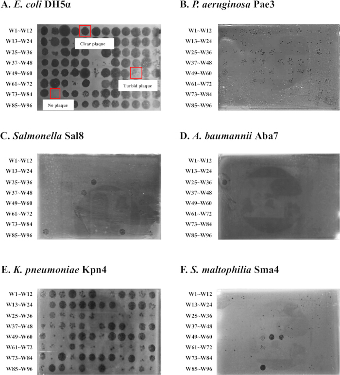
Figure 1: Representative images of phage plaques formed on six different host strains. Representative spot outcomes are indicated as C (clear plaque), T (turbid plaque), and − (no plaque). Plaques were observed on LB soft agar lawns seeded with E. coli DH5α (A), P. aeruginosa Pae3 (B), Salmonella Sal8 (C), A. baumannii Aba7 (D), K. pneumoniae Kpn4 (E), and S. maltophilia Sma4 (F) after application of environmental concentrates using the automated spot-on-lawn method. For activity scoring, both C and T were considered active, whereas − was considered inactive. Please click here to view a larger version of this figure.
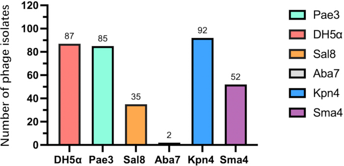
Figure 2: Number of phage isolates obtained against each host strain. The bar graph shows the total count of phages recovered from 96 environmental samples using the six indicated host bacteria. The highest number was obtained against K. pneumoniae Kpn4 (92 isolates), followed by E. coli DH5α (87 isolates), P. aeruginosa Pae3 (85 isolates), S. maltophilia Sma4 (52 isolates), Salmonella Sal8 (35 isolates), and A. baumannii Aba7 (2 isolates). Please click here to view a larger version of this figure.
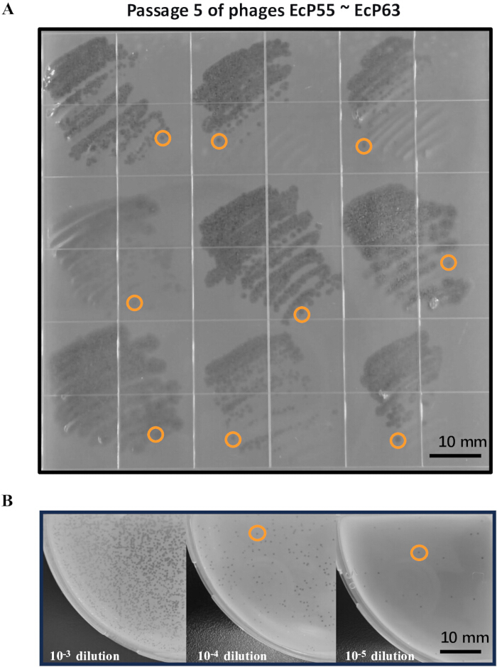
Figure 3: Purification and validation of phage clones. (A) Representative images of single plaques obtained after five successive streak-plate passages (P5) on host lawns. Circles indicate representative single plaques selected for picking. (B) Double-layer agar plate validation showing morphologically consistent plaques at dilutions of 10-3, 10-4, and 10-5. Please click here to view a larger version of this figure.
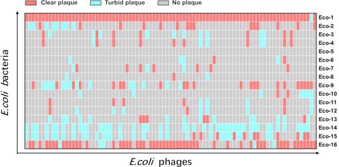
Figure 4: Host range heatmap of 87 E. coli phages against 16 tested E. coli strains. Red squares indicate clear plaques; turquoise squares indicate turbid plaques; and grey squares indicate no lysis. Please click here to view a larger version of this figure.
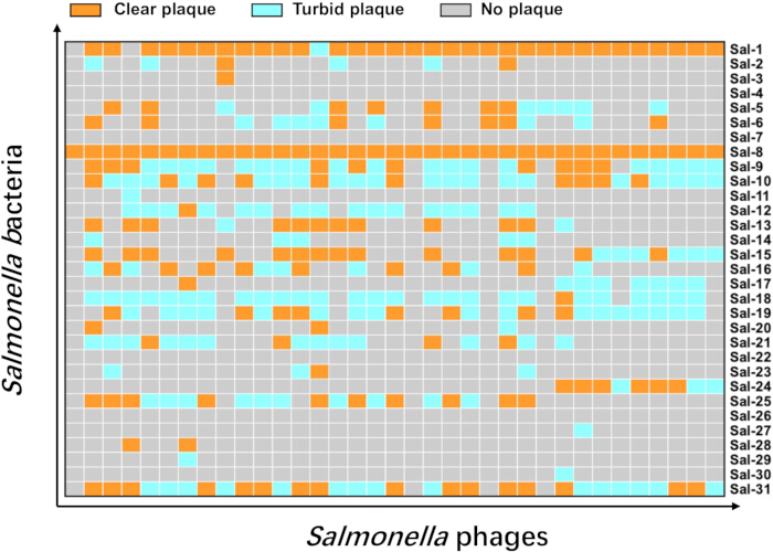
Figure 5: Host range heatmap of 35 Salmonella phages against 31 tested Salmonella strains. Orange squares indicate clear plaques; turquoise squares indicate turbid plaques; and grey squares indicate no lysis. Please click here to view a larger version of this figure.
| Phages | Plaque forming abilitya | |||||||||||||||
| Eco-1 | Eco-2 | Eco-3 | Eco-4 | Eco-5 | Eco-6 | Eco-7 | Eco-8 | Eco-9 | Eco-10 | Eco-11 | Eco-12 | Eco-13 | Eco-14 | Eco-15 | Eco-16 | |
| EcP1 | C | C | - | - | - | - | - | - | - | - | - | - | - | - | - | C |
| EcP2 | T | - | - | - | - | - | - | - | C | T | T | T | - | - | - | C |
| EcP3 | C | - | - | - | - | - | - | - | C | T | - | - | T | T | C | C |
| EcP4 | C | - | - | - | - | - | - | - | C | T | - | - | C | T | C | C |
| EcP5 | C | - | - | - | - | - | - | - | C | T | - | - | - | T | T | C |
| EcP6 | C | C | - | - | - | - | - | - | C | - | - | - | - | T | C | C |
| EcP7 | C | - | - | - | - | - | - | - | - | - | - | - | - | - | - | C |
| EcP8 | C | - | C | C | - | - | - | - | - | - | - | - | - | - | - | - |
| EcP9 | C | - | - | - | - | - | C | - | - | C | - | - | - | - | - | C |
| EcP10 | C | - | C | C | - | C | - | - | C | - | C | C | T | T | - | C |
| EcP11 | C | - | - | - | - | - | - | - | - | - | - | - | - | - | - | C |
| EcP12 | C | C | - | - | - | T | - | - | C | C | - | - | T | T | C | C |
| EcP13 | C | - | - | - | - | - | - | - | - | - | - | - | - | - | - | T |
| EcP14 | C | - | - | - | - | - | - | - | - | - | - | - | - | T | T | - |
| EcP15 | C | - | - | - | - | - | - | - | - | - | - | - | - | T | T | - |
| EcP16 | C | - | T | T | - | - | - | - | - | - | - | - | - | - | - | C |
| EcP17 | C | - | T | T | - | - | - | - | - | T | - | - | - | - | - | C |
| EcP18 | C | C | - | - | - | - | - | - | C | - | - | - | - | T | C | C |
| EcP19 | C | C | - | - | - | - | - | - | C | - | - | - | T | T | C | T |
| EcP20 | C | C | - | - | - | - | - | - | C | - | - | - | T | T | T | C |
| EcP21 | C | - | C | C | - | - | - | - | - | - | T | T | - | - | - | - |
| EcP22 | C | - | - | - | - | - | - | - | - | - | - | - | - | T | C | - |
| EcP23 | C | - | - | - | - | - | - | - | C | - | - | - | - | T | T | C |
| EcP24 | C | - | - | - | - | - | - | - | - | - | - | - | - | T | T | - |
| EcP25 | C | - | - | - | - | - | - | - | - | - | - | - | - | T | C | - |
| EcP26 | C | - | - | - | - | - | - | - | - | - | - | - | - | T | T | - |
| EcP27 | C | - | - | - | - | - | - | - | - | - | - | - | - | T | T | C |
| EcP28 | C | - | - | - | - | - | - | - | - | - | - | - | - | T | T | - |
| EcP29 | C | - | - | - | - | - | - | - | - | - | - | - | - | T | T | - |
| EcP31 | C | - | - | - | - | - | - | - | - | - | - | - | - | T | C | - |
| EcP32 | C | - | - | - | - | C | - | - | - | - | - | - | T | - | - | C |
| EcP33 | C | - | - | - | - | - | C | - | - | - | - | - | - | T | T | - |
| EcP34 | C | - | T | T | - | - | - | T | - | - | T | T | C | - | - | - |
| EcP35 | C | - | - | - | - | - | - | - | - | - | - | - | C | T | - | - |
| EcP36 | C | - | T | T | - | - | - | - | - | - | T | T | T | - | C | - |
| EcP37 | C | - | - | - | - | - | C | - | - | - | - | - | - | - | - | C |
| EcP38 | C | C | - | - | - | - | - | - | C | - | - | - | - | T | C | C |
| EcP39 | C | C | - | - | - | - | - | - | - | - | - | - | - | - | - | C |
| EcP40 | C | C | - | - | - | - | - | - | C | - | - | C | - | T | T | C |
| EcP41 | C | - | T | C | - | T | T | - | C | - | C | - | T | T | - | C |
| EcP42 | C | - | - | - | - | T | - | - | T | - | - | - | - | - | - | C |
| EcP43 | C | C | - | - | - | - | - | - | - | - | - | - | - | - | - | C |
| EcP44 | C | - | T | C | - | - | - | C | - | - | - | - | - | - | - | C |
| EcP45 | C | - | - | - | - | - | - | - | - | - | - | - | - | - | - | C |
| EcP46 | C | - | - | - | - | - | - | - | T | - | - | - | T | T | T | C |
| EcP47 | C | - | - | - | - | - | - | - | - | - | - | - | - | - | - | C |
| EcP48 | C | C | - | - | - | - | - | - | C | - | - | - | - | T | T | C |
| EcP49 | C | - | - | - | - | - | - | - | - | - | - | - | - | - | - | C |
| EcP50 | C | - | - | - | - | - | C | - | - | - | - | - | - | - | - | C |
| EcP51 | C | - | - | - | - | - | - | - | - | - | - | - | - | - | - | C |
| EcP52 | C | - | T | - | - | - | T | - | - | - | - | - | C | T | - | - |
| EcP53 | C | C | - | - | - | - | - | - | T | - | - | - | C | T | T | C |
| EcP54 | C | - | C | C | - | - | C | - | - | - | - | - | C | - | - | C |
| EcP55 | C | - | - | - | - | - | - | - | - | - | - | - | - | - | - | C |
| EcP56 | C | - | - | - | - | - | - | - | T | T | - | - | - | T | T | - |
| EcP57 | C | - | - | - | - | - | - | - | T | - | - | - | - | - | - | C |
| EcP58 | C | - | - | - | - | - | C | - | - | - | - | - | - | - | - | C |
| EcP59 | C | - | - | - | - | - | - | - | C | - | C | T | - | - | - | C |
| EcP60 | C | C | - | - | - | - | - | - | C | - | - | - | - | T | T | C |
| EcP61 | C | - | - | - | - | - | - | - | - | - | - | - | - | - | - | - |
| EcP62 | C | - | C | C | - | - | - | - | C | - | C | T | - | - | - | C |
| EcP63 | C | - | - | - | - | - | - | - | - | - | - | - | - | T | T | - |
| EcP64 | C | - | - | - | - | - | - | - | - | - | - | - | - | T | T | - |
| EcP65 | C | - | - | - | - | - | - | - | - | - | - | - | - | T | T | T |
| EcP66 | C | - | T | - | - | - | - | - | - | - | - | - | - | T | - | T |
| EcP67 | C | - | C | C | - | - | - | - | - | - | - | - | - | - | T | - |
| EcP68 | C | - | T | T | - | - | - | - | - | - | - | - | - | - | - | - |
| EcP69 | C | - | - | - | - | - | - | - | T | - | - | - | T | - | - | - |
| EcP70 | C | - | - | - | - | - | - | - | - | - | - | - | - | T | T | C |
| EcP71 | C | - | - | - | - | - | - | - | - | - | - | - | - | T | T | - |
| EcP72 | C | C | - | - | - | - | - | - | C | T | - | - | T | T | - | C |
| EcP73 | C | - | - | - | - | - | C | - | T | - | - | T | - | - | - | C |
| EcP74 | C | T | - | - | - | - | T | - | C | - | - | - | - | T | T | C |
| EcP75 | C | - | - | - | - | T | - | - | T | - | - | - | - | - | - | C |
| EcP76 | C | T | - | - | - | - | - | T | T | - | - | - | T | - | - | C |
| EcP77 | C | T | - | - | - | - | - | - | T | - | - | - | T | T | T | C |
| EcP78 | C | - | - | - | - | - | - | - | - | - | - | - | - | - | - | T |
| EcP79 | C | T | - | - | - | - | - | - | C | - | - | - | - | T | T | C |
| EcP80 | C | T | - | - | - | - | - | - | T | - | - | - | - | T | T | C |
| EcP81 | C | - | T | - | - | - | - | - | T | - | C | C | T | - | - | C |
| EcP82 | C | T | - | - | - | - | - | - | T | - | - | - | - | T | T | C |
| EcP83 | C | - | C | C | - | - | - | - | T | - | - | - | - | - | - | - |
| EcP84 | C | C | - | - | - | - | - | - | - | - | - | - | - | - | - | C |
| EcP85 | C | - | - | - | - | - | - | - | - | - | - | - | - | T | T | - |
| EcP86 | C | T | - | - | - | - | - | - | C | - | - | - | - | T | T | C |
| EcP87 | C | T | - | - | - | - | - | - | - | - | - | - | - | - | - | C |
| EcP88 | C | - | - | - | - | - | - | - | - | - | - | - | - | T | T | - |
Table 1: The host range of 87 E. coli phages (EcP1 ~ EcP29, EcP31 ~ EcP88) against 16 E. coli strains (Eco-1 ~ Eco-16). Each row represents a phage; each column represents a host strain. Plaque-forming ability was determined by the clarity of the lysis zone: “-”: no plaque; “T”: turbid plaque; “C”: clear plaque. The data are derived from spot-on-lawn screening of environmental samples. Host coverage was defined as the number of tested host strains lysed by at least one phage (“C” or “T”), divided by the total number of tested host strains. Please click here to download this Table.
| Phages | Plaque forming abilitya | ||||||||||||||||||||||||||||||
| Sal-1 | Sal-2 | Sal-3 | Sal-4 | Sal-5 | Sal-6 | Sal-7 | Sal-8 | Sal-9 | Sal-10 | Sal-11 | Sal-12 | Sal-13 | Sal-14 | Sal-15 | Sal-16 | Sal-17 | Sal-18 | Sal-19 | Sal-20 | Sal-21 | Sal-22 | Sal-23 | Sal-24 | Sal-25 | Sal-26 | Sal-27 | Sal-28 | Sal-29 | Sal-30 | Sal-31 | |
| SalP3 | C | - | - | - | - | - | - | C | T | T | - | - | - | - | T | - | - | - | - | - | - | - | - | T | - | - | - | - | - | - | T |
| SalP4 | C | - | - | - | - | - | - | C | T | T | - | - | - | - | T | - | T | T | T | - | - | - | - | T | - | - | - | - | - | - | C |
| SalP5 | C | - | - | - | - | - | - | C | T | T | - | - | - | - | T | - | T | T | T | - | - | - | - | C | - | - | - | - | - | - | C |
| SalP6 | C | - | - | - | T | C | - | C | T | T | - | - | - | - | C | - | T | T | T | - | - | - | - | C | - | - | - | - | - | - | T |
| SalP7 | C | - | - | - | - | - | - | C | T | C | - | - | - | - | T | - | T | T | T | - | - | - | - | C | - | - | - | - | - | - | T |
| SalP8 | C | - | - | - | - | - | - | C | - | T | - | - | - | - | T | - | - | - | T | - | - | - | - | T | - | - | - | - | - | - | T |
| SalP9 | C | - | - | - | - | - | - | C | C | C | - | - | - | - | T | - | T | T | T | - | - | - | - | C | - | - | - | - | - | - | T |
| SalP10 | C | - | - | - | T | T | - | C | C | C | - | - | - | - | C | T | T | T | T | - | - | - | - | C | - | - | T | - | - | - | T |
| SalP11 | C | - | - | - | T | - | - | C | C | C | - | - | T | - | - | - | T | C | C | - | T | - | - | C | - | - | - | - | - | T | C |
| SalP12 | C | - | - | - | T | - | - | C | - | - | - | - | - | - | - | - | - | - | - | - | - | - | - | - | - | - | - | - | - | - | - |
| SalP13 | C | - | - | - | T | T | - | C | C | T | - | T | C | T | C | C | - | T | C | - | T | - | T | - | C | - | - | - | - | - | C |
| SalP14 | C | C | - | - | C | C | - | C | T | T | - | T | C | T | C | - | - | T | T | T | C | - | - | - | C | - | - | - | - | - | C |
| SalP15 | C | - | - | - | C | C | - | C | - | - | - | - | - | - | - | - | - | - | - | - | - | - | - | - | - | - | - | - | - | - | - |
| SalP16 | C | - | - | - | - | - | - | C | T | T | - | T | - | - | - | T | - | T | T | - | T | - | - | - | T | - | - | - | - | - | C |
| SalP17 | C | - | - | - | - | - | - | C | T | T | - | T | - | - | - | C | - | T | C | - | - | - | - | - | C | - | - | - | - | - | C |
| SalP18 | C | T | - | - | C | C | - | C | T | T | - | T | C | - | C | - | - | T | - | - | C | - | - | - | T | - | - | - | - | - | T |
| SalP19 | C | - | - | - | - | - | - | C | - | - | - | - | - | - | - | - | - | - | - | - | - | - | - | - | - | - | - | - | - | - | - |
| SalP20 | C | - | - | - | - | - | - | C | C | C | - | T | - | - | - | C | - | T | C | - | - | - | - | - | C | - | - | - | - | - | C |
| SalP21 | C | - | - | - | C | T | - | C | T | T | - | T | - | - | - | - | - | T | T | - | - | - | - | - | T | - | - | - | - | - | T |
| SalP22 | C | - | - | - | - | - | - | C | C | T | - | T | C | - | C | T | - | T | T | - | T | - | - | - | C | - | - | - | - | - | T |
| SalP23 | C | T | - | - | C | C | - | C | T | T | - | - | C | - | C | - | - | - | - | - | T | - | - | - | T | - | - | - | - | - | T |
| SalP24 | T | - | - | - | T | T | - | C | C | C | - | T | C | - | C | - | - | T | T | C | T | - | C | - | C | - | - | - | - | - | C |
| SalP25 | C | - | - | - | - | T | - | C | T | T | - | T | C | T | C | C | - | T | C | - | T | - | T | - | - | - | - | - | - | - | C |
| SalP26 | C | - | - | - | - | T | - | C | T | T | - | - | C | T | C | T | - | T | C | - | C | - | - | - | T | - | - | - | - | - | T |
| SalP27 | C | - | - | - | - | - | - | C | T | T | - | T | - | - | - | T | - | T | T | - | - | - | - | - | T | - | - | - | - | - | C |
| SalP28 | C | - | - | - | - | T | - | C | T | C | - | T | - | - | - | C | - | T | C | - | - | - | - | - | T | - | - | - | - | - | C |
| SalP29 | C | C | C | - | T | - | - | C | - | - | - | - | T | - | C | - | - | - | - | - | - | - | - | - | - | - | - | - | - | - | T |
| SalP30 | C | - | - | - | - | - | - | C | T | C | - | T | - | - | - | C | - | T | T | - | T | - | - | - | C | - | - | - | - | - | C |
| SalP31 | C | - | - | - | - | - | - | C | T | T | - | C | - | - | - | - | C | T | T | - | T | - | - | - | T | - | - | C | T | - | T |
| SalP32 | C | - | - | - | - | - | - | C | T | C | - | T | - | - | - | C | - | T | T | - | T | - | - | - | T | - | - | - | - | - | T |
| SalP33 | C | T | - | - | C | C | - | C | T | T | - | T | C | - | C | - | - | T | - | - | C | - | - | - | T | - | - | - | - | - | T |
| SalP34 | - | - | - | - | - | - | - | C | C | T | T | T | C | - | C | T | - | T | T | - | T | - | - | - | C | - | - | C | - | - | C |
| SalP35 | C | - | - | - | C | - | - | C | C | T | - | - | - | - | - | C | - | T | C | - | T | - | T | - | C | - | - | - | - | - | C |
| SalP36 | C | T | - | - | - | C | - | C | C | C | - | - | C | T | C | T | - | T | - | C | T | - | - | - | C | - | - | - | - | - | C |
| SalP37 | - | - | - | - | - | - | - | C | - | - | - | - | - | - | - | - | - | - | - | - | - | - | - | - | - | - | - | - | - | - | - |
Table 2: The host range of 35 Salmonella phages (SalP3 ~ SalP37) against 31 Salmonella strains (Sal-1 ~ Sal-31). Each row represents (SalP3~SalP37) against 31 Salmonella strains (Sal-1~Sal-31). Each row represents a phage; each column represents a host strain. Plaque-forming ability was determined by the clarity of the lysis zone: “-”: no plaque; “T”: turbid plaque; “C”: clear plaque. The data are derived from spot-on-lawn screening of environmental samples. Host coverage was defined as the number of tested host strains lysed by at least one phage (“C” or “T”), divided by the total number of tested host strains. Please click here to download this Table.
Supplementary Figure 1: Layout of instrument deck plates for automated spot screening. P1: tip box; P3: 96-deep-well plate with environmental concentrates; P4–P12: host-containing soft agar plates. Please click here to download this file.
Supplementary Figure 2: Automated liquid-handling parameters for high-throughput spot screening. Liquid class settings for 50 µL pipette tips, including tip-on parameters, aspiration speed, dispensing speed, and dispensing height above the agar surface. Please click here to download this file.
Supplementary Figure 3: Screenshot of the “Open” window for program file selection. The user selects the pre-saved protocol file from the program interface. Please click here to download this file.
Supplementary Figure 4: Screenshot of the run interface showing program verification and execution. The user clicks “Run” after successful “Check” verification. Please click here to download this file.
المناقشة
Several steps in this protocol require careful attention to ensure optimal outcomes. First, we recommend overnight incubation at 4 °C following PEG 8000 addition for precipitation; incomplete dissolution may result in suboptimal phage precipitation and reduced recovery rates. Second, host strains should be harvested at mid-to-late logarithmic phase to ensure maximal susceptibility to phage infection, as stationary-phase cells often exhibit reduced receptor expression and altered membrane composition that impede phage adsorption15. Finally, we recommend a minimum of five successive streaking passages to reduce the risk of mixed phage populations and to promote genetic homogeneity. Purity should be confirmed by a conventional double-layer agar assay to verify plaque morphology prior to propagation and host-range analysis.
To improve reproducibility, we provide troubleshooting guidance for common issues in phage isolation and plaque assays. Weak or turbid plaques may reflect intrinsic phage lytic capacity or suboptimal host/titer conditions; broader environmental sampling may help isolate stronger phages, while mid-exponential hosts, higher phage input, and optimized overlay/incubation can improve condition-dependent cases. Low recovery of phages from environmental water may result from poor adsorption, low host density, or buffer issues; pre-enrichment, optimized adsorption, minimal processing, and inhibitor removal are recommended. Irregular spotting may reflect uneven bacterial lawns, inconsistent plate drying, variable spot volumes, or non-level plates, and can be minimized by standardizing lawn density, plate drying, and spotting volumes. Mixed plaques should be addressed by repeated single-plaque picking, serial streak purification, and purity confirmation. These measures can reduce batch-to-batch variability and improve reproducibility across laboratories. Medium selection is also an important consideration in phage enrichment. LB is widely used for many Gram-negative bacteria, including Escherichia coli, Klebsiella, and Pseudomonas, but it is not optimal for all target hosts; for example, Listeria species grow poorly in LB and generally require enriched or selective media such as Listeria Special Broth13. Selective media can also suppress non-target bacteria, thereby reducing competition and enhancing the recovery of phages specific to the target host. Such trade-offs should be considered when designing high-throughput enrichment workflows, and the choice of medium may need to be adapted to the target pathogen.
Despite its advantages in throughput and efficiency, this protocol has several limitations. First, spot screening provides only a qualitative assessment of phage activity. The presence or absence of plaques indicates infective potential but does not directly quantify lytic efficiency or burst size. Precise infectivity quantification requires subsequent liquid-culture lysis kinetics or one-step growth curve analysis16. Moreover, the 5 µL spotting volume may limit detection of extremely low-titer phages. Second, the current workflow does not include genetic characterization of isolated phages, necessitating downstream whole-genome sequencing and bioinformatic analysis to assess their therapeutic safety and exclude lysogenic phages17. Whole-genome sequencing will also enable deduplication of potentially redundant phages, which is particularly relevant given the observed host-range overlap. Third, PEG precipitation may introduce recovery bias for different phage types, and its efficiency should be compared with other concentration methods, such as ultrafiltration or ultracentrifugation18. Fourth, automated liquid-handling robots may theoretically drop pipette tips, which has implications for biological safety and containment. In our enclosed system, tips remained securely attached during operation and were released only into the waste container at the programmed step; no unintended drop events were observed. The workstation is enclosed, UV-decontaminated, and routine calibration, matched tips, and inspection of tip ejection further reduce this risk.
Conventional phage isolation workflows typically employ single-host enrichment followed by repeated double-layer agar plating for each host-sample combination, a process that is labor-intensive and material-consuming19. High-throughput alternatives such as turbidity-based or dye-based microplate assays enable simultaneous screening of numerous samples but suffer from false positives caused by non-phage-mediated bacterial lysis and still require subsequent plaque confirmation. Our integrated approach combines the specificity of plaque-based detection with the throughput of automated liquid handling, enabling screening against diverse host panels without the need for repeated double-layer plating. The use of 96-deep-well format storage further facilitates large-scale processing and systematic archiving of phage libraries. Compared with these existing methods, our protocol offers a practical balance between throughput, reliability, and operational simplicity that is readily adoptable by standard microbiology laboratories.
This protocol is particularly valuable for researchers engaged in constructing phage biobanks, preparing customized phage cocktails for personalized therapy, and monitoring phage diversity in ecological and clinical settings. The ability to simultaneously screen environmental samples against multiple pathogens within a single experimental setup significantly accelerates the discovery of phages with novel host ranges and lytic properties. Future integration of automated image analysis for plaque detection and scoring will enable objective, high-throughput quantification and reduce reliance on manual observation. Combining whole-genome sequencing with machine learning approaches may further predict phage host range and accelerate rational cocktail design.
The method described herein is protected by a granted patent (Patent No. ZL202311255800.3), which covers the integrated workflow for high-throughput isolation and purification of multiple host phages from environmental samples, underscoring its novelty and potential for translational applications, including customized phage cocktail development for clinical therapy and industrial biocontrol. In summary, this protocol represents a practical and scalable approach that will substantially accelerate the discovery and characterization of phage resources, providing robust support for combating antimicrobial-resistant bacterial infections.
الإفصاحات
The method described in this manuscript is covered by granted patent ZL202311255800.3 (March 8, 2024), titled “A High-Throughput Method for the Isolation and Purification of Multi-Host Phages.” Wan, X. F., Lu, H. Z., Duan, X. K., Han, S. H., Shen, J. Y., Zheng, M. B., and Zhou, Y. are listed as inventors. The patent is co-assigned to Shenzhen National Clinical Research Center for Infectious Diseases and Shenzhen Third People's Hospital (Institute of Hepatology). Duan, X. K., is employed by PhageNovoTech Co., Ltd. The remaining authors declare no competing financial interests.
The authors used Deepseek for language editing and grammar checking and reviewed and edited the resulting content. The authors take full responsibility for the final publication.
شكر وتقدير
This study was supported by National Natural Science Foundation of China (No. 82473753, No. 82070420); National Key R&D Program of China (No. 2025YFC3408400); Shenzhen key laboratory for infectious diseases; Shenzhen Clinical Medical Center for Emerging infectious diseases (LCYSSQ20220823091203007); Guangdong Key Lab for Diagnosis & Treatment of Emerging Infectious Diseases, Shenzhen Third People's Hospital, Shenzhen, China; Shanghai Science and Technology Innovation Action Plan in 2022 (No. 22N31900800), Shenzhen Science and Technology Program (No. KJZD20240903100500001), Shenzhen Natural Science Foundation (JCYJ20250604143818024), Zhong Nanshan Medical Foundation of Guangdong Province (ZNZXS-2024010), and Shenzhen High-level Hospital Construction Fund.
المواد
| الاسم | الشركة | رقم فهرسي | التعليقات |
|---|---|---|---|
| 96-deep-well plates | NEST | 503162 | |
| 96-well cell culture plate, flat bottom, tissue culture-treated, sterile | NEST | 701001 | 127.76 mm × 85.48 mm |
| Agar | Solarbio | A8190 | |
| Cell and Tissue Culture Plates | Biofil | TCP001001 | 128 mm × 85.7 mm |
| Class II, Type A2 biological safety cabinet | Thermo Fisher Scientific | 1389 | |
| LB nutrient agar | Solarbio | L8290 | |
| Luria-Bertani broth (LB) | Solarbio | L8291 | |
| NEMO full-function liquid handling system | Biohander | version 1.2.2.3 | |
| Pasteur Pipette | NEST | 318212 | |
| Petri dishes | Biosharp | BS-90-D1 | 90 mm |
| Phosphate-buffered saline (PBS) | Solarbio | G4207 | |
| Polyethylene glycol (PEG) 8000 | Biofroxx | 1363GR500 | |
| Sodium chloride (NaCl) | Solarbio | S8210 | |
| Square petri dishes | Biosharp | BS-100-SD | 100 mm × 100 mm |
| Syringe filters | Millipore | SLGPR33RB | 0.22 μm |
| Tecan tips | Haier Biomedical | 121157 | 50 μL |
المراجع
- Antimicrobial Resistance Collaborators. Global burden of bacterial antimicrobial resistance in 2019: a systematic analysis. Lancet. 2022;399(10325):629–55.
- Singh K, Biswas A, Chakrabarti AK, Dutta S. Phage therapy as a protective tool against pathogenic bacteria: how far we are? Curr Pharm Biotechnol. 2023;24(10):1277–90.
- Chanishvili N. Phage therapy—history from Twort and d'Herelle through Soviet experience to current approaches. Adv Virus Res. 2012;83:3–40.
- Strathdee SA, Hatfull GF, Mutalik VK, Schooley RT. Phage therapy: from biological mechanisms to future directions. Cell. 2023;186(1):17–31.
- Onallah H, et al. Protocol for phage matching, treatment, and monitoring for compassionate bacteriophage use in non-resolving infections. STAR Protoc. 2024;5(2):102949.
- Pirnay JP, et al. Personalized bacteriophage therapy outcomes for 100 consecutive cases: a multicentre, multinational, retrospective observational study. Nat Microbiol. 2024;9(6):1434–53.
- Sarhan WA, Azzazy HM. Phage approved in food, why not as a therapeutic? Expert Rev Anti Infect Ther. 2015;13(1):91–101.
- Gao L, et al. Isolation and characterization of a lytic vibriophage OY1 and its biocontrol effects against Vibrio spp. Front Microbiol. 2022;13:830692.
- Wommack KE, Colwell RR. Virioplankton: viruses in aquatic ecosystems. Microbiol Mol Biol Rev. 2000;64(1):69–114.
- Kortright KE, Chan BK, Koff JL, Turner PE. Phage therapy: a renewed approach to combat antibiotic-resistant bacteria. Cell Host Microbe. 2019;25(2):219–32.
- Gill JJ, Hyman P. Phage choice, isolation, and preparation for phage therapy. Curr Pharm Biotechnol. 2010;11(1):2–14.
- Yu P, et al. Isolation of polyvalent bacteriophages by sequential multiple-host approaches. Appl Environ Microbiol. 2016;82(3):808–15.
- Martinez-Soto CE, et al. PHIDA: a high throughput turbidimetric data analytic tool to compare host range profiles of bacteriophages isolated using different enrichment methods. Viruses. 2021;13(11):2120.
- Kropinski AM, et al. Enumeration of bacteriophages by double agar overlay plaque assay. In: Clokie MRJ, Kropinski AM, editors. Bacteriophages: methods and protocols. Volume 1: isolation, characterization, and interactions. Totowa, NJ: Humana Press; 2009. p. 69–76.
- Jiang L, et al. Growth stage-related capsular polysaccharide translocon Wza in Vibrio splendidus modifies phage vB_VspM_VS2 susceptibility. Commun Biol. 2024;7(1):1338.
- Vaiyapuri M, et al. Comparison of single and multi-host enrichment approach for harnessing lytic phages against antimicrobial-resistant E. coli: repurposing the enrichment step. Biologia. 2021;76(3):1041–52.
- Hyman P. Phages for phage therapy: isolation, characterization, and host range breadth. Pharmaceuticals (Basel). 2019;12(1):35.
- Hjelmsø MH, et al. Evaluation of methods for the concentration and extraction of viruses from sewage in the context of metagenomic sequencing. PLoS One. 2017;12(1):e0170199.
- Olsen NS, Hendriksen NB, Hansen LH, Kot W. A new high-throughput screening method for phages: enabling crude isolation and fast identification of diverse phages with therapeutic potential. Phage (New Rochelle). 2020;1(3):137–48.
إعادة الطباعة والأذونات
تم نشر هذه المقالة
الفيديو قريباً