This protocol describes ultrasound-guided high-voltage long-duration pulsed radiofrequency of the suprascapular nerve in patients with persistent hemiplegic shoulder pain, including patient selection, ultrasound localization, sensory and motor confirmation, radiofrequency parameter settings, and post-procedural assessment.
مقالة منهجية
Ultrasound-Guided High-Voltage Long-Duration Pulsed Radiofrequency of the Suprascapular Nerve for Persistent Hemiplegic Shoulder Pain
في هذه المقالة
ملخص
الملخص
Hemiplegic shoulder pain (HSP) is a common complication after stroke that can interfere with upper-limb rehabilitation. Persistent cases may respond inadequately to conventional conservative treatments or suprascapular nerve blocks. This article describes a standardized ultrasound-guided high-voltage long-duration pulsed radiofrequency (HL-PRF) protocol targeting the suprascapular nerve. Ultrasound is used to identify the suprascapular notch, surrounding anatomical structures, and adjacent vessels. A radiofrequency cannula is advanced to the target under real-time guidance, and its position is confirmed by sensory and motor stimulation. HL-PRF is then delivered at 99 V for 6 min while the temperature is maintained at or below 42 °C. The clinical outcomes presented here are illustrative observations from two patients with persistent HSP and do not represent findings from a controlled clinical study. In both cases, lower resting and movement-evoked pain scores and increased shoulder passive range of motion were observed at the 1-month follow-up. Pain scores at the 3-month telephone follow-up remained below baseline values. No procedure-related adverse events were observed or documented in either case during the available follow-up period. These cases are included to illustrate the clinical application and potential outcomes of the protocol rather than to establish efficacy. This protocol provides a detailed procedural framework for ultrasound-guided HL-PRF of the suprascapular nerve in patients with persistent HSP.
المقدمة
Hemiplegic shoulder pain (HSP) is one of the most common and disabling complications following a stroke, with an incidence rate ranging from 16% to 84%, typically manifesting within the first six months post-stroke1,2,3. The pathological mechanism of HSP is highly complex, involving biomechanical alterations, motor dysfunction such as hemiplegic muscle spasticity, and both central and peripheral nervous system sensitization4,5,6,7,8. Severe resting and movement-evoked pain not only restricts the maximum passive range of motion (PROM) of the shoulder joint but also directly impedes the overall motor function recovery of the hemiplegic upper limb7. Currently, conventional conservative treatments for persistent HSP, including physical therapy, transcutaneous electrical nerve stimulation, and oral analgesics, often yield limited efficacy9,10,11,12. Among invasive interventions, the suprascapular nerve (SSN) has emerged as an ideal analgesic target because it provides approximately 70% of the sensory innervation to the shoulder joint13. However, alternative techniques have notable drawbacks; simple local anesthetic nerve blocks provide only short-term relief, while repeated corticosteroid injections carry potential side effects such as hyperglycemia and tendon rupture10,14. Pulsed radiofrequency (PRF) has garnered attention in the wider body of literature as a non-destructive neuromodulation technique that releases short, high-frequency electrical currents followed by a silent cooling phase, keeping the local tissue temperature at or below 42 oC15,16. Because it avoids protein denaturation, PRF does not destroy myelin sheaths or axonal structures, circumventing the risks of neuritis or long-term motor deficits associated with conventional thermal radiofrequency ablation17,18.
Suprascapular nerve PRF has previously been investigated for hemiplegic shoulder pain, including in randomized controlled studies, supporting its use as an interventional option for this population8,19. More recently, high-voltage PRF targeting the suprascapular nerve has been described in patients with chronic shoulder pain20,21,22. The 99 V and 6 min settings used in the present protocol were selected from previously published high-voltage and long-duration PRF approaches, including studies of other peripheral neuralgia indications23,24, while maintaining the electrode-tip temperature at or below 42 °C. These parameters were adopted to provide a standardized procedural framework for suprascapular nerve HL-PRF rather than being derived from a parameter-optimization study. Accordingly, the primary purpose of this Methods Article is to provide a standardized, ultrasound-guided procedural framework adapted specifically to patients with persistent HSP. The protocol emphasizes patient selection, anatomical localization of the suprascapular notch and adjacent structures, sensory and motor confirmation of cannula position, standardized procedural monitoring, and structured clinical outcome assessment in the post-stroke rehabilitation setting. This protocol is intended for rehabilitation physicians, pain physicians, and other clinicians experienced in musculoskeletal ultrasound and percutaneous radiofrequency procedures, working in clinical settings with appropriate ultrasound and radiofrequency equipment, sterile procedural facilities, and patient monitoring capabilities.
البروتوكول
All procedures described in this protocol followed the guidelines of the institutional human research ethics committee (Ethics Approval Number: WZY2026-LW-021-01). Written informed consent was obtained from all patients before the procedures.
NOTE: The representative clinical outcomes reported in this article are derived from two patients with persistent hemiplegic shoulder pain who underwent the protocol described below. These cases are presented as illustrative clinical examples and do not constitute a controlled clinical study. The primary purpose of this article is to describe the procedural steps required to perform ultrasound-guided high-voltage long-duration pulsed radiofrequency of the suprascapular nerve.
1. Patient selection and preparation
- Confirm that the patient is at least 18 years old and has imaging-confirmed ischemic or hemorrhagic stroke with subsequent hemiplegia, regardless of muscle tone status.
- Confirm that at least 1 month has elapsed since stroke onset and that affected-side shoulder pain has persisted for at least 4 weeks despite conventional rehabilitation/physical modalities and/or stepwise analgesic treatment.
- Confirm movement-evoked pain intensity of at least 4 on the Numerical Rating Scale (NRS) during passive external rotation of the affected shoulder to the maximum tolerated range. For patients with mild aphasia or cognitive impairment who can reliably use the Faces Pain Scale-Revised (FPS-R), require an FPS-R score of at least 4.
- Confirm some limitation in passive range of motion of the affected shoulder.
NOTE: Conventional treatments include oral or topical analgesics, local injection therapy, acupuncture, and routine rehabilitation interventions. - Exclude patients who underwent peripheral nerve radiofrequency intervention involving the affected shoulder within the previous 6 months or previous surgery involving the affected shoulder or cervical region.
- Exclude patients with traumatic or non-stroke neurological conditions causing paralysis of the affected shoulder, or with another clearly identifiable orthopedic or neurological cause of shoulder or cervical pain, such as shoulder dislocation, fracture, severe rotator cuff tear, or cervical radiculopathy.
- Exclude patients with severe cognitive impairment, severe aphasia, or psychiatric disorders that prevent them from following basic instructions or providing a reliable pain assessment; severe cardiac, hepatic, or renal dysfunction that precludes the procedure; or local skin infection at the planned puncture site.
NOTE: Do not exclude a patient solely because verbal communication is impaired if the patient can reliably indicate pain using the FPS-R. Previous acupuncture or local injection therapy alone is not an exclusion criterion. - Before the procedure, reconfirm eligibility and the absence of new contraindications.
- Review and document current analgesic medications and concomitant rehabilitation treatments. Record any recent changes.
- Confirm that the patient can understand and respond to the instructions required for sensory and motor stimulation testing. Reinspect the planned puncture site for local skin infection before proceeding.
- Position the patient in a seated or lateral decubitus position and expose the affected shoulder and cervicothoracic region.
- Attach a neutral electrode pad to the patient's affected upper limb.
- Disinfect the affected shoulder and surrounding skin with povidone-iodine. Use the disposable sterile medical underpad (90 cm × 200 cm) as a sterile drape to establish the procedural field.
CAUTION: Povidone-iodine may irritate the skin, eyes, or mucous membranes. Wear gloves, avoid splashing or direct contact with eyes and mucous membranes, and follow institutional chemical-safety procedures.
2. Ultrasound localization of the suprascapular nerve
- Select a low-frequency curvilinear ultrasound transducer (e.g., 2–5 MHz) and cover it with a sterile ultrasound probe cover. Place the transducer parallel to the middle and lateral one-third of the scapular spine on the affected side to perform a short-axis scan.
- Adjust the imaging depth (typically 5–7 cm) and gain to visualize the trapezius, supraspinatus, suprascapular notch, superior transverse scapular ligament, and underlying bony acoustic shadow (see Figure 1).
- (Critical step) Identify the suprascapular notch and superior transverse scapular ligament before cannula insertion. When the SSN is not directly visualized, use the osseofibrous space formed by the notch and ligament as the primary target landmark.
- Activate color Doppler mode and identify the suprascapular artery. If the artery is not displayed, observe the suprascapular notch in real time for arterial pulsation and use the pulsation as an additional vascular landmark. Plan the subsequent cannula trajectory to avoid the vessel.
3. Cannula placement and stimulation testing
- Use a 22 G, 10 cm radiofrequency cannula with a 10 mm active tip. Under real-time ultrasound guidance, use an in-plane approach and slowly advance the cannula from the medial to the lateral side of the transducer toward the suprascapular notch.
CAUTION: Percutaneous cannula insertion presents sharps and blood-exposure hazards. Wear gloves and appropriate personal protective equipment, handle the cannula and stylet using standard sharps precautions, and avoid recapping or manipulating contaminated sharps unnecessarily. - Under continuous real-time ultrasound guidance, advance the cannula tip through the superior transverse scapular ligament into the suprascapular notch until the tip contacts the bony floor. Retract the cannula slightly and position the tip adjacent to the suprascapular nerve (see Figure 2).
- (Critical step) Maintain continuous visualization of the cannula tip during transligamentous advancement and confirm bony-floor contact before slight retraction. Do not advance the cannula when the tip cannot be visualized.
- Set the sensory stimulation frequency to 50 Hz and gradually increase the stimulation voltage. A concordant shoulder sensation, such as a dull ache, pain, or paresthesia, at <0.5 V supports proximity of the cannula tip to the suprascapular nerve.
- Set the motor stimulation frequency to 2 Hz and gradually increase the voltage. Observe for rhythmic contractions of the supraspinatus or infraspinatus at <0.5 V; such a response supports placement of the cannula tip near motor fibers of the suprascapular nerve (see Supplementary Video 1).
- (Critical step) Interpret the sensory and motor stimulation thresholds together with the ultrasound-confirmed anatomical position. Do not proceed to HL-PRF until the stimulation findings support cannula placement adjacent to the SSN.
NOTE: If the expected sensory and motor responses are not obtained, carefully adjust the depth, direction, or rotational orientation of the cannula and repeat the stimulation tests. Avoid rapid or forceful cannula movements.
4. High-voltage long-duration pulsed radiofrequency treatment
- After satisfactory sensory and motor stimulation responses have been obtained, perform negative aspiration to confirm that there is no blood return. Then select the pulsed radiofrequency mode on the radiofrequency generator.
- Set the target temperature limit to a maximum of 42 °C.
NOTE: Maintaining the electrode-tip temperature at or below 42 °C is intended to limit the risk of thermal neurolysis and motor injury. - Set the radiofrequency voltage to 99 V, treatment duration to 6 min, pulse frequency to 2 Hz, and preset pulse width to 20 ms (see Figure 3).
NOTE: The 99 V and 6 min parameters were selected a priori based on previously published high-voltage and long-duration PRF approaches. Alternative voltage or duration settings were not systematically tested during protocol development or in the two illustrative cases presented here. - Start HL-PRF delivery. Monitor verbal and nonverbal signs of discomfort and treatment tolerance throughout the 6 min procedure. Observe for rhythmic contractions of the supraspinatus or infraspinatus during treatment.
- If intolerable pain or strong muscle contraction occurs, pause HL-PRF delivery or reduce the voltage. Resume gradual titration toward the target voltage only after the symptoms have subsided and the patient can tolerate further stimulation.
- After the 6 min radiofrequency treatment is complete, slowly withdraw the stylet.
- Prepare 2 mL of 0.375% ropivacaine by mixing 1 mL of 0.75% ropivacaine hydrochloride injection with 1 mL of 0.9% sodium chloride. Attach the syringe containing the prepared solution to the cannula.
CAUTION: Ropivacaine can cause local-anesthetic systemic toxicity if administered intravascularly or in excessive dose. Verify the drug, concentration, and volume; wear gloves; and follow institutional medication-safety and monitoring procedures. - Aspirate to confirm that there is no blood return.
- Slowly inject the prepared 2 mL ropivacaine solution through the cannula while monitoring the patient.
NOTE: The ropivacaine injection is administered after completion of HL-PRF to reduce immediate post-procedural discomfort. - Withdraw the radiofrequency cannula slowly.
- Apply local pressure to the puncture site for 3 min to prevent bleeding. Cover the puncture wound with a sterile adhesive wound dressing.
- Immediately discard used cannulas, stylets, needles, and syringes in an approved sharps container. Dispose of contaminated probe covers, dressings, underpads, and other clinical waste according to institutional biosafety and medical-waste policies.
5. Outcome assessment and safety monitoring
- Assess clinical outcomes at baseline and at the 1-month post-procedural follow-up. Record resting pain intensity using the 0–10 Numerical Rating Scale (NRS). Assess movement-evoked pain during passive external rotation of the affected shoulder to the maximum tolerated range and record the corresponding NRS score.
- Measure passive range of motion (PROM) of the affected shoulder in the supine position using an electronic goniometer. Record abduction, external rotation, forward flexion, and adduction in degrees.
- Assess upper-limb motor function using the Fugl-Meyer Assessment-Upper Extremity (FMA-UE) at baseline and at the 1-month follow-up.
- Have the clinical outcome assessments performed by a physician who is not involved in the HL-PRF procedure. Document whether the assessor is blinded to treatment status.
NOTE: In the two representative cases presented here, assessments were performed by a physician who was not involved in the intervention but was aware that the patients had undergone HL-PRF. - At the 3-month telephone follow-up, reassess resting and movement-evoked pain by patient report. PROM and FMA-UE are not reassessed during the telephone follow-up.
- Monitor and document procedure-related adverse events, including neuritis, hematoma, infection, new sensory or motor deficits, intolerable stimulation-related pain, thermal injury, and the need for additional rescue analgesia or other rescue pain interventions.
NOTE: Figure 4 presents a schematic of the six-step workflow for ultrasound-guided suprascapular nerve HL-PRF protocol described above.
النتائج
The Representative Results section presents clinical observations from n = 2 patients with persistent hemiplegic shoulder pain who underwent ultrasound-guided high-voltage long-duration pulsed radiofrequency (HL-PRF; 99 V, 6 min) of the suprascapular nerve. These cases are provided as illustrative examples of the application of the protocol rather than as evidence from a controlled clinical study. The two patients had markedly different baseline upper-limb motor function, as assessed using the Fugl-Meyer Assessment-Upper Extremity (FMA-UE), allowing the observed clinical outcomes to be illustrated across different levels of baseline motor impairment (see Table 1).
Case 1: Higher baseline upper-limb motor function
Case 1 was a 45-year-old man with left hemiplegia following intracerebral hemorrhage who underwent HL-PRF 76 days after stroke onset. Shoulder pain had persisted for approximately 50 days despite conventional treatment, meeting the criteria for persistent HSP described above. At baseline, the FMA-UE score was 47. The NRS score was 2 for resting pain and 5 for movement-evoked pain. Shoulder PROM was 123.6 ° for abduction, 67.0 ° for external rotation, 151.2° for forward flexion, and 40.7° for adduction.
Concomitant rehabilitation before and after HL-PRF included occupational therapy, physical agent modalities (microwave therapy and medium-frequency electrical stimulation), acupuncture, and tuina (Chinese therapeutic massage), generally performed 5 times per week for 30 min per session for each modality. Because shoulder pain was aggravated during occupational therapy, occupational therapy was temporarily suspended for approximately 1 week before HL-PRF and was resumed after the procedure; the other rehabilitation interventions were continued. The patient was taking celecoxib 200 mg twice daily before HL-PRF and discontinued it on post-procedural day 2.
At the 1-month post-procedural follow-up, the resting NRS score changed from 2 at baseline to 0, and the movement-evoked NRS score changed from 5 to 1. Shoulder PROM increased from 123.6° to 156.5° for abduction and from 67.0° to 79.3° for external rotation, while forward flexion changed from 151.2° to 151.5° and adduction from 40.7° to 42.4°. The FMA-UE score increased from 47 to 56. Supplementary Video 2 and Supplementary Video 3 illustrate the observed changes in shoulder movement between baseline and the 1-month follow-up. At the 3-month telephone follow-up, the patient reported a resting NRS score of 0 and a movement-evoked pain score of 2. These findings describe the clinical course observed in this individual case and should not be interpreted as evidence of treatment efficacy.
Case 2: Lower baseline upper-limb motor function
Case 2 was a 56-year-old man with left hemiplegia following ischemic stroke who underwent HL-PRF 46 days after stroke onset. Shoulder pain had persisted for approximately 30 days despite conventional treatment, meeting the criteria for persistent HSP described above. Baseline motor function was markedly impaired, with manual muscle testing grade 1 and an FMA-UE score of 5. The NRS score was 4.5 for resting pain and 7.5 for movement-evoked pain. Shoulder PROM was 95.0° for abduction, 60.7° for external rotation, 114.4° for forward flexion, and 16.1° for adduction.
Concomitant rehabilitation included occupational therapy, physical agent modalities (microwave therapy and medium-frequency electrical stimulation), acupuncture, and tuina (Chinese therapeutic massage). The rehabilitation regimen was maintained without substantial change before and after HL-PRF, with each modality generally performed 5 times per week for approximately 30 min per session. The patient was taking celecoxib 200 mg twice daily before HL-PRF and discontinued it on post-procedural day 6. Neither patient received antispasticity medication during the peri-procedural period.
At the 1-month post-procedural follow-up, the resting NRS score changed from 4.5 at baseline to 1.5, and the movement-evoked NRS score changed from 7.5 to 3.5. Shoulder PROM increased from 95.0° to 110.1° for abduction, from 60.7° to 86.4° for external rotation, from 114.4° to 126.2° for forward flexion, and from 16.1° to 29.9° for adduction. The FMA-UE score changed from 5 to 6. At the 3-month telephone follow-up, the patient reported a resting NRS score of 2.5 and a movement-evoked pain score of 4. Thus, pain scores remained below their baseline values at the telephone follow-up, whereas the change in upper-limb motor function at 1 month was limited. These observations illustrate the clinical course of a patient with markedly impaired baseline upper-limb motor function and do not establish a causal treatment effect.
Interpretation of representative outcomes
Across the two representative cases, lower resting and movement-evoked pain scores and greater shoulder PROM were observed at the 1-month follow-up compared with baseline. Changes in FMA-UE differed between the two patients: Case 1 increased from 47 to 56, whereas Case 2 increased from 5 to 6. In these two representative cases, changes in pain and passive shoulder mobility occurred alongside different patterns of upper-limb motor recovery despite different levels of baseline motor impairment.
These observations should be interpreted descriptively. The presentation includes only two patients, without a control group, and both patients received concomitant rehabilitation and underwent changes in analgesic medication during follow-up. Spontaneous neurological recovery after stroke may also have contributed to the observed changes. Therefore, these cases do not establish the efficacy, reproducibility, or independent treatment effect of HL-PRF. Rather, they illustrate how the protocol can be implemented clinically and how pain, PROM, and upper-limb motor outcomes can be documented following the procedure.
Safety observations
No procedure-related neuritis, hematoma, infection, new sensory or motor deficit, intolerable stimulation-related pain, or thermal injury was observed or documented in either representative case during the available follow-up period. Neither patient required any additional rescue analgesia or other rescue pain intervention after the procedure.
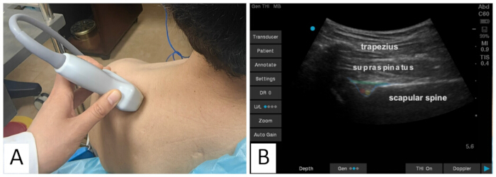
Figure 1: Ultrasound positioning and anatomical visualization of the suprascapular nerve. (A) Patient positioning and transducer placement. The patient is in a seated position with the affected shoulder fully exposed. A convex ultrasound transducer is placed transversely, parallel to the middle and lateral one-third of the scapular spine. (B) The corresponding short-axis ultrasound image. The trapezius muscle, supraspinatus muscle, and scapular spine are indicated by text labels. The suprascapular notch is outlined by a blue semi-transparent line, and the superior transverse scapular ligament is outlined by a green semi-transparent line above it. Together, they form an osseofibrous tunnel containing the suprascapular artery (red semi-transparent dot) and the accompanying suprascapular nerve (yellow semi-transparent dot). Please click here to view a larger version of this figure.
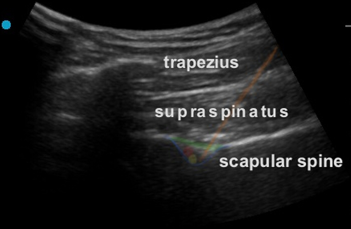
Figure 2: Schematic of the ultrasound-guided in-plane cannula trajectory. The anatomical structures and corresponding color annotations are identical to those described in Figure 1. The orange line illustrates the cannula trajectory using an in-plane approach. The cannula is advanced from the medial to the lateral side toward the suprascapular notch under continuous real-time ultrasound guidance. The cannula tip is advanced through the superior transverse scapular ligament until it contacts the bony floor of the suprascapular notch and is then retracted slightly to position the tip adjacent to the suprascapular nerve. Sensory stimulation is performed first, followed by motor stimulation. After satisfactory sensory and motor responses have been obtained, negative aspiration is performed to confirm the absence of blood return before HL-PRF delivery. Nonessential ultrasound-system interface labels were removed from the image for clarity. Please click here to view a larger version of this figure.
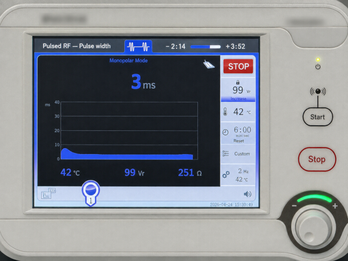
Figure 3: Interface of the radiofrequency generator displaying treatment parameters. The edited image shows representative real-time parameters during HL-PRF delivery. The protocol uses a target voltage of 99 V, a total treatment duration of 6 min, a maximum temperature limit of 42 °C, a pulse frequency of 2 Hz, and a preset pulse width of 20 ms. The pulse width displayed in this representative image is 3 ms; this value represents the real-time device display during treatment rather than the preset pulse-width setting. Manufacturer branding has been blurred, non-English interface labels and buttons have been translated into English, and the nonessential CQM label has been removed. Abbreviations: HL-PRF, high-voltage long-duration pulsed radiofrequency; RF, radiofrequency; ms, milliseconds; V, volts; Vr, device-specific voltage readout; °C, degrees Celsius; Hz, hertz; Ω, ohms; min, minutes; sec, seconds. Please click here to view a larger version of this figure.
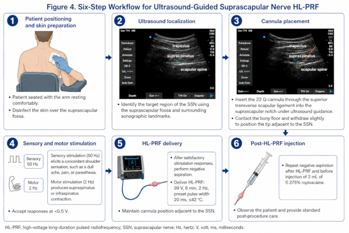
Figure 4: Six-step workflow for ultrasound-guided suprascapular nerve high-voltage long-duration pulsed radiofrequency. The schematic summarizes the six principal steps of the procedure. (1) The patient is positioned in a seated position with the affected upper limb resting comfortably, followed by skin preparation over the suprascapular region. (2) Ultrasound is used to identify the target region of the suprascapular nerve (SSN) using the suprascapular fossa and surrounding sonographic landmarks. (3) A 22 G, 10 cm radiofrequency cannula with a 10 mm active tip is advanced under ultrasound guidance through the superior transverse scapular ligament into the suprascapular notch. After contacting the bony floor, the cannula is withdrawn slightly to position the tip adjacent to the SSN. (4) Sensory stimulation is performed at 50 Hz and motor stimulation at 2 Hz. A concordant shoulder sensation during sensory stimulation and contraction of the supraspinatus or infraspinatus during motor stimulation at < 0.5 V support appropriate cannula positioning. (5) After satisfactory stimulation responses are obtained, negative aspiration is performed, followed by HL-PRF delivery at 99 V for 6 min, with a pulse frequency of 2 Hz, a preset pulse width of 20 ms, and a maximum electrode-tip temperature of 42 °C. (6) After completion of HL-PRF, negative aspiration is repeated before injection of 2 mL of 0.375% ropivacaine, followed by routine post-procedural observation and care. Abbreviations: HL-PRF = high-voltage long-duration pulsed radiofrequency; SSN = suprascapular nerve; Hz = hertz; V = volt; ms = milliseconds. Please click here to view a larger version of this figure.
| A. Baseline characteristics | Case 1 | Case 2 | |
| Age (years) | 45 | 56 | |
| Sex | Male | Male | |
| Stroke type | Intracerebral hemorrhage | Ischemic stroke | |
| Affected side | Left | Left | |
| Time since stroke onset (days) | 76 | 46 | |
| HSP duration (days) | ~50 | ~30 | |
| Baseline analgesic | Celecoxib 200 mg BID | Celecoxib 200 mg BID | |
| B. Clinical outcomes - Case 1 | BL | 1 mo | 3 mo |
| FMA-UE score | 47 | 56 | — |
| NRS, resting pain | 2 | 0 | 0 |
| NRS, movement-evoked pain | 5 | 1 | 2 |
| PROM, abduction (°) | 123.6 | 156.5 | — |
| PROM, external rotation (°) | 67 | 79.3 | — |
| PROM, forward flexion (°) | 151.2 | 151.5 | — |
| PROM, adduction (°) | 40.7 | 42.4 | — |
| B. Clinical outcomes - Case 2 | BL | 1 mo | 3 mo |
| FMA-UE score | 5 | 6 | — |
| NRS, resting pain | 4.5 | 1.5 | 2.5 |
| NRS, movement-evoked pain | 7.5 | 3.5 | 4 |
| PROM, abduction (°) | 95 | 110.1 | — |
| PROM, external rotation (°) | 60.7 | 86.4 | — |
| PROM, forward flexion (°) | 114.4 | 126.2 | — |
| PROM, adduction (°) | 16.1 | 29.9 | — |
Table 1. Baseline clinical characteristics and outcomes of the two representative cases. The table summarizes baseline demographic and clinical characteristics and clinical outcomes assessed at baseline, 1 month, and 3 months after HL-PRF. Outcomes include resting and movement-evoked pain assessed using the NRS, upper-limb motor function assessed using the FMA-UE, and shoulder PROM in abduction, external rotation, forward flexion, and adduction. The 3-month follow-up was conducted by telephone; therefore, only pain scores were obtained at this time point, and an em dash (—) indicates variables that were not assessed at the 3-month telephone follow-up. Abbreviations: BL = baseline; FMA-UE = Fugl-Meyer Assessment-Upper Extremity; mo = month; HL-PRF = high-voltage long-duration pulsed radiofrequency; HSP = hemiplegic shoulder pain; NRS = Numerical Rating Scale; PROM = passive range of motion; BID = twice daily.
Supplementary Video 1: Motor testing during ultrasound-guided cannula placement. This video demonstrates the motor response during physiological testing. At a stimulation frequency of 2 Hz and a voltage <0.5 V, rhythmic contractions of the supraspinatus and infraspinatus are observed, supporting placement of the cannula tip near motor fibers of the suprascapular nerve. Please click here to download this file.
Supplementary Video 2: Pre-treatment shoulder motor function (Case 1). This video shows the baseline upper-limb motor function of the representative patient (Case 1) prior to the HL-PRF intervention. The patient exhibits pain-related protective behaviors and marked limitation of passive shoulder range of motion, particularly during abduction and external rotation. Please click here to download this file.
Supplementary Video 3: Post-treatment shoulder motor function (Case 1). This video shows the same patient at the 1-month post-procedural follow-up and illustrates the observed changes in shoulder movement compared with the baseline recording shown in Supplementary Video 2. Please click here to download this file.
المناقشة
Several procedural steps are important for consistent implementation of this protocol. The key success-determining steps are labeled CRITICAL STEP in the Protocol, including ultrasound target identification, transligamentous cannula advancement, and interpretation of stimulation thresholds. Ultrasound is used to identify the suprascapular notch, the target region of the suprascapular nerve, and adjacent vascular structures while guiding cannula advancement. Cannula position is subsequently assessed using sensory and motor stimulation before HL-PRF delivery. The radiofrequency parameters are then applied according to the predefined settings, including a target voltage of 99 V, treatment duration of 6 min, pulse frequency of 2 Hz, preset pulse width of 20 ms, and a maximum temperature limit of 42 °C20. The 99 V and 6 min settings were informed by published high-voltage and long-duration PRF studies23,24. Together, these steps provide a standardized approach to anatomical targeting, physiological confirmation, and parameter delivery. The present report did not directly evaluate electric-field distribution, the depth of neuromodulation, or neural remodeling.
When the target nerve is not clearly distinguishable on ultrasound, localize the target region using the surrounding anatomical landmarks. In clinical practice, the SSN is often poorly visualized; therefore, identify the osseofibrous space formed by the suprascapular notch and the superior transverse scapular ligament as the primary target landmark20,21. When color Doppler does not clearly display flow in the small suprascapular artery, use real-time observation of subtle vascular pulsation within the suprascapular notch as an additional anatomical cue for identifying the region adjacent to the SSN.
If the target sensory and motor stimulation thresholds (e.g., sensory stimulation at <0.5 V) are not achieved during pre-treatment physiological testing, adjust the depth, direction, or rotational orientation of the cannula tip and repeat the stimulation tests24. Avoid rapid or forceful cannula movements. When further fine adjustment is required, use gentle axial rotation of the cannula before reassessing cannula position and stimulation thresholds, because electric-field distribution around the active tip varies with electrode geometry and orientation25,26.
Despite its potential clinical utility, this protocol has several important limitations. The primary constraint is its high operator dependence; successfully executing the procedure requires advanced proficiency in both functional musculoskeletal ultrasound and interventional pain techniques27,28. Second, the specific clinical characteristics of stroke survivors introduce unique procedural challenges. Cognitive impairment and aphasia may complicate patient cooperation and verbal feedback during sensory stimulation testing. Patients with mild communication or cognitive impairment may still be eligible when they can follow basic procedural instructions and provide a reliable pain response, including through the FPS-R when appropriate. In contrast, patients with severe impairment that prevents reliable communication or cooperation with sensory and motor testing are not suitable for this protocol8,29. Furthermore, severe limb spasticity and anatomical contractures often hinder optimal patient positioning and obscure clear ultrasound visualization30. Additionally, central sensitization—manifesting as hyperalgesia or allodynia—is common in this population, which may decrease patient tolerance during electrical stimulation and complicate the procedure31. Another notable limitation is the current paucity of long-term follow-up data. Due to logistical constraints, the extended 3-month follow-up was conducted solely via telephone, capturing only subjective pain assessments (NRS scores). The lack of comprehensive, in-person physical examinations prevents a complete objective evaluation of long-term functional recovery, such as sustained improvements in PROM or motor function scales. Consequently, the durability of its neuromodulatory effects on neural plasticity remains elusive, and it is unknown whether hemiplegic shoulder pain might recur as patients progress into increasingly intensive motor rehabilitation phases8,29,32. No alternative voltage or duration settings were systematically tested during protocol development or in the two illustrative cases presented here. The settings of 99 V and 6 min were selected a priori based on previously published HL-PRF approaches rather than on a comparative parameter-optimization process. Therefore, the present report does not establish 99 V for 6 min as the optimal parameter combination for suprascapular nerve HL-PRF in patients with HSP. Future controlled studies comparing different voltage and duration settings are needed to determine the optimal treatment parameters and to evaluate their relationship with clinical outcomes and safety.
An additional limitation is the presence of concomitant rehabilitation and changes in analgesic medication. Both patients continued multimodal rehabilitation around the time of HL-PRF, and celecoxib was discontinued on post-procedural day 2 in Case 1 and day 6 in Case 2. In addition, occupational therapy in Case 1 had been temporarily suspended because of pain and was resumed after the procedure. Therefore, the observed changes in pain, PROM, and upper-limb function cannot be attributed solely to HL-PRF. These representative cases should be interpreted as illustrations of the clinical application of the protocol rather than as evidence of the independent treatment effect of HL-PRF. Because the 3-month assessment was conducted by telephone, movement-evoked pain at this time point was based on patient report rather than standardized examiner-performed passive external rotation, and PROM and FMA-UE could not be reassessed.
The present protocol should be considered an HSP-specific application and procedural standardization of an existing PRF strategy rather than a newly developed radiofrequency technique. Suprascapular nerve PRF has previously been investigated in patients with HSP, including in randomized controlled studies8,29. More recently, high-voltage PRF of the suprascapular nerve has been described for chronic shoulder pain, and an exploratory randomized pilot trial has compared high-voltage (100 V) with standard-voltage (45 V) PRF20,21,22. These studies reflect increasing clinical interest in high-voltage SSN PRF but do not establish a universally optimal voltage or treatment duration. A recent systematic review of suprascapular nerve radiofrequency also reported potential improvements in pain and function, although the certainty of the available evidence remained low33. Accordingly, the principal contribution of the present Methods Article lies in the detailed ultrasound-guided adaptation of this approach for patients with persistent HSP, including patient selection, anatomical targeting, sensory and motor confirmation of cannula position, procedural monitoring, and structured outcome assessment within the post-stroke rehabilitation setting.
In conclusion, this article provides a standardized ultrasound-guided procedural framework for applying HL-PRF to the suprascapular nerve in patients with persistent HSP. In the two representative cases, lower pain scores and greater shoulder PROM were observed at the 1-month follow-up, with no documented procedure-related adverse events during the available follow-up period. However, the small number of cases, concomitant rehabilitation, changes in analgesic medication, absence of a control group, and potential spontaneous neurological recovery preclude causal conclusions regarding efficacy or long-term safety. Further controlled studies are required to evaluate the clinical effectiveness, safety, and optimal treatment parameters of this approach.
الإفصاحات
The authors declare that they have no conflicts of interest.
شكر وتقدير
This work was supported by the Wenzhou Basic Public Welfare Research Project (Grant No. Y2023128) and the Zhejiang Provincial Basic Public Welfare Research Program (Grant No. LGF20H170001). No non-author contributors provided technical assistance, figure preparation, or other support requiring acknowledgment.
المواد
| الاسم | الشركة | رقم فهرسي | التعليقات |
|---|---|---|---|
| Disposable adhesive wound dressing | Zhende Medical Co., Ltd. | 6 cm × 7 cm; Registration No. 20182140234 | |
| Disposable sterile preparation kit (including povidone-iodine, cotton balls, forceps, and gauze) | Winner Medical Co., Ltd. | 602-002125-06 | |
| Disposable sterile medical underpad (used as sterile drape), 90 cm × 200 cm | Zhende Medical Co., Ltd. | 90 cm × 200 cm; Registration No. 20192140262 | |
| Electronic joint goniometer (0–180°, digital) | HAOLIANG Measuring Tools | 82311-00 | |
| Faces Pain Scale-Revised (FPS-R) | International Association for the Study of Pain (IASP) | Faces Pain Scale-Revised (FPS-R) clinical assessment tool | |
| Neutral electrode pad | Leonhard Lang GmbH | RS25 | |
| Radiofrequency cannula, 22 G, 10 cm, 10 mm active tip | Beijing NeoScience Co., Ltd. | CC422A | |
| Radiofrequency generator | Beijing NeoScience Co., Ltd. | CR2010E | |
| Ropivacaine hydrochloride injection (0.75%) | Qilu Pharmaceutical Co., Ltd. | 5J0023C70 | |
| Sodium chloride injection (0.9%; diluent) | China Otsuka Pharmaceutical Co., Ltd. | DB01-1.0701. | |
| Sterile ultrasound gel | Guangzhou Guangong Technology Development Co., Ltd. | 20 g sachet; GZFDA No. 20160194 | |
| Sterile ultrasound probe cover | Zhejiang Chunan Renhe Medical Supplies Industry and Trade Co., Ltd. | A1475EP | |
| Syringe (5 mL) | Shanghai Kindly Enterprise Development Group Co., Ltd. | 5 mL; Registration No. 20163140518 | |
| Ultrasound system with color Doppler capability | FUJIFILM Sonosite, Inc. | SonoSite SII | |
| Ultrasound transducer, curvilinear (5–2 MHz) | FUJIFILM Sonosite, Inc. | C60 |
المراجع
- Koog YH, Jin SS, Yoon K, Min BI. Interventions for hemiplegic shoulder pain: systematic review of randomised controlled trials. Disabil Rehabil. 2010;32(4):282-91.
- Lindgren I, Jönsson AC, Norrving B, Lindgren A. Shoulder pain after stroke: a prospective population-based study. Stroke. 2007;38(2):343-48.
- Tan PL, Wee TC, Lim AT. Poststroke shoulder pain. Singapore Med J. 2024;65(8):449-53.
- Wilson RD, Chae J. Hemiplegic shoulder pain. Phys Med Rehabil Clin N Am. 2015;26(4):641-55.
- Zeilig G, et al. Hemiplegic shoulder pain: evidence of a neuropathic origin. Pain. 2013;154(2):263-71.
- Jones AKP, Brown CA. Post-stroke shoulder pain: nociceptive or neuropathic? Pain. 2013;154(2):189.
- Chang MC. The effects of ultrasound-guided corticosteroid injection for the treatment of hemiplegic shoulder pain on depression and anxiety in patients with chronic stroke. Int J Neurosci. 2017;127(11):958-64.
- Alanbay E, et al. Effectiveness of suprascapular nerve pulsed radiofrequency treatment for hemiplegic shoulder pain: a randomized-controlled trial. Pain Physician. 2020;23(3):245-52.
- Hernández-Ortíz AR, et al. Changes in muscle tone, function, and pain in the chronic hemiparetic shoulder after dry needling within or outside trigger points in stroke patients: a crossover randomized clinical trial. Pain Med. 2020;21(11):2939-47.
- Buchbinder R, Green S, Youd JM. Corticosteroid injections for shoulder pain. Cochrane Database Syst Rev. 2003;2003(1):CD004016.
- Huang YC, et al. Effects of kinesio taping on hemiplegic hand in patients with upper limb post-stroke spasticity: a randomized controlled pilot study. Eur J Phys Rehabil Med. 2019;55(5):551-57.
- Ghozy S, et al. Efficacy of kinesio taping in treatment of shoulder pain and disability: a systematic review and meta-analysis of randomised controlled trials. Physiotherapy. 2020;107:176-88.
- Manchikanti L. Role of neuraxial steroids in interventional pain management. Pain Physician. 2002;5(2):182-99.
- Vorster W, et al. The sensory branch distribution of the suprascapular nerve: an anatomic study. J Shoulder Elbow Surg. 2008;17(3):500-2.
- Munglani R. The longer term effect of pulsed radiofrequency for neuropathic pain. Pain. 1999;80(1-2):437-9.
- Chua NHL, Vissers KC, Sluijter ME. Pulsed radiofrequency treatment in interventional pain management: mechanisms and potential indications-a review. Acta Neurochir (Wien). 2011;153(4):763-71.
- Chang MC. Efficacy of pulsed radiofrequency stimulation in patients with peripheral neuropathic pain: a narrative review. Pain Physician. 2018;21(3):E225–E234.
- Cahana A, Vutskits L, Muller D. Acute differential modulation of synaptic transmission and cell survival during exposure to pulsed and continuous radiofrequency energy. J Pain. 2003;4(4):197-202.
- Picelli A, et al. A retrospective case series of ultrasound-guided suprascapular nerve pulsed radiofrequency treatment for hemiplegic shoulder pain in patients with chronic stroke. J Pain Res. 2018;11:1115-20.
- García-Amigo J, et al. Effectiveness of high-voltage radiofrequency on suprascapular nerve for the treatment of chronic shoulder pain. Interv Pain Med. 2026;5(2):100752.
- García Amigo J, Cabaleiro Burguillos B, Cordero García C. High voltage pulsed radiofrequency of the suprascapular nerve for the treatment of chronic shoulder pain. PM R. 2025;17(7):865-867.
- Costa JRP, et al. High versus low-voltage pulsed radiofrequency of the suprascapular nerve for chronic shoulder pain: a randomized pilot clinical trial. Interv Pain Med. 2026;5(3):100805.
- Luo F, et al. High voltage pulsed radiofrequency for the treatment of refractory neuralgia of the infraorbital nerve: a prospective double-blinded randomized controlled study. Pain Physician. 2017;20(4):271-279.
- Wang CL, Song T. The clinical efficacy of high-voltage long-duration pulsed radiofrequency treatment in pudendal neuralgia: a retrospective study. Neuromodulation. 2022;25(8):1372-77.
- Bogduk N. Pulsed radiofrequency. Pain Med. 2006;7(5):396-407.
- Cosman ER Jr, Cosman ER Sr. Electric and thermal field effects in tissue around radiofrequency electrodes. Pain Med. 2005;6(6):405-24.
- Na D, et al. Pulsed radiofrequency neuromodulation for post-stroke shoulder pain in patients with hemorrhagic stroke. J Korean Neurosurg Soc. 2024;67(5):568-77.
- Ergonenc T, Beyaz SG. Effects of ultrasound-guided suprascapular nerve pulsed radiofrequency on chronic shoulder pain. Med Ultrason. 2018;20(4):461–466.
- Kim TH, Chang MC. Comparison of the effectiveness of pulsed radiofrequency of the suprascapular nerve and intra-articular corticosteroid injection for hemiplegic shoulder pain management. J Integr Neurosci. 2021;20(3):687-693.
- Kalichman L, Ratmansky M. Underlying pathology and associated factors of hemiplegic shoulder pain. Am J Phys Med Rehabil. 2011;90(9):768-80.
- Roosink M, et al. Somatosensory symptoms and signs and conditioned pain modulation in chronic post-stroke shoulder pain. J Pain. 2011;12(4):476-85.
- Yang C, et al. The management of hemiplegic shoulder pain in stroke subjects undergoing pulsed radiofrequency treatment of the suprascapular and axillary nerves: a pilot study. Ann Palliat Med. 2020;9(5):3357-65.
- Sebastián Infante J, Blackburn N, Felipe Vargas J. Radiofrequency of suprascapular nerve as an analgesic strategy for chronic pain management. Systematic review and meta-analysis. Rev Esp Anestesiol Reanim (Engl Ed). 2024;71(9):678-86.