CT-derived skeletal muscle quantity and quality may help characterize treatment resilience after pancreaticoduodenectomy. Current evidence supports cautious, pathology-specific risk stratification rather than causal or intervention-guiding conclusions.
مقالة مراجعة
Body Composition and Treatment Resilience After Pancreaticoduodenectomy
في هذه المقالة
ملخص
الملخص
Pancreaticoduodenectomy remains a cornerstone of curative-intent treatment for periampullary malignancies. Postoperative recovery, receipt of adjuvant therapy, and long-term outcomes vary even among patients with comparable tumor stage and surgical care. Host-related factors, including body composition, may contribute to this variability. Body mass index does not distinguish skeletal muscle from adipose tissue, whereas computed tomography (CT)-derived muscle area and attenuation provide separate measures of muscle quantity and quality. In this review, treatment resilience denotes the capacity to progress through surgery and planned postoperative or adjuvant care without clinically consequential interruption. Reported indicators include postoperative complications and recovery, time to adjuvant treatment, grade 3 or higher toxicity, dose reduction or delay, early discontinuation, and treatment completion; thresholds vary among studies. Retrospective studies associate adverse muscle phenotypes with some of these outcomes, but results are heterogeneous and do not establish causality. Associations with survival also vary by measurement method, tumor type, and treatment delivery. This narrative review summarizes CT-based assessment, perioperative and treatment-delivery outcomes, tumor-specific considerations, and the limited evidence for prehabilitation and nutritional interventions. Body-composition measures may complement established clinical and pathological factors, but prospective validation and standardized methods are required before they can direct treatment.
المقدمة
Pancreaticoduodenectomy remains a cornerstone of curative-intent treatment for pancreatic head, ampullary, distal bile duct, and duodenal cancers. Postoperative morbidity remains substantial despite advances in surgical technique, perioperative care, and centralization1. These malignancies share an operation but differ in biology, adjuvant-treatment strategies, and prognosis2,3,4. Accordingly, tumor type, lymph-node status, resection margin, histological subtype, and perineural or lymphovascular invasion remain central to outcome assessment.
Host-related factors may also modify surgical recovery and the ability to receive subsequent treatment. Nutritional status and physical reserve are clinically relevant but incompletely characterized. Body mass index (BMI) cannot distinguish lean tissue from adipose tissue and may obscure muscle depletion in patients with obesity or cancer cachexia5.
CT images obtained for routine oncologic care permit opportunistic assessment of skeletal muscle. Total skeletal muscle area at the third lumbar vertebral level, usually normalized to height squared as skeletal muscle index, estimates muscle quantity6. Mean muscle attenuation in Hounsfield units estimates radiological muscle quality and intramuscular lipid infiltration. These measures are related, but not interchangeable, and CT-defined low muscle quantity alone does not satisfy the consensus clinical definition of sarcopenia, which also requires impaired muscle strength.7 Reproducibility depends on image selection, segmentation, software, acquisition protocol, contrast phase, and cutoff definition8,9,10.
This narrative review examines body composition in relation to perioperative outcomes, adjuvant-treatment delivery, and survival after pancreaticoduodenectomy. Treatment resilience is used as an organizing concept rather than a validated composite endpoint. It encompasses postoperative complications and recovery, time to adjuvant treatment, grade 3 or higher treatment toxicity, dose reduction or delay, early discontinuation, and completion of the planned regimen. Because definitions and thresholds differ across studies, each measure is interpreted as reported rather than combined. Figure 1 summarizes the proposed associations and their tumor-related modifiers.
Literature search and scope
We performed a targeted PubMed search from database inception through September 21, 2026, using combinations of pancreaticoduodenectomy, pancreatoduodenectomy, periampullary, pancreatic cancer, body composition, skeletal muscle, sarcopenia, myosteatosis, muscle attenuation, postoperative complications, adjuvant chemotherapy, toxicity, treatment completion, survival, prehabilitation, and nutrition. We considered English-language adult studies reporting CT-derived muscle quantity or quality in pancreaticoduodenectomy, periampullary, or pancreatic cancer populations. Systematic reviews, consensus or methodological articles, and intervention studies were included to address measurement and clinical translation. Studies from broader pancreatic or gastrointestinal oncology were used only when directly relevant and are identified as such.
Reference lists of relevant reviews and included articles were screened for additional studies. Because this was a focused narrative review, study selection was not conducted as a systematic review, no formal risk-of-bias tool or meta-analysis was applied, and selective-citation bias cannot be excluded. We therefore emphasize population, design, and methodological limitations rather than grading the evidence with subjective consistency labels.
مراجعة ومنظور
Limitations of conventional anthropometric measures in pancreatic surgery
Body mass index is a simple anthropometric measure, but it provides limited information about the relative amounts of skeletal muscle and adipose tissue. In cancer populations, cachexia, systemic inflammation, obesity, and age-related muscle loss may coexist; therefore, similar BMI values can represent different body-composition phenotypes5.
Patients with normal or elevated BMI may have low muscle quantity, sometimes described as sarcopenic obesity when excess adiposity and sarcopenia coexist. Conversely, a low BMI does not by itself establish poor muscle quality or impaired strength. BMI, CT-derived muscle quantity, CT-derived muscle attenuation, and functional measures should therefore be treated as distinct, complementary constructs.
Table 1 summarizes the principal measures, reported outcomes, clinical interpretation, and limitations. CT-based measures can add information to BMI, but no single body-composition metric has been shown to direct perioperative or adjuvant-treatment decisions.
Imaging-based skeletal muscle assessment: Concepts and methodology
CT-based body-composition analysis uses images already acquired for staging or surgical planning and therefore does not need to add radiation exposure. Dual-energy X-ray absorptiometry and bioelectrical impedance estimate whole-body composition, while magnetic resonance imaging can quantify regional muscle and fat without ionizing radiation. CT is particularly practical in pancreatic surgery because abdominal scans are routinely available and provide anatomical and attenuation data, although it does not directly measure muscle strength or physical performance6,8.
Muscle quantity is commonly measured by segmenting all visible skeletal muscles on a single axial image at the third lumbar vertebral level. The cross-sectional area can be normalized to height squared to calculate skeletal muscle index6. Psoas area or psoas muscle index is simpler, but samples only one muscle group and is not interchangeable with total L3 skeletal muscle area10. Muscle quality is commonly estimated from mean CT attenuation in Hounsfield units; lower attenuation is interpreted as greater lipid infiltration or myosteatosis. Reduced muscle quantity and low attenuation can coexist, but either phenotype may occur alone. In research based only on CT, the term low muscle quantity is more precise than sarcopenia unless muscle strength has also been assessed7.
A reproducible workflow should specify the interval between CT and surgery, vertebral level, muscle groups, attenuation thresholds, normalization method, software and version, and whether segmentation was manual, semiautomated, or automated. Studies should also report scanner voltage, slice thickness, reconstruction method, and contrast phase because these factors can alter attenuation and, to a lesser extent, measured area8,9. Sex-specific and population-specific cutoffs should be reported rather than assumed to be universal. These technical sources of variation limit direct comparison among studies.
Body composition and short-term surgical outcomes
Postoperative morbidity after pancreaticoduodenectomy includes delayed gastric emptying, pancreatic fistula, infectious complications, and prolonged hospitalization. Systematic reviews and retrospective cohorts have examined whether low muscle quantity or attenuation identifies patients at higher risk, but definitions and results vary11,12.
Some cohorts report associations between low muscle measures and major complications, pancreatic fistula, or delayed recovery after pancreaticoduodenectomy13,14,15. Other analyses show attenuated or outcome-specific associations after adjustment. These observational findings support risk-marker evaluation but do not show that low muscle quantity or attenuation causes postoperative morbidity.
Skeletal muscle participates in glucose disposal, protein metabolism, and inflammatory signaling. These mechanisms provide biological plausibility for an association with recovery, but mechanistic observations should not be equated with clinical causality. Nutritional status, inflammation, comorbidity, tumor burden, and treatment selection may confound reported associations.
Influence of body composition on adjuvant therapy delivery
Adjuvant-treatment recommendations differ by pathological subtype and risk profile. For example, evidence and regimen selection for ampullary adenocarcinoma are not identical to those for pancreatic ductal adenocarcinoma16. Postoperative complications can delay recovery and are associated with lower use of adjuvant chemotherapy after resection for pancreatic cancer17.
Pancreaticoduodenectomy-specific retrospective studies suggest that selected muscle measures may be associated with the time to adjuvant treatment, toxicity, dose modification, or treatment completion. In a cohort of periampullary malignancies, higher Hounsfield-unit average calculation, but not psoas muscle index, was associated with several adjuvant-treatment outcomes18. In resected pancreatic ductal adenocarcinoma, sarcopenia was associated with completion of S-1 adjuvant chemotherapy19. Evidence linking muscle density to grade 3 or higher toxicity in metastatic pancreatic cancer provides a broader oncologic context but should not be directly generalized to postoperative populations20.
These measures may help characterize treatment resilience, but they are not validated as stand-alone treatment-selection tools. Associations may reflect nutritional status, inflammation, performance status, postoperative complications, and tumor type. Prospective studies using prespecified endpoints are needed before CT-derived measures are used to modify regimens or supportive care.
Body composition and long-term survival: Evidence and uncertainty
Associations between body composition and long-term survival are heterogeneous. Low muscle attenuation was associated with worse survival after pancreaticoduodenectomy for nonpancreatic periampullary cancer in one cohort15, while low muscle quantity was associated with overall survival in a pancreatic cancer cohort21. Effect estimates vary after adjustment for stage, nodal status, margin status, and adjuvant-treatment receipt.
Differences in anatomical landmarks, segmentation methods, cutoff definitions, populations, covariates, and follow-up contribute to inconsistency. Body composition should therefore complement rather than replace established pathological and treatment-related prognostic factors.
Low muscle measures may coexist with hypoalbuminemia, systemic inflammation, and adverse immune-nutritional indices. These factors have also been associated with outcomes after pancreaticoduodenectomy in specific tumor populations22,23. Their overlap complicates causal interpretation and raises the possibility of residual confounding.
Tumor-specific interpretation
Pancreatic ductal adenocarcinoma, ampullary adenocarcinoma, distal cholangiocarcinoma, and duodenal adenocarcinoma differ in natural history and adjuvant management. Current body-composition studies rarely provide adequately powered, directly comparative estimates across all four pathological subtypes. Findings from nonpancreatic periampullary cancer15, pancreatic ductal adenocarcinoma19,21, or ampullary cancer22,23 should therefore be interpreted within those populations rather than generalized to all patients undergoing pancreaticoduodenectomy.
Intervention evidence
Prehabilitation and nutritional optimization are potential strategies, not established consequences of an adverse CT phenotype. A small randomized trial of prehabilitation before pancreaticoduodenectomy did not reduce overall or major postoperative complications, although delayed gastric emptying was less frequent24. A systematic review found that pancreatic-cancer prehabilitation studies were small and heterogeneous25, and a review of randomized nutritional interventions around pancreaticoduodenectomy reported mixed evidence across regimens and outcomes26. No trial has shown that selecting or adapting an intervention according to CT-derived muscle quantity or attenuation improves treatment completion or survival.
Methodological challenges and clinical translation
Most available studies are retrospective and often single-center, with risks of selection bias, inconsistent adjustment, and limited causal inference. There is no consensus on the optimal anatomical landmark, acquisition protocol, segmentation approach, or cutoff for low muscle quantity or attenuation. These limitations complicate study comparison and clinical implementation.
A single preoperative measurement also cannot capture changes during postoperative recovery or systemic treatment. Longitudinal studies remain limited. Automated or semiautomated segmentation may improve scalability, but technical harmonization and external validation are required before routine use.
Future studies should prespecify acquisition and segmentation methods, report continuous as well as categorized measures, use pathology-specific analyses, and evaluate incremental value beyond established clinical models. Prospective multicenter validation should precede treatment algorithms based on body composition.
الاستنتاجات
CT-derived muscle quantity and attenuation may complement established clinical and pathological factors when characterizing risk after pancreaticoduodenectomy. The evidence is predominantly retrospective, methodologically heterogeneous, and uneven across pancreatic, ampullary, distal bile duct, and duodenal cancers. Associations with postoperative outcomes, adjuvant-treatment delivery, and survival should therefore not be interpreted as causal. Standardized imaging methods, pathology-specific prospective validation, and trials showing that targeted interventions improve patient-important outcomes are required before body-composition measures can guide treatment.
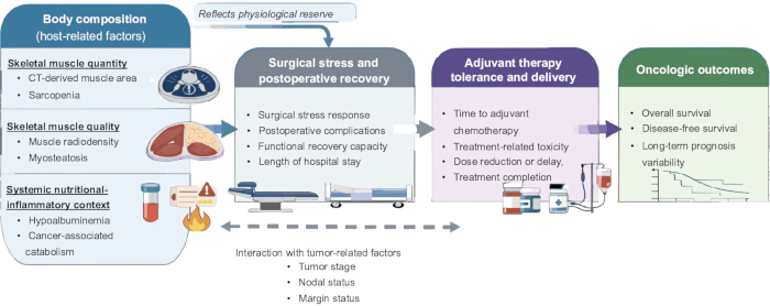
Figure 1: Conceptual framework linking CT-derived body composition with outcomes after pancreaticoduodenectomy. Muscle quantity, muscle quality, and the nutritional-inflammatory context may be associated with postoperative recovery, which can influence the timing and completion of adjuvant therapy. Associations with overall and disease-free survival remain heterogeneous and are modified by tumor type, stage, nodal status, margin status, and treatment delivery. Arrows indicate hypothesized or observed associations rather than proven causal effects. Please click here to view a larger version of this figure.
| Domain | Indicator or measure | Reported findings | Potential clinical relevance | Evidence context and limitations |
| Assessment limitations | Body mass index (BMI) | BMI does not distinguish skeletal muscle from adipose tissue; patients with similar BMI may have different muscle phenotypes. | Useful as a general anthropometric measure but insufficient alone for body-composition assessment. | Does not measure muscle quantity, attenuation, strength, or performance; associations are confounded by obesity, cachexia, and comorbidity. |
| Muscle quantity | Total L3 skeletal muscle area or skeletal muscle index (SMI); psoas area or psoas muscle index (PMI) | Low muscle quantity has been associated with selected postoperative and survival outcomes. | May complement clinical risk assessment when measurement and population are specified. | L3 total muscle and psoas-only measures are not interchangeable. Cutoffs, normalization, and terminology vary; CT-defined low quantity alone is not the consensus clinical definition of sarcopenia. |
| Muscle quality | Mean muscle attenuation or skeletal muscle density in Hounsfield units; myosteatosis | Low attenuation reflects greater intramuscular lipid infiltration and has been associated with selected recovery, treatment-delivery, and survival outcomes. | Provides information complementary to muscle area; it is not established as a superior or causal marker. | Strongly affected by contrast phase, scanner and reconstruction parameters, segmentation, and cutoff definitions. Evidence is mainly retrospective. |
| Short-term surgical outcomes | Low muscle quantity and/or low muscle attenuation | Systematic reviews and cohorts report associations with major complications, pancreatic fistula, length of stay, or delayed recovery, but findings differ by endpoint and method. | May help identify patients who warrant broader perioperative assessment. | Predominantly retrospective and heterogeneous; adjusted associations are inconsistent and do not establish causality. |
| Adjuvant-treatment initiation | Muscle attenuation or psoas-based measures; postoperative recovery | Selected pancreaticoduodenectomy cohorts report associations with time to adjuvant treatment; postoperative complications are also associated with lower receipt of adjuvant chemotherapy. | May flag risk of delayed or omitted postoperative treatment. | Limited retrospective evidence; definitions of timely initiation vary and tumor populations are not uniform. |
| Adjuvant-treatment tolerance and completion | Muscle attenuation, low muscle quantity, and study-specific sarcopenia definitions | Selected studies report associations with toxicity, dose modification, discontinuation, or treatment completion. | May complement performance status and nutritional assessment before and during treatment. | Pancreaticoduodenectomy-specific evidence is limited and regimen-specific. Evidence from metastatic pancreatic cancer should not be generalized directly to postoperative populations. |
| Long-term survival | Preoperative muscle quantity or attenuation | Some studies report associations with overall survival, whereas effects may attenuate after adjustment for stage, nodal status, margin status, and adjuvant treatment. | Potential adjunct to tumor- and treatment-based prognostic models. | Results are heterogeneous across measurement methods and pathological populations; residual confounding is likely. |
| Nutritional-inflammatory context | Muscle measures with albumin, inflammatory markers, or immune-nutritional indices | Adverse muscle phenotypes often coexist with malnutrition and systemic inflammation. | Supports integrated host assessment rather than interpretation of a single CT measure. | Correlated host factors complicate attribution of independent effects; evidence is population-specific and observational. |
| Tumor-specific interpretation | Body composition considered with tumor type, stage, nodal status, margin status, and treatment delivery | Pancreatic, ampullary, distal bile duct, and duodenal cancers differ in biology, treatment, and prognosis. | Body composition may complement but cannot replace pathology-specific staging and treatment factors. | Direct, adequately powered comparisons across all pathological subtypes are lacking; findings should not be generalized across periampullary malignancies. |
| Methodological and implementation issues | Opportunistic CT assessment using prespecified acquisition, segmentation, normalization, and quality-control procedures | Routine CT permits assessment without additional radiation, but methods and cutoffs remain insufficiently standardized. | Feasible for research and potentially scalable with automated segmentation. | Report CT-to-surgery interval, vertebral level, muscle groups, software, contrast phase, scanner parameters, cutoffs, and reliability. Clinical decision thresholds are not validated. |
| Interventions and future research | Longitudinal CT measures; prehabilitation; nutritional optimization | Small trials and reviews report mixed effects on postoperative outcomes; no study has shown that CT-guided intervention selection improves treatment completion or survival. | Potential research targets for multimodal supportive care. | Prospective pathology-specific validation and intervention trials with prespecified patient-important outcomes are required. |
| Interpretive principle | Body-composition measures used alongside established clinical and pathological factors | Reported associations describe risk markers rather than proven determinants of outcome. | Supports cautious, multidisciplinary interpretation. | The evidence base is mainly retrospective and methodologically heterogeneous; no formal evidence-grading framework was applied in this narrative review. |
Table 1: Body-composition measures and reported outcomes after pancreaticoduodenectomy. The table summarizes measurement approaches, reported associations, potential clinical relevance, and population- or method-specific limitations.
الإفصاحات
The authors declare no conflicts of interest.
المراجع
- Cameron JL. One thousand consecutive pancreaticoduodenectomies and beyond: a personal series. Am J Surg. 2007;194(4 Suppl):S11-S15.
- Williams JL, et al. Association of histopathologic phenotype of periampullary adenocarcinomas with survival. JAMA Surg. 2017;152(1):82-88.
- Quero G, et al. The impact of the histological classification of ampullary carcinomas on long-term outcomes after pancreaticoduodenectomy: a single tertiary referral center evaluation. Langenbecks Arch Surg. 2022;407(7):2811-2821.
- Chen JW, et al. Predicting patient survival after pancreaticoduodenectomy for malignancy: histopathological criteria based on perineural infiltration and lymphovascular invasion. HPB (Oxford). 2010;12(2):101-108.
- Martin L, et al. Cancer cachexia in the age of obesity: skeletal muscle depletion is a powerful prognostic factor, independent of body mass index. J Clin Oncol. 2013;31(12):1539-1547.
- Mourtzakis M, et al. A practical and precise approach to quantification of body composition in cancer patients using computed tomography images acquired during routine care. Appl Physiol Nutr Metab. 2008;33(5):997-1006.
- Cruz-Jentoft AJ, et al. Sarcopenia: revised European consensus on definition and diagnosis. Age Ageing. 2019;48(1):16-31.
- Tolonen A, et al. Methodology, clinical applications, and future directions of body composition analysis using CT images: a review. Eur J Radiol. 2021;145:109943.
- Lortie J, et al. The effect of computed tomography parameters on sarcopenia and myosteatosis assessment: a scoping review. J Cachexia Sarcopenia Muscle. 2022;13(6):2807-2819.
- Pigneur F, et al. Psoas muscle index is not representative of total skeletal muscle index in patients with cancer. J Cachexia Sarcopenia Muscle. 2023;14(4):1613-1620.
- Bundred J, Kamarajah SK, Roberts KJ. Body composition assessment and sarcopenia in patients with pancreatic cancer: a systematic review and meta-analysis. HPB (Oxford). 2019;21(12):1603-1612.
- Zhang QH, et al. Sarcopenia adversely impacts clinical outcomes in patients undergoing pancreaticoduodenectomy: a systematic review and meta-analysis. World J Gastrointest Surg. 2024;16(6):1857-1870.
- Duan K, et al. Skeletal muscle depletion and nutrition support affected postoperative complications in patients undergoing pancreaticoduodenectomy. Eur J Clin Nutr. 2021;75(8):1218-1226.
- Jang M, et al. Predictive value of sarcopenia and visceral obesity for postoperative pancreatic fistula after pancreaticoduodenectomy analyzed on clinically acquired CT and MRI. Eur Radiol. 2019;29(5):2417-2425.
- van Rijssen LB, et al. Skeletal muscle quality is associated with worse survival after pancreatoduodenectomy for periampullary, nonpancreatic cancer. Ann Surg Oncol. 2017;24(1):272-280.
- Jin Z, et al. Prognostic factors and benefits of adjuvant therapy after pancreatoduodenectomy for ampullary adenocarcinoma: Mayo Clinic experience. Eur J Surg Oncol. 2018;44(5):677-683.
- Merkow RP, et al. Postoperative complications reduce adjuvant chemotherapy use in resectable pancreatic cancer. Ann Surg. 2014;260(2):372-377.
- Hajibandeh S, et al. Psoas muscle variables and adjuvant chemotherapy-related and survival outcomes following pancreaticoduodenectomy for periampullary malignancy. World J Gastrointest Surg. 2026;18(2):114743.
- Takagi K, et al. Impact of sarcopenia on S-1 adjuvant chemotherapy and prognosis in patients with pancreatic cancer. Biosci Trends. 2023;17(4):310-317.
- Kim IH, et al. Clinical significance of skeletal muscle density and sarcopenia in patients with metastatic pancreatic cancer receiving first-line chemotherapy. BMC Cancer. 2021;21:77.
- Peng YC, et al. Preoperative sarcopenia is associated with poor overall survival in pancreatic cancer patients following pancreaticoduodenectomy. Eur Radiol. 2021;31(4):2472-2481.
- Jiang J, et al. The impact of preoperative and postoperative malnutrition on outcomes for ampullary carcinoma after pancreaticoduodenectomy. Front Oncol. 2021;11:748341.
- Sun C, et al. Preoperative prognostic nutritional index predicts long-term outcomes of patients with ampullary adenocarcinoma after curative pancreatoduodenectomy. World J Gastrointest Surg. 2024;16(5):1291-1300.
- Ausania F, et al. Prehabilitation in patients undergoing pancreaticoduodenectomy: a randomized controlled trial. Rev Esp Enferm Dig. 2019;111(8):603-608.
- Bundred JR, et al. Prehabilitation prior to surgery for pancreatic cancer: a systematic review. Pancreatology. 2020;20(6):1243-1250.
- Takagi K, et al. Current evidence of nutritional therapy in pancreatoduodenectomy: systematic review of randomized controlled trials. Ann Gastroenterol Surg. 2019;3(6):620-629.