This protocol describes procedures to detect changes in phosphorylation modifications of target proteins using Phos-tag gel electrophoresis in clinical samples and in vitro kinase assays.
Method Article
Unveiling Phosphorylation Modification Using Phos-tag SDS-PAGE Gel Electrophoresis and In Vitro Kinase Assay
Opens in a new tab
In This Article
Summary
Loading...
$$\rightleftharpoonup{xx}$$
$$\longleftharp{xx}$$,
$$\longrightharp{xx}$$,
Abstract
Loading...
$$\rightleftharpoonup{xx}$$
$$\longleftharp{xx}$$,
$$\longrightharp{xx}$$,
Phosphorylation is a classic post-translational modification that regulates protein function. Proteins typically contain multiple potential phosphorylation sites, which can be modified by various kinases at different locations. Studying phosphorylation changes of target proteins in disease often requires phospho-specific antibodies. However, commercial options may be limited to a single site or entirely unavailable. Here, a method is described to detect changes in phosphorylation modifications of target proteins in clinical samples using the Phos-tag gel electrophoresis and to identify detailed phosphorylation sites through in vitro kinase assays. Phosphorylated proteins bound to Phos-tag exhibit slower migration rates in SDS-PAGE gel electrophoresis, enabling semi-quantitative analysis of phosphorylation changes in disease tissues based on the mean pixel intensity of the slowly migrating bands. By combining Phos-tag SDS-PAGE with immunoblotting using pan-specific antibodies against multiple candidate proteins, researchers can efficiently identify target proteins with phosphorylation changes. Following the screening of candidate kinases, in vitro kinase assays are performed with the target protein, and the resulting phosphorylated products are subjected to mass spectrometry for precise site identification. This method does not require specific phosphorylated protein antibodies, allowing large-scale screening of tissue samples to identify altered phosphorylation states in disease. Furthermore, the identified phosphorylation sites can be used to develop specific antibodies for quantitative and localization analysis in disease tissues, providing deeper insights into their functional roles.
Introduction
Loading...
$$\rightleftharpoonup{xx}$$
$$\longleftharp{xx}$$,
$$\longrightharp{xx}$$,
Phosphorylation is a key post-translational modification for modulating the subcellular localization, kinase activity, and transcriptional activity of proteins. More than 500 protein kinase genes have been found in the human genome, which are involved in signal transduction, gene expression, and protein-protein interaction1. Phosphorylation and dephosphorylation are under the control of various kinases, which are quickly reversible to regulate cell adhesion, cell proliferation, and cell differentiation2. However, abnormal protein phosphorylation modifications are responsible for many human diseases, including tumorigenesis, autoimmune disorders, neurodegeneration, and subfertility3,4,5.
Phosphoproteomic profiling of clinical specimens yields multiple candidate differentially phosphorylated proteins6. To reveal the key phosphorylation modifications in human diseases, an easy and efficient method to detect the phosphorylation status of specific target proteins is necessary. Phosphorylation at different sites could lead to distinct functional alterations in the protein. Hence, using phosphorylation-specific antibodies targeting particular amino acid sites is undoubtedly the best method for detecting changes in protein phosphorylation. However, well-characterized phospho-specific antibodies are absent for many proteins. In addition, a more suitable approach is required to identify novel potential phosphorylation sites. Phosphorylation at specific amino acid residues often alters a protein's migration rate in gel electrophoresis, and slower-migrating bands can reflect changes in phosphorylation status7. However, these mobility shifts are usually subtle, making them difficult to distinguish. Phos-tag is a synthesized chemical compound designed from an alkaline phosphatase catalytic domain as a model, which can capture phosphorylated Ser/Thr/Tyr and His/Asp/Lys residues. Therefore, phos-tag conjugation effectively amplifies the migration rate differences between phosphorylated and non-phosphorylated proteins, while also distinguishing variations in the number of phosphorylation sites8.
This Phos-tag-based methodology for detecting target protein phosphorylation changes in tissue samples offers distinct advantages over conventional approaches. Crucially, it eliminates the requirement for phospho-specific antibodies, instead utilizing standard WB-grade protein-specific antibodies for detection. The phosphorylated and non-phosphorylated forms of target proteins are resolved as distinct bands during Phos-tag SDS-PAGE, enabling semi-quantitative assessment of phosphorylation level alterations in disease states through comparative band intensity analysis. The workflow in this protocol includes: (1) Phos-tag SDS-PAGE separation of protein samples, (2) immunoblot screening for phosphorylated targets, (3) in vitro kinase assays with candidate kinases verified by phospho-Ser/Thr antibodies. Subsequent mass spectrometry analysis (conducted by an external service platform) identifies phosphorylation sites to enable site-specific antibody development; the detailed methodology is not included in the current protocol. Notably, this approach was successfully applied to detect Nur77 phosphorylation changes in clinical samples, identifying Mst1 kinase as the upstream regulator of Nur77 phosphorylation and developing a site-specific phospho-antibody for clinical detection of phosphorylation at threonine 366 of Nur779. Therefore, this approach proves particularly valuable during preliminary research phases when investigating multiple candidate proteins, as it allows large-scale screening of phosphorylation status changes across numerous targets using commercially available antibodies.
Protocol
Loading...
$$\rightleftharpoonup{xx}$$
$$\longleftharp{xx}$$,
$$\longrightharp{xx}$$,
The Institutional Review Boards at Nanjing Drum Tower Hospital approved the human research (2013-408081-01). The current protocol is established using endometrial tissue as a representative model. With appropriate optimization, this methodology can be extended to other clinical specimens, including but not limited to tumor tissues for protein extraction and subsequent analyses. Endometrial biopsy is performed in the mid-secretory phase from fertile women (Control group) and women with recurrent embryo implantation failure undergoingin vitro fertilization-embryo transfer (Disease group). All patients provided informed consent prior to the sampling procedure. The reagents and the equipment used are listed in the Table of Materials.
1. Preparation of samples
- Rinse the obtained fresh tissue samples twice with pre-cooled PBS to remove blood cells, and separate approximately 100 mg of tissue into a 1.5 mL centrifuge tube.
- Place the tissue in a 2 mL round-bottom grinding tube containing 1 mL of tissue protein lysis buffer (150.0 mmol/L NaCl, 1.0% NP-40, 50.0 mmol/L Tris pH 8.0, 0.5% sodium deoxycholate, protease inhibitor cocktail, and phosphatase inhibitor cocktail 2 and 3).
- Set the speed of the high-speed tissue disperser to level 5 (out of 6 levels), and ground the tissue for 10 s, followed by 30 s of incubation on ice. Repeat this process 5 times to obtain tissue homogenate.
- Lyse the homogenate by slow rotation on a shaker at 4 °C for 30 min, then centrifuge at 4 °C and 13,000 × g for 10 min, and collect the supernatant.
- Determine the protein concentration using the BCA method.
- Take a total of 300 µg of protein, adjust to an equal volume, and mix with 4× Laemmli protein sample buffer. Denature the mixture by heating it at 95 °C for 8 min, cooling it to room temperature, and then loading it for further analysis.
NOTE: Before step 2, conventional Western blotting analyses are recommended to detect the protein of interest using total antibodies to evaluate the expression levels.
2. Gel preparation of Phos-tag SDS-PAGE gel
- Prepare 100 mL 30% w/v Acrylamide/ N, N'-methylene-bisacrylamide Mixed Solution by adding 29.0 g of Acrylamide and 1.0 g of N, N'-methylene-bisacrylamide.
NOTE: Keep at 4 °C in the dark. - Prepare 1.5 mol/L Tris-HCl Solution (pH 8.8), 0.5 mol/L Tris-HCl Solution (pH 6.8), 10% w/v SDS Solution, and 10% w/v Ammonium Persulfate Solution as used in conventional SDS-PAGE.
- Prepare 5.0 mmol/L Phos-tag Solution and 10 mmol/L MnCl2 Solution specific for Phos-tag SDS-PAGE gel.
- Prepare resolving gel solution, in case of preparation of the 10 mL solution with 6 w/v% polyacrylamide gel with 50 µM Phos-tag Solution and 100 µM MnCl2, by mixing 2 mL 30% (w/v) Acrylamide/Bis-acrylamide mixed solution, 2.5 mL 1.5 mol/L Tris/HCl Solution (pH 8.8), 0.1 mL 5.0 mmol/L Phos-tag solution, 0.1 mL 10 mmol/L MnCl2 solution, 0.1 mL 10% (w/v) SDS solution, 10 µL of TEMED, and 5.14 mL of distilled Water.
- Gently vortex the prepared resolving gel solution to ensure homogeneity, then place the gel solution tube in a vacuum desiccator and apply a vacuum (0.1 MPa) for 2 min until no visible bubbles remain.
- Add 50 µL of 10% ammonium persulfate and mix gently but thoroughly.
- Immediately pipette the solution into the assembled gel cassette until it reaches ~1 cm below the top edge of the short plate. Avoid introducing bubbles.
- Carefully layer 0.5 mL distilled water on top of the gel to exclude air and ensure a flat interface. Allow polymerization at room temperature for 20-30 min.
- At the same time, prepare a standard Tris-HCl buffered stacking (4.5% w/v polyacrylamide gel) gel solution by mixing 0.6 mL 30% (w/v) Acrylamide/Bis-acrylamide mixed solution, 1 mL 0.5 mol/L Tris-HCl Solution (pH 6.8), 40 µL 10% (w/v) SDS solution, 4 µL TEMED and 2.33 mL distilled Water. Gently vortex and degas.
- Ensure the gel has completely solidified (tilt the gel cassette to observe the tilting of the upper liquid layer while confirming the immobility of the intermediate interface). Carefully pour off the water overlay, then drain completely.
- Add 20 µL of 10% ammonium persulfate into the prepared stacking gel solution, mix gently, and immediately pipette it on top of the resolving gel.
- Carefully place a 10- or 15-well comb into the stacking gel solution, avoiding bubbles. Allow the stacking gel to polymerize at room temperature for 10 min.
NOTE: Always prepare the gel just before use. Please optimize the Phos-tagged and acrylamide concentrations for each protein of interest.
3. Electrophoresis of Phos-tag SDS-PAGE
- Assemble the electrophoresis equipment and fill the electrode chambers with conventional Tris/Glycine/SDS Running Buffer.
- Load 30 µg protein sample into each well using a micropipette.
- Perform electrophoresis under constant current conditions (30 mA/gel) until the bromophenol blue reaches the bottom of the resolving gel.
- Soak the gels in methanol-free SDS-PAGE Transfer Buffer with 5 mM EDTA for 10 min twice to remove bivalent cations.
- Transfer the proteins onto the PVDF membrane at 90 V for 90 min using Wet/Tank Blotting Systems.
4. Immunoblotting and analysis
- Block the membrane with 5% solution of skim milk or bovine serum albumen powder (diluted in 1×Tris-Borate-Sodium Tween-20 solution, TBST) at room temperature for 1 h.
- Dilute the primary antibody against the target protein with an appropriate ratio and incubate with the membrane at 4 °C overnight.
- Wash the membrane 3 times with TBST, 10 min per wash, at room temperature with gentle agitation.
- Incubate the membrane with an HRP-conjugated secondary antibody at room temperature for 1 h.
- Wash the membrane 3 times with TBST, 10 min per wash, at room temperature with gentle agitation.
- Incubate with the enhanced chemiluminescence substrate in the dark for 1 min, followed by exposure.
- Capture the images using an automated gel documentation system, and quantify the target bands (slowly migrating phosphorylated target protein and rapidly migrating non-phosphorylated target protein) for grayscale intensity using ImageJ software.
NOTE: Western blots of Phos-Tag gels cannot be used to make molecular weight assignments. - Calculate the ratio of slowly migrating phosphorylated bands to rapidly migrating non-phosphorylated bands for each sample, and analyze the statistical difference of phosphorylation percentage between groups.
5. Immunoprecipitation of the target protein
- Construct expression plasmids encoding FLAG-tagged (or alternative epitope tags including but not limited to HA, GFP, GST, or His) wild-type or site-specific phospho-mutants of the target protein.
- Transfect HEK293 cells with expression plasmids using Lipofectamine 3000 according to the manufacturer's protocol.
- Lyse cells in 1 mL tissue protein lysis buffer as described in step 1 to extract protein supernatant.
- Incubate the supernatant with 20 µL of Anti-FLAG M2 affinity gel overnight at 4 °C with rotation.
- Centrifuge (1000 × g, 3 min), collect agarose beads, and wash with 10 column volumes of TBS to remove nonspecifically bound proteins.
- Dissolve FLAG peptide in TBS to a final concentration of 100 µg/mL.
- Elute the bound FLAG fusion protein from the anti-FLAG M2 affinity gel by competitive elution with five one-column volumes of FLAG peptide solution (use 100 µL solution for 20 µL agarose beads) for 5-10 min with gentle mixing.
- Centrifuge (1000 × g, 3 min) and collect supernatant.
NOTE: If the kinase is known, proceed to step 6; otherwise, first identify potential kinases through yeast two-hybrid screening or bioinformatics prediction (e.g., using PhosphositePlus or Group-based Prediction System) before continuing to step 6.
6. In vitro kinase assay to identify phosphorylated amino acids
- Prepare kinase buffer (30 µL per reaction): 40 mM HEPES pH7.6, 10 mM MgCl2, 1 mM EGTA, 1 mM DTT, 2.5 mM β-glycerophosphate, 200 µM ATP, 100 ng of active kinase, immunoprecipitated FLAG fusion protein.
- Incubate at 30 °C for 30 min in a thermomixer with gentle agitation.
- Stop reactions by adding 10 µL 4× Laemmli buffer, and denature at 95 °C for 5 min.
- Perform conventional SDS-PAGE electrophoresis using immunoprecipitated FLAG fusion protein reacted with or without active kinase.
- Probe with Anti-phospho-Thr antibody and Anti-phospho-Ser antibody to identify phosphorylated amino acids. Probe with Anti-Flag antibody as control (see Table of Materials).
- Complete the immunoblotting and analysis as described in step 4.
Results
Loading...
$$\rightleftharpoonup{xx}$$
$$\longleftharp{xx}$$,
$$\longrightharp{xx}$$,
The preliminary phosphoproteomic profiling of human samples from the control group and the disease group identified Nur77 as a candidate differentially phosphorylated protein. To evaluate the efficacy of Phos-tag SDS-PAGE in detecting Nur77 protein phosphorylation, a Mn2+-Phos-tag SDS-PAGE experiment was performed. After total protein extraction from tissue samples, 30 µg of protein was loaded and separated by Mn2+-Phos-tag gel electrophoresis, followed by transfer onto PVDF membrane. Immunoblotting was performed using a Nur77 primary antibody followed by an HRP-conjugated anti-rabbit secondary antibody, with detection by ECL chemiluminescence. Two distinct bands were observed (Figure 1A). The faster-migrating band (lower) represents non-phosphorylated Nur77; the slower-migrating band (upper) represents phosphorylated Nur77. Semi-quantitative analysis demonstrated a significantly reduced phosphorylated-to-non-phosphorylated Nur77 ratio in the infertile group compared to controls (Figure 1B), indicating impaired Nur77 phosphorylation in the disease state.
To validate the reliability of this Phos-tag-based approach, mass spectrometry analysis was used to further reveal Mst1-mediated phosphorylation at threonine 366 (T366) of Nur77 as a candidate modification site (Figure 2A). By developing a phosphorylation-specific antibody targeting Nur77-T366, we subsequently found that Nur77-T366 phosphorylation was indeed significantly reduced in the disease group (Figure 2B). These findings collectively demonstrate that the phosphorylation changes detected by Phos-tag SDS-PAGE are highly reproducible and biologically relevant.
An in vitro kinase assay was conducted for Nur77 phosphorylation site identification. Immunoprecipitated Nur77 wild-type or 366 Thr-to-Ala (T366A) mutation protein was incubated with activated Mst1 kinase. Subsequent immunoblotting using phospho-threonine or phospho-serine antibodies demonstrated that Mst1 phosphorylated threonine (but not serine) residues in wild-type Nur77, whereas neither threonine nor serine phosphorylation was detected in the T366A mutant (Figure 3). These results indicate that Thr366 is the specific phosphorylation site targeted by Mst1 in Nur77.

Figure 1: Phos-tag SDS-PAGE analysis of phosphorylated Nur77. (A) Phos-tag SDS-PAGE analysis of Nur77 and phosphorylated Nur77 protein in human endometrium from infertile women with recurrent implantation failure and fertile controls. (B) Semi-quantitative analysis of phosphorylated-to-non-phosphorylated Nur77 ratio.* P < 0.05, Student's t-test. Please click here to view a larger version of this figure.
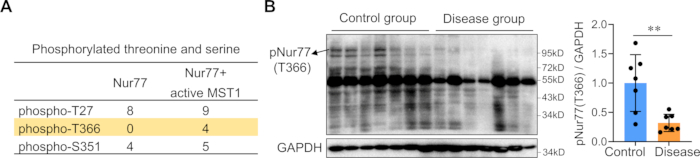
Figure 2: Verification of Nur77 phosphorylation changes. (A) The identified number of peptides containing phosphorylated threonine and serine in the Nur77 plus active Mst1 group and Nur77 alone group using liquid chromatography-tandem mass spectrometry analysis showed the differential phosphorylation of Nur77 at Thr366. (B) Western blot analysis of phospho-Nur77-Thr366 protein level in human endometrium from infertile women with recurrent implantation failure and fertile controls.*P < 0.05, Student's t-test. Please click here to view a larger version of this figure.

Figure 3: Detection of Nur77 phosphorylation site. An in vitro phosphorylation assay was conducted by incubating precipitated FLAG-Nur77 wild-type or 366 Thr-to-Ala (T366A) mutation protein with recombinant active Mst1 for 30 min at 30 °C. Western blot analysis was performed with anti-FLAG, anti-phospho-threonine (pThr), anti-phospho-Serine (pSer), and anti-MST1 antibodies. Please click here to view a larger version of this figure.
Supplementary Figure 1: Phos-tag SDS-PAGE analysis of phosphorylated Nur77 in 10% gel. Phos-tag SDS-PAGE analysis of Nur77 and phosphorylated Nur77 protein in two endometrial samples from fertile controls using a 10 w/v% polyacrylamide gel with 50 µM Phos-tag Solution and 100 µM MnCl2. Please click here to download this File.
Supplementary Figure 2: Phos-tag SDS-PAGE analysis of phosphorylated Nur77 using a CST antibody for Nur77. Phos-tag SDS-PAGE analysis of human endometrium from infertile women with recurrent implantation failure and fertile controls using a CST antibody for Nur77. Please click here to download this File.
Discussion
Loading...
$$\rightleftharpoonup{xx}$$
$$\longleftharp{xx}$$,
$$\longrightharp{xx}$$,
The Phos-tag-based approach for detecting phosphorylation changes of target proteins in clinical tissues offers significant advantages for protein phosphorylation research. Phosphorylated proteins exhibit reduced electrophoretic mobility due to Phos-tag binding, enabling high-throughput screening of the phosphorylation status of candidate proteins without requiring phospho-specific antibodies. This is particularly valuable for initial large-scale profiling of multiple candidate proteins. Proteins with altered phosphorylation modifications can be prioritized for subsequent in vitro kinase assay to identify specific phosphorylation sites.
While the sample preparation for Phos-tag SDS-PAGE shares similarities with conventional Western blotting, several critical technical considerations must be noted. EDTA-containing reagents must be avoided during sample processing as EDTA chelates Mn2+ or Zn2+ ions essential for Phos-tag function. However, it's essential to incubate the gel in transfer buffer containing 10 mM EDTA after electrophoresis but prior to membrane transfer, as this removes residual Mn2+ or Zn2+ ions that would otherwise severely impair protein transfer efficiency.
The preparation of Phos-tag SDS-PAGE gel requires careful optimization. Phos-tag alters protein migration in a non-linear fashion, making traditional protein markers unreliable for molecular weight estimation. For the detection of Nur77 in conventional SDS-PAGE, 8%-10% polyacrylamide gel is applied. However, reducing the gel concentration to 6% significantly improved the resolution of phosphorylated and non-phosphorylated forms of Nur77 (Supplementary Figure 1). In addition, Zn2+ exhibits distinct binding affinity for the phosphate group compared to Mn2+, although the underlying mechanisms remain unclear. In cases where Mn2+-Phos-tag electrophoresis consistently produces suboptimal results for certain proteins, Zn2+ substitution may improve resolution, particularly for high-molecular-weight proteins10.
There are several limitations to the Phos-tag-based approach for detecting phosphorylation changes. Not all antibodies suitable for conventional Western blotting perform well in Phos-tag systems. In our experience with phosphorylated Nur77 detection, an ABCAM antibody that works well in conventional Western blotting for Nur77 failed to detect phosphorylated forms in Phos-tag gels. After comparative testing of commercial antibodies from several suppliers, we found that a CST antibody for Nur77 successfully revealed both phospho- and non-phospho-isoforms (Supplementary Figure 2). This suggests that phosphorylation modifications at specific amino acid residues may sterically hinder antibody-epitope binding, consequently preventing the antibody from recognizing both phosphorylated and non-phosphorylated protein forms11. We have not detected proteins other than Nur77 using Phos-tag SDS-PAGE. Consequently, researchers may need to screen multiple antibodies for optimal detection. However, commercially available antibodies may not always be obtainable for certain targets, potentially limiting the method's universality.
Following the detection of altered phosphorylation levels of target proteins using phos-tag SDS PAGE in diseased tissues, an in vitrokinase assay can be employed to identify specific phosphorylation sites12. Notably, the methodological details of how Mst1 was identified as the kinase responsible for Nur77 phosphorylation were not fully elaborated in this protocol. In brief, we first identified multiple Nur77-interacting proteins, including several phosphokinases, through yeast two-hybrid screening13. Bioinformatics analysis suggested Mst1 as a potential kinase for Nur77, and subsequent Western blotting revealed that Mst1 expression was significantly downregulated in the disease group, implicating it as the upstream kinase mediating the reduced phosphorylation of Nur77. As described in our previous study, decreased phosphorylation of Nur77 in the infertile endometrium was mediated by Mst1 kinase at threonine 366 (T366) of Nur77. Subsequently, a phospho-specific antibody against Nur77-T366 was generated for Western blot analysis of extensive clinical samples to demonstrate decreased Nur77-T366 phosphorylation in endometrial tissues from infertile patients. These findings support the potential clinical utility of Nur77-T366 phosphorylation as one of the biomarkers of endometrial receptivity in fertility assessments. Although bioinformatics tools for predicting kinase-substrate interactions and phosphorylation sites, such as PhosphositePlus, Group-based Prediction System, and NetKSA, have become increasingly sophisticated and abundant14, in vitro kinase assays remain indispensable for validation. These computational approaches, while powerful for preliminary screening, often yield false-positive predictions due to the complexity of phosphorylation networks and contextual dependencies in cellular signaling. Experimental verification through in vitro kinase assays provides direct biochemical evidence of kinase activity toward the substrate, while phospho site-specific antibodies enable effective detection of phosphorylation events.
In summary, phos-tag SDS-PAGE Gel Electrophoresis combined with in vitro kinase assay serves as a powerful tool enabling researchers to efficiently and cost-effectively screen for altered protein phosphorylation patterns in diseased tissues. This approach facilitates subsequent mechanistic investigations and supports the development of specific phospho-antibodies for clinical tissue detection applications.
Disclosures
Loading...
$$\rightleftharpoonup{xx}$$
$$\longleftharp{xx}$$,
$$\longrightharp{xx}$$,
The authors declare no potential conflicts of interest.
Acknowledgements
Loading...
$$\rightleftharpoonup{xx}$$
$$\longleftharp{xx}$$,
$$\longrightharp{xx}$$,
This work was supported by the National Natural Science Foundation of China (82271698) and the Natural Science Foundation of Jiangsu Province (BK20231117).
Materials
| Name | Company | Catalog Number | Comments |
|---|---|---|---|
| 10% SDS Solution | Sangon Biotech | B548118 | |
| 10x Tris/Glycine/SDS | BIO-RAD | 1610732 | |
| 4× Laemmli protein sample buffer | BIO-RAD | 1610747 | |
| Acrylamide | Sangon Biotech | A601032 | |
| Ammonium persulfate | Sangon Biotech | A100486 | |
| Anti-FLAG M2 affinity gel | Millipore | A2220 | |
| Enhanced chemiluminescence kit | ThermoFisher | 32106 | |
| FLAG antibody | CST | 14793 | |
| FLAG Peptide | Millipore | F3290 | |
| HRP-conjugated secondary antibody | Bioworld Technology | BS13278 | |
| Lipofectamine 3000 | ThermoFisher | L3000015 | |
| Mini Trans-Blot Cell | BIO-RAD | 1703930 | |
| Mini-PROTEAN Tetra Vertical Electrophoresis Cell | BIO-RAD | 1658005 | |
| Mst1 antibody | CST | 3682 | |
| N, N'-methylene-bisacrylamide | Sangon Biotech | A600025 | |
| N,N,N',N'-Tetramethylethylenediamine(TEMED) | Sangon Biotech | A610508 | |
| Nur77 antibody | CST | 3960 | |
| Phosphatase Inhibitor Cocktail 2 | Sigma | P5726 | For the inhibition of tyrosine protein phosphatases, acid and alkaline phosphatases |
| Phosphatase Inhibitor Cocktail 3 | Sigma | P0044 | For the inhibition of protein phosphatase 2A, alkaline phosphatases, protein phosphatases 1 and 2A |
| Phospho-serine antibody | Sigma | P5747 | |
| Phospho-threonine antibody | Sigma | P6623 | |
| Phos-tag Acrylamide | Wako | 304-93526 | For the preparation of Phos-tag SDS PAGE gel |
| Pierce BCA Protein Assay Kit | Thermo | 23227 | |
| Protease Inhibitor Cocktail | Sigma | S8830 | EDTA-Free, tablet, for the inhibition of serine, cysteine, aspartic and metalloproteases |
| PVDF membrane | Roche | 3010040001 | |
| ThermoMixer | Eppendorf | 2231001127 | |
| Tissue Disperser | IKA | T 10 | To obtain tissue homogenate |
| Tris-HCl Buffer (0.5M, pH 6.8) | Sangon Biotech | B546020 | |
| Tris-HCl Buffer (1.5M, pH 8.8) | Sangon Biotech | B546019 | |
| Vacuum desiccator | ThermoFisher | 5311-0250 | To degas the gel solution |
| β-Mercaptoethanol | Sigma | M3148 |
References
Loading...
$$\rightleftharpoonup{xx}$$
$$\longleftharp{xx}$$,
$$\longrightharp{xx}$$,
- The protein kinase complement of the human genome. Science (New York, N.Y.). 298 (5600), 1912-1934 (2002).">Manning, G., Whyte, D. B., Martinez, R., Hunter, T., Sudarsanam, S. The protein kinase complement of the human genome. Science (New York, N.Y.). 298 (5600), 1912-1934 (2002).
- Regulation of mitochondrial functions by protein phosphorylation and dephosphorylation. Cell Biosci. 6, 25(2016).">Lim, S., et al. Regulation of mitochondrial functions by protein phosphorylation and dephosphorylation. Cell Biosci. 6, 25(2016).
- Role of protein phosphorylation in cell signaling, disease, and the intervention therapy. MedComm. 3 (4), e175(2022).">Pang, K., et al. Role of protein phosphorylation in cell signaling, disease, and the intervention therapy. MedComm. 3 (4), e175(2022).
- Molecular Pathways: Targeting protein tyrosine phosphatases in cancer. Clin Cancer Res. 23 (9), 2136-2142 (2017).">Bollu, L. R., Mazumdar, A., Savage, M. I., Brown, P. H. Molecular Pathways: Targeting protein tyrosine phosphatases in cancer. Clin Cancer Res. 23 (9), 2136-2142 (2017).
- Tau protein kinases: involvement in Alzheimer's disease. Ageing Res Rev. 12 (1), 289-309 (2013).">Martin, L., et al. Tau protein kinases: involvement in Alzheimer's disease. Ageing Res Rev. 12 (1), 289-309 (2013).
- Integrating the analysis of human biopsies using post-translational modifications proteomics. Protein Sci. 33 (4), e4979(2024).">Bhardwaj, S., et al. Integrating the analysis of human biopsies using post-translational modifications proteomics. Protein Sci. 33 (4), e4979(2024).
- Phosphoprotein profiling by PA-GeLC-MS/MS. J Proteome Res. 7 (7), 2812-2824 (2008).">Kristjansdottir, K., Wolfgeher, D., Lucius, N., Angulo, D. S., Kron, S. J. Phosphoprotein profiling by PA-GeLC-MS/MS. J Proteome Res. 7 (7), 2812-2824 (2008).
- Phosphate-binding tag, a new tool to visualize phosphorylated proteins. Mol Cell Proteomics. 5 (4), 749-757 (2006).">Kinoshita, E., Kinoshita-Kikuta, E., Takiyama, K., Koike, T. Phosphate-binding tag, a new tool to visualize phosphorylated proteins. Mol Cell Proteomics. 5 (4), 749-757 (2006).
- Mst1-mediated phosphorylation of Nur77 improves the endometrial receptivity in human and mice. EBioMedicine. 88, 104433(2023).">Cai, X., et al. Mst1-mediated phosphorylation of Nur77 improves the endometrial receptivity in human and mice. EBioMedicine. 88, 104433(2023).
- Improved Phos-tag SDS-PAGE under neutral pH conditions for advanced protein phosphorylation profiling. Proteomics. 11 (2), 319-323 (2011).">Kinoshita, E., Kinoshita-Kikuta, E. Improved Phos-tag SDS-PAGE under neutral pH conditions for advanced protein phosphorylation profiling. Proteomics. 11 (2), 319-323 (2011).
- An ultra-specific avian antibody to phosphorylated tau protein reveals a unique mechanism for phosphoepitope recognition. J Biol Chem. 287 (53), 44425-44434 (2012).">Shih, H. H., et al. An ultra-specific avian antibody to phosphorylated tau protein reveals a unique mechanism for phosphoepitope recognition. J Biol Chem. 287 (53), 44425-44434 (2012).
- Identification of cyclin-dependent kinase 1 specific phosphorylation sites by an in vitro kinase assay. J Vis Exp. (135), e57674(2018).">Cui, H., Loftus, K. M., Noell, C. R., Solmaz, S. R. Identification of cyclin-dependent kinase 1 specific phosphorylation sites by an in vitro kinase assay. J Vis Exp. (135), e57674(2018).
- Mapping protein-protein interactions using yeast two-hybrid assays. Cold Spring Harb Protoc. 2015 (5), 442-452 (2015).">Mehla, J., Caufield, J. H., Uetz, P. Mapping protein-protein interactions using yeast two-hybrid assays. Cold Spring Harb Protoc. 2015 (5), 442-452 (2015).
- Prediction of kinase-substrate associations using the functional landscape of kinases and phosphorylation sites. Pac Symp Biocompu. 28, 73-84 (2023).">Ayati, M., Yilmaz, S., Blasco Tavares Pereira Lopes, F., Chance, M., Koyuturk, M. Prediction of kinase-substrate associations using the functional landscape of kinases and phosphorylation sites. Pac Symp Biocompu. 28, 73-84 (2023).
Reprints and Permissions
Request permission to reuse the text or figures of this JoVE article
Request Permission