The video demonstrates the generation of recombinant arenaviruses in Vero cells by delivering host polymerase–driven plasmids encoding viral proteins and antigenomic RNA segments. It presents the initiation of viral gene expression by host enzymes, the formation of replication complexes, the progression of genome synthesis and ribonucleoprotein assembly, and the formation of recombinant viral particles.
Article de méthode
Rescue of Recombinant Arenaviruses in Virus-Susceptible Mammalian Cells
35 vues
⸱
29 octobre 2026
Dans cet article
Résumé
Protocole
1. Arenavirus Rescue Transfection
Our rescue system was based on the use of both polymerase II protein expression plasmids encoding the nucleoprotein (NP) and RNA-dependent RNA polymerase (L), the viral trans-acting factors required for RNA replication and gene expression of the arenavirus genome, and plasmids capable of directing intracellular synthesis, via the cellular RNA polymerase I (pol-I), of the S and L antigenome RNA species. In our studies, we used the pCAGGs protein expression plasmid, which uses the chicken β-actin promoter and polyadenylation (pA) signal sequences, and the human pol-I plasmid, which uses the human polymerase I promoter and murine terminator sequences (Figure 1). The S and L RNA segments are cloned into the hpol-I plasmid in an antigenomic orientation to allow for the generation of genomic RNA segments upon transcription by hpol-I. For the rescue of wild-type recombinant lymphocytic choriomeningitis virus (rLCMV) and Candid#1 (rCandid#1), the pCAGGs NP and L from each virus were co-transfected with their respective human pol-I S and L RNA segment plasmids (Figure 2). Generation of recombinant trisegmented LCMV (r3LCMV) and Candid#1 (r3Candid#1) followed a similar protocol, but the S segment was split into two different pol-I S plasmids, each encoding a distinctive reporter gene: in one, the NP open reading frame (ORF) was replaced with the green fluorescent protein (GFP) reporter gene (hpol-I S GFP/GP) and, in the other, the viral glycoprotein precursor (GPC) ORF was replaced with the Gaussia luciferase (Gluc) reporter gene (hpol-I S NP/Gluc). A schematic representation of the protocol is illustrated in Figure 3. The following transfection and infection protocol was established for 6-well plates.
- Opti-MEM-Lipofectamine 2000 (LPF2000) mixture: Prepare 250 μl of Opti-MEM media and, depending on the virus rescue, 10 - 12 μg of LPF2000 (1 μg/μl) per transfection (Table 1). A ratio of 2.5 μg LPF2000 per μg of DNA is recommended. Incubate for 5 - 10 min at room temperature (RT). Meanwhile, prepare the plasmid transfection mixture in a separate microcentrifuge tube (step 1.2).
- Plasmid DNA transfection mixture: Prepare the plasmid transfection cocktail in a microcentrifuge tube using the recommended amounts for each virus rescue (see Table 1). Bring the final volume to 50 μl with Opti-MEM.
- Opti-MEM-LPF2000-DNA plasmid mixture: Add 250 μl of the Opti-MEM-LPF2000 mixture (step 1.1) into the plasmid DNA transfection mixture (step 1.2). Incubate this mixture for 20 - 30 min at RT. Meanwhile, prepare and count 1 x 10⁶ Vero cells per transfection reaction. Vero cells will be transfected in suspension.
- Preparation of Vero cells: Vero cells are cultured in 100 mm dishes. Before starting, bring the 1x phosphate-buffered saline (PBS), Dulbecco's modified Eagle medium (DMEM) 10% fetal bovine serum (FBS) 1% penicillin-streptomycin (PS) media, and trypsin-ethylenediaminetetraacetic acid (EDTA) mixture to 37 °C.
- Wash the cells twice with 5 mL of 1x PBS.
- Trypsinize cells with 1 mL of trypsin-EDTA and wait until cells detach (approximately 5 min). Gentle tapping may be required to fully detach the cells from the dishes.
- Carefully resuspend the cells in 10 mL of DMEM containing 10% FBS 1% PS in a 15 mL centrifuge tube.
- Centrifuge the cells for 5 min at 1,000 revolutions per minute (rpm) in a centrifuge.
- Resuspend the cells in 10 mL of DMEM containing 10% FBS and 1% PS. Count the cells using a hemocytometer and adjust the concentration to 1 x 10⁶ cells/mL. It was observed that 0.5 - 1 x 10⁶ Vero cells per transfection produced the best results.
- Mix LPF2000/DNA and cells: After a 20 - 30 min incubation at room temperature (step 1.3), add 1 mL of Vero cells (1 x 10⁶) (step 1.4) to the Opti-MEM-LPF2000-DNA plasmid mixture and incubate for 5 min at room temperature.
- Seed LPF2000/DNA-cell mix into 6-well plates: Add the DNA-LPF2000-Vero mixture (step 1.5) into the wells of the 6-well plate. Gently shake the 6-well plate and incubate the transfected cells overnight in the incubator at 37 °C and 5% CO2.
Change transfection medium: Approximately 16 - 24 hr post-transfection, remove the transfection media, add 2 mL of infection media and incubate the transfected cells for an additional 48 hr.
Table 1: Lipofectamine/plasmid DNA concentration for arenavirus rescue. Recommended DNA and LPF2000 concentrations for generation of recombinant wild-type and trisegmented arenaviruses in Vero cells are indicated.
Plasmid / Reagent Wild-type viral rescue Trisegmented viral rescue pCAGGs NP (μg) 0.8 0.8 pCAGGs L (μg) 1.0 1.0 hpPOLI L (μg) 1.4 1.4 hpPOLI S (μg) 0.8 - hpPOLI S GPc/GFP (μg) - 0.8 hpPOLI S NP/Gluc (μg) - 0.8 Total DNA (μg) 4 4.8 Lipofectamine 2000 (μg)* 10 12 *Lipofectamine 2000 is prepared in 250 μL of Opti-MEM, in a separate microcentrifuge tube.
Résultats
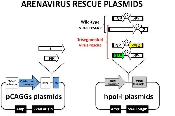
Figure 1. Arenavirus rescue plasmids. Diagram of the plasmids used for the generation of recombinant arenavirus in Vero cells. Protein expression pCAGGs plasmid uses the chicken β-actin promoter and the rabbit β-globin polyadenylation (pA) signal sequences to direct the synthesis of viral L and NP. CMV-IE enhancer: cytomegalovirus immediate early enhancer. Intron: chicken β-actin intron sequence. The hpol-I plasmids use the human polymerase I promoter and murine polymerase terminator sequences to direct the synthesis of the viral RNA segments. Both pCAGGs and hpol-I plasmids are Ampicillin resistant (Ampr).
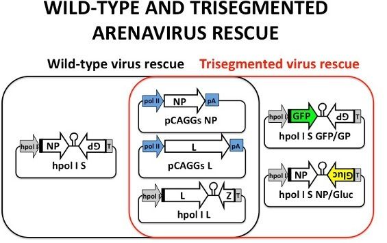
Figure 2. Wild-type and trisegmented arenavirus rescue. In the black oval are the plasmids required for the generation of recombinant wild-type arenavirus: pCAGGs NP and L, and hpol-I S and L. NP and L are required to initiate viral transcription and replication. The hpol-I S and L direct intracellular synthesis, via pol-I, of S and L antigenomic RNA species. In the red oval are the plasmids required for the generation of trisegmented arenaviruses: pCAGGs NP and L, as well as the hpol-I L plasmid, which are the same as those described for wild-type arenavirus rescue. The hpol-I S is separated into two plasmids: in one plasmid, NP is replaced with GFP (hpol-I S GFP/GP), and in the other plasmid, the GP is replaced with the Gluc gene (hpol-I S NP/Gluc).
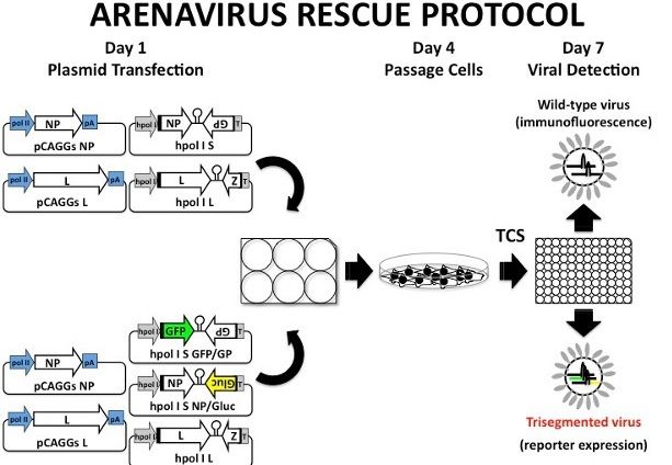
Figure 3. Arenavirus rescue protocol. Arenavirus rescue is done in Vero cells using a 6-well plate format. Briefly, Vero cells are transfected in suspension on Day 1 using Lipofectamine 2000. At 24 hr post-transfection, the media is replaced with fresh infectious media. Transfected cells are incubated for an additional 48 hr and then scaled-up into a 100 mm dishes (Day 4). Vero cells in the 100 mm dishes are incubated for an additional 72 hr. On Day 7 post-transfection, tissue culture supernatants (TCS) are collected for viral detection by either immunofluorescence assay (rLCMV and rCandid#1) or fluorescence microscopy (r3LCMV and r3Candid#1) following infection in fresh Vero cells. If the viral titers are lower than expected, TCS can be used to infect fresh Vero cells to amplify the virus.
Matériaux
| Nom | Entreprise | Numéro de catalogue | Commentaires |
|---|---|---|---|
| Vero E6 cells | American Type Culture Collection (ATCC) | CRL-1586 | |
| hpol-I L plasmid | |||
| Dulbecco's modified Eagle medium (DMEM) | |||
| Fetal bovine serum (FBS) | |||
| Penicillin-streptomycin (PS), 100× | |||
| Opti-MEM | |||
| Sodium chloride (NaCl) | |||
| Potassium chloride (KCl) | |||
| Disodium hydrogen phosphate heptahydrate (Na₂HPO₄·7H₂O) | |||
| Potassium dihydrogen phosphate (KH₂PO₄) | |||
| Double distilled water (ddH₂O) | |||
| rCandid#1 | |||
| rLCMV (Armstrong 53b) |
