Ce protocole décrit un modèle de rongeur sans masque ni suture des paupières de myopie induite par des injections sous-conjonctivales de TGF-β2, qui active l'axe NLRP3/IL-1β-MMP2 provoquant une élongation axiale, tandis qu'un régime fractionné d'enregistrement biométrique minimise les artefacts liés à l'anesthésie pour des données de haute fidélité.
Article de méthode
Établissement d'un modèle de myopie chez le rongeur induit par le TGF-β à l'aide d'injections sous-conjonctivales
Dans cet article
Résumé
Résumé
La myopie est un problème majeur de santé publique mondiale, caractérisé par un allongement axial excessif et un remodelage de la matrice extracellulaire (ECM) de la sclère. Bien que les modèles expérimentaux soient essentiels pour étudier ces mécanismes, les méthodes classiques de défocalisation optique chez les petits rongeurs présentent des limites techniques. Ici, un modèle pharmacologique de myopie standardisable a été mis en place et évalué par des injections sous-conjonctivales répétées de facteur de croissance transformant bêta (TGF-β). Pour assurer une délivrance ciblée, une technique chirurgicale de stabilisation bimanuelle a été développée afin d'évertir la paupière supérieure, permettant à une aiguille de calibre 33 de viser la conjonctive palpébrale selon un angle faible de 20° à 30°. Grâce à un criblage in vivo chez le rat, le TGF-β2 (500 pg/œil) s'est révélé un facteur majeur de croissance postérieure de l'œil. Une titration subséquente de la réponse à la dose chez la souris a révélé un profil en forme de U inversé. Dans le groupe principal de validation, la dose la plus faible (250 pg/œil) a induit un allongement axial marqué (0,197 ± 0,012 mm) et des décalages réfractifs myopiques (−1,58 ± 0,63 D). Au niveau moléculaire, l'administration répétée de TGF-β2 a activé la cascade de l'inflammasome de la famille des récepteurs de type NOD contenant un domaine pyrine 3 (NLRP3) / interleukine-1β (IL-1β) au niveau rétinien et a augmenté l'expression de la métalloprotéinase 2 de la matrice (MMP2) chez les deux espèces. Associé à un protocole de mesure biométrique réparti sur deux jours, qui minimise les artefacts liés à l'anesthésie, ce protocole fournit une plateforme fiable et reproductible pour évaluer des interventions anti-myopie.
Introduction
La myopie est devenue un problème majeur de santé publique mondiale, sa prévalence devant toucher près de 50 % de la population mondiale d'ici 20501,2. Le principal facteur pathologique à l'origine de la myopie progressive est l'allongement axial excessif de l'œil, qui provoque un remodelage actif de la matrice extracellulaire et une dégénérescence tissulaire, entraînant un amincissement structural des tuniques oculaires postérieures3. Ces modifications dynamiques comprennent à la fois un remodelage structural (caractérisé par la dégradation de la matrice extracellulaire sclérale, la perte de collagène et l'amincissement des couches postérieures) et un remodelage fonctionnel (marqué par une rigidité biomécanique réduite, une perfusion choroïdienne altérée et une signalisation inflammatoire anormale entre la rétine et l'épithélium pigmentaire rétinien), qui augmentent collectivement le risque, tout au long de la vie, de complications menaçant la vision et de déficiences visuelles irréversibles, notamment la maculopathie myopique, le décollement de rétine, le glaucome et la cataracte prématurée4,5,6,7. Malgré les lourdes conséquences cliniques et socio-économiques de cette affection, les mécanismes moléculaires précis situés en amont et régissant la croissance oculaire anormale restent incomplètement élucidés. Par conséquent, l'identification des voies de signalisation responsables de l'allongement axial et le développement de modèles animaux reproductibles et standardisés constituent des étapes essentielles pour découvrir de nouvelles cibles thérapeutiques et mieux contrôler l'épidémie mondiale de myopie.
Bien que la myopie induite par privation de forme (FDM) et la myopie induite par lentille (LIM) constituent des modèles de référence pour l'étude du défaut de mise au point optique et de l'emmétropisation8,9,10,11, la mise en œuvre de ces modèles de défocalisation optique chez les petits rongeurs — en particulier chez la souris — présente des défis techniques importants. Le comportement actif de toilette des souris entraîne fréquemment un détachement des lunettes, nécessitant des mesures correctives stressantes telles que l'utilisation de colliers de protection12De plus, les frottements mécaniques, la suture des paupières et les modifications du microenvironnement sous l'occlusif provoquent couramment des complications cornéennes secondaires, telles qu'une opacité ou une ulcération cornéenne.13,14,15Bien que les modèles FDM et LIM restent des paradigmes fondamentaux pour l'étude du flou visuel, des complications oculaires secondaires au niveau de la surface oculaire—telles que l'opacité cornéenne, le frottement mécanique ou l'irritation due à la suture des paupières—peuvent interférer avec les mesures biométriques optiques.15Ces artefacts procéduraux compromettent la stabilité du film lacrymal et la transparence cornéenne, entraînant des taux d'exclusion expérimentale plus élevés et un bruit de mesure accru13,16. En revanche, l'induction pharmacologique contourne les obstacles physiques, préservant la clarté cornéenne et facilitant une biométrie optique et ultrasonore fiable et reproductible. Dans la myopie, une élongation oculaire excessive est provoquée par une remodélisation altérée de la matrice extracellulaire (ECM) de la sclère postérieure.17, où le facteur de croissance transformant-bêta (TGF-β) les isoformes agissent comme des régulateurs clés de la synthèse du collagène et des protéoglycanes18,19. Des données émergentes suggèrent que le TGF-β la signalisation peut interagir avec une inflammation oculaire de bas grade et une dégradation de la matrice médiée par la métalloprotéinase 2 (MMP2)20, pouvant contribuer au remodelage de la sclérotique et à l'élongation axiale21,22,23,24. Ceci est soutenu par des taux élevés de facteur de croissance transformant bêta 2 (TGF-β2), l'interleukine-6 (IL-6) et la MMP2 rapportés dans l'humeur aqueuse des yeux myopes25,26,27, ainsi que l'activation du TGF-β / facteur nucléaire kappa B (NF-κB) / axe MMP2 dans les modèles expérimentaux20,21. Bien que ces résultats reflètent une signalisation moléculaire intraoculaire dynamique au cours de la progression de la myopie, les contributions anatomiques relatives aux marqueurs de l'humeur aqueuse—qu'elles proviennent d'une sécrétion antérieure locale, d'un flux depuis un dépôt sous-conjonctival ou d'une diffusion postérieure par le vitré—demeurent à élucider complètement. Toutefois, il convient de reconnaître l'étendue et les limites inhérentes à ce modèle pharmacologique. En raison de l'administration sous-conjonctivale de TGF-β la livraison contourne le défaut de focalisation optique et les voies de traitement visuel, ce modèle est particulièrement adapté à l'analyse des cascades biochimiques en aval — spécifiquement TGF-β2 / Récepteur de type NOD, famille pyrine, domaine contenant 3 (NLRP3) / interleukine-1β (IL-1β) / la remodelisation de la MEC sclérale médiée par la MMP2 — et le criblage d'agents thérapeutiques candidats ciblant les voies inflammatoires ou de dégradation de la matrice. En revanche, elle n'est pas adaptée à l'étude de la transduction rétinienne des signaux visuels en amont, de la signalisation optique de la rétine à la sclérotique ou des mécanismes d'emmetropisation induits par la lumière environnementale, domaines pour lesquels les modèles traditionnels de myopie induite par défaut de mise au point (FDM) ou par lentille (LIM) restent indispensables.
Pour combler ces lacunes méthodologiques, un modèle de myopie a été établi et validé à l'aide d'injections sous-conjonctivales répétées de TGF-β chez le rat et la souris, offrant une approche améliorée qui minimise les traumatismes mécaniques et l'irritation de la surface oculaire. En évitant l'utilisation d'obturateurs physiques, ce protocole permet un contrôle précis de la posologie pharmacologique et des délais expérimentaux, tout en réduisant considérablement les facteurs de confusion comportementaux associés aux dispositifs fixés sur la tête. Dans cette étude, l'activité de trois isoformes humaines recombinantes de TGF-β a été évaluée (ces dernières présentant une réactivité croisée fonctionnelle validée dans les tissus de rongeurs, soutenue par une conservation de la séquence28) chez le rat, et la cinétique de réponse dose-dépendante non linéaire du TGF-β2 a été cartographiée chez la souris. De manière importante, ce régime standardisé a induit efficacement une myopie phénotypique — caractérisée par un allongement axial concomitant et des décalages réfractifs myopiques — ainsi que l'activation de l'inflammasome de la famille des récepteurs de type NOD contenant un domaine pyrine 3 (NLRP3) / interleukine-1β (IL-1β) et de l'axe MMP2. En définitive, ce protocole fournit un cadre viable et défini au niveau moléculaire pour cibler le remodelage scléral en aval, tout en précisant ses limites opérationnelles spécifiques.
Protocole
Toutes les expériences sur les animaux ont été réalisées conformément aux lignes directrices et aux règlements du Comité institutionnel de soins et d'utilisation des animaux (IACUC) de l'Université de médecine de Chine (CMU). Toutes les procédures étaient conformes à la déclaration de l'Association pour la recherche en vision et en ophtalmologie (ARVO) concernant l'utilisation des animaux dans la recherche ophtalmologique et visuelle. Une approbation complète du CMU-IACUC a été obtenue avant le début de toutes les expériences. Les animaux ont été euthanasiés à l'issue de l'expérience strictement conformément aux directives du Comité institutionnel de soins et d'utilisation des animaux (IACUC) de l'Université de médecine de Chine (CMU) et aux réglementations locales en matière de bien-être animal.
1. Animaux et approbation éthique
- Obtenir des rats mâles Brown Norway (BN) (âgés de 3 à 4 semaines, pesant 60 à 80 g) et des souris mâles C57BL/6 (B6) (âgées de 3 à 4 semaines, pesant 15 à 20 g) auprès d'un fournisseur approprié d'animaux de laboratoire.
- Héberger tous les animaux dans un environnement contrôlé (cycle lumière/obscurité de 12 h, 22 °C ± 2 °C, humidité relative de 50 % ± 10 %) avec un accès ad libitum à une nourriture standard et à de l'eau.
- Prévoir un minimum de 3 jours d'acclimatation avant d'entreprendre toute procédure expérimentale.
- Répartir aléatoirement les rats ou les souris en groupes de traitement : groupe témoin recevant le véhicule et groupe recevant une injection de TGF-β2. Utiliser un minimum de N = 5 animaux par cohorte afin d'assurer une puissance statistique adéquate.
REMARQUE : Un plan expérimental inter-sujets a été utilisé. Les deux yeux d'un même animal ont reçu le même traitement (soit un contrôle bilatéral au véhicule [1–2 µL BSS], soit un traitement actif bilatéral avec rhTGF-β2).- Mesurer les paramètres oculaires bilatéraux (longueur axiale et erreur réfractive) pour chaque animal.
REMARQUE : S'assurer d'obtenir l'approbation écrite du CMU-IACUC et que tout le personnel effectuant les procédures est correctement formé et certifié.
- Mesurer les paramètres oculaires bilatéraux (longueur axiale et erreur réfractive) pour chaque animal.
2. Préparation de la solution de TGF-β2
REMARQUE : Effectuez toutes les étapes de reconstitution et de dilution à l'intérieur d'une hotte de sécurité biologique certifiée en utilisant des techniques strictes d'asepsie.
- Reconstituer le TGF-β2 humain recombinant lyophilisé dans du glycérol stérile à 50 % contenant 0,1 % d'albumine de sérum bovin (BSA) afin d'obtenir une solution mère à 100 µg/mL.
- Préparer les solutions de travail en réalisant des dilutions sériées de la solution mère dans une solution saline tamponnée stérile (BSS).
- Pour les cohortes de rats (dose de 500 pg), diluer la solution mère à une concentration finale de 250 ng/mL (250 pg/µL) pour un volume d'injection de 2 µL par œil.
- Pour les cohortes de souris destinées à l'étude de la relation dose-réponse, préparer trois solutions de travail indépendantes aux concentrations de 250 ng/mL, 375 ng/mL et 500 ng/mL (250, 375 et 500 pg/µL) pour un volume d'injection de 1 µL par œil.
- Préparer des aliquotes unidose de 15 à 20 µL dans des tubes microcentrifuges stériles, les congeler rapidement dans l'azote liquide, puis les conserver à -20 °C. Décongeler les aliquotes sur de la glace juste avant utilisation.
ATTENTION : L'azote liquide peut provoquer de graves brûlures par le froid et présente un risque d'asphyxie. Porter des équipements de protection individuelle cryogéniques adaptés et le manipuler dans un endroit bien ventilé conformément aux procédures de sécurité institutionnelles. - Préparer simultanément la solution témoin (véhicule) en utilisant une solution saline tamponnée stérile (BSS) supplémentée avec une matrice équivalente de glycérol à 50 % et de BSA à 0,1 %.
3. Procédure d'injection sous-conjonctivale
- Placer les animaux dans une chambre d'induction contenant 3–4 % d'isoflurane pour l'induction anesthésique. Ensuite, transférer l'animal vers un embout nasal délivrant 1,5–2 % d'isoflurane pour le maintien de l'anesthésie.
ATTENTION : L'isoflurane est un anesthésique volatil. Utiliser un système d'aspiration approprié et le manipuler conformément aux procédures institutionnelles de santé et sécurité au travail. - Vérifier une profondeur anesthésique adéquate en confirmant l'absence d'un réflexe de retrait à la pincée des orteils.
- Positionner l'animal en décubitus ventral sous un spot à fibre optique. Saisir délicatement, soulever et légèrement éverser la paupière supérieure à l'aide de microforceps à pointes fines afin d'exposer complètement la conjonctive palpébrale et le fornix conjonctival (Figure 1B).
- Pré-humidifier une microsyringe en verre de 10 µL et une aiguille de calibre 33 deux à trois fois avec la solution de travail pour éliminer l'espace mort. Charger le volume cible (2 µL pour les rats ou 1 µL pour les souris) et purger toutes les microbulles.
ATTENTION : Manipuler les aiguilles et autres objets tranchants avec précaution et les jeter dans un conteneur pour déchets tranchants homologué, conformément aux réglementations institutionnelles. - Orienter l'aiguille 33 G avec le biseau vers le haut (biseau vers le haut) et viser le site d'insertion désigné sur la surface conjonctivale palpébrale exposée (Figure 1B,C).
- Insérer la pointe de l'aiguille à travers l'épithélium conjonctival selon un angle faible de 20°–30° (Figure 1C).
- Faire progresser délicatement l'aiguille à travers l'espace sous-muqueux étroit, au-delà du fornix conjonctival, jusqu'au domaine sous-conjonctival palpébral (Figure 1C).
ÉTAPE CRITIQUE (structures à éviter) : Éviter strictement de pénétrer les glandes de Meibomius sous-jacentes ou de perforer accidentellement des structures oculaires plus profondes. Confirmer visuellement un positionnement superficiel correct sous le spot en s'assurant que le biseau de l'aiguille reste visible sous la surface conjonctivale translucide tout au long de l'avancement. - Injecter lentement la solution pendant 5 à 10 s. Maintenir l'aiguille en place pendant 5 s supplémentaires avant de la retirer lentement afin d'éviter tout reflux du liquide.
REMARQUE : Une administration sous-conjonctivale correcte est confirmée par une élévation localisée immédiate de la conjonctive et une décoloration (formation d'une bulle).- Si aucune bulle ne se forme ou si une fuite importante survient, effectuer une seule réinjection dans l'espace conjonctival palpébral inférieur et noter l'écart dans le journal expérimental.
REMARQUE : Limiter strictement les réinjections à une tentative par œil et par séance ; exclure les animaux présentant un échec persistant de l'administration de l'analyse longitudinale.
- Si aucune bulle ne se forme ou si une fuite importante survient, effectuer une seule réinjection dans l'espace conjonctival palpébral inférieur et noter l'écart dans le journal expérimental.
- Nettoyer la cavité de la microsyringe entre chaque injection animale au sein du même groupe en rinçant trois fois avec de l'éthanol stérile à 70 %, puis cinq fois avec une solution saline stérile.
- Examiner la pointe de l'aiguille 33 G sous grossissement toutes les 3 à 5 injections, et remplacer immédiatement l'aiguille si la pointe s'émousse ou se courbe.
- Injecter d'abord tous les animaux contrôles véhicules, avant de manipuler ou d'injecter les cohortes traitées, afin d'éliminer tout risque de contamination croisée pharmacologique. Répéter le cycle de nettoyage de la seringue avant de passer aux groupes traités.
- Rincer le corps et le piston de la seringue avec une solution de nettoyage spécifique pour microsyringe, puis avec de l'eau ultrapure et de l'éthanol absolu après chaque séance expérimentale, et les conserver au sec.
- Répéter les injections sous-conjonctivales une fois par semaine pendant 3 semaines consécutives (jours 0, 7 et 14). Réaliser les évaluations biométriques longitudinales au moment de la ligne de base (jours −1 et 0) et à l'aboutissement (jours 20 et 21), comme décrit dans la section 4.
- Placer l'animal dans une cage de récupération chauffée, garnie de serviettes en papier, après l'injection. Surveiller l'animal en continu jusqu'à ce que la récupération complète en décubitus sternal et une activité ambulatoire normale soient rétablies.
- Examiner les yeux au microscope stéréoscopique après la récupération afin d'exclure toute hémorragie intraoculaire, opacité cornéenne ou traumatisme mécanique aigu. Surveiller les animaux quotidiennement pour détecter tout signe de détresse ou d'infection localisée.
4. Méthodes de validation du modèle
- Évaluation du défaut de réfraction oculaire
- Placer les rongeurs sur un tapis chauffant à commande thermostatique afin de maintenir une température corporelle physiologique et prévenir la formation de cataractes anesthésiques. Maintenir les animaux sous une anesthésie légère à l'isoflurane (1–1,5 %) au moyen d'un cône nasal pour assurer une profondeur anesthésique constante.
- Instiller 1 goutte de Mydrin-P (tropicamide à 0,5 % et chlorhydrate de phényléphrine à 0,5 %) dans chaque œil 10 à 15 minutes avant la mesure afin d'induire une cycloplégie et une dilatation pupillaire complètes.
- Placer l'animal en décubitus sternal sous un éclairage ambiant tamisé et indirect. Appliquer continuellement du BSS stérile jusqu'au moment précis de l'acquisition optique afin de préserver la transparence cornéenne.
- Mesurer la réfraction oculaire à l'aide d'un photoréfractomètre infrarouge automatisé dans un intervalle strict de 5 à 10 minutes après l'induction anesthésique. Chez les rats BN, écarter délicatement les paupières manuellement afin d'exposer complètement la pupille dilatée pendant l'acquisition.
- Acquérir trois mesures optiques indépendantes par œil et faire la moyenne des valeurs pour déterminer l'erreur de réfraction de base ou finale.
ÉTAPE CRITIQUE (Durée & ; Séquence) : Réaliser la photoréfraction le jour 1 de la séquence d'évaluation (jour –1 pour la valeur de référence ; jour 20 pour le point final) dans une lumière tamisée, avant toute procédure au contact. Effectuer la photoréfraction après une échographie en contact ou l'application de gel acoustique perturbe gravement l'optique cornéenne, disperse la lumière infrarouge et rend inexactes les mesures automatisées de la réfraction.
- Mesure de la longueur axiale par échographie en mode B à haute fréquence
- Le jour suivant (jour 0 pour la valeur initiale ou jour 21 pour le point final), évaluer la longueur axiale oculaire sous anesthésie légère à l'isoflurane (1–1,5 %) à l'aide d'une sonde d'échographie linéaire portative de 16 MHz étalonnée selon la vitesse acoustique des tissus de rongeur (1532–1550 m/s).
- Pour les souris B6 (interface cornéenne directe) : appliquer une couche protectrice de base de gel de carbomère Ginpol (0,2 %) directement sur la cornée, suivie d'une couche de gel acoustique standard.
REMARQUE : Veiller à ce que la couche de gel recouvre entièrement la surface cornéenne sans introduire de bulles d'air piégées, qui provoquent une atténuation acoustique. - Pour les rats BN (interface transpaupiérale) : positionner l'animal avec les paupières fermées, appliquer du gel échographique sur la surface externe de la paupière, puis effectuer les mesures de manière transpaupiérale.
REMARQUE : La mesure transpalpébrale élimine tout contact direct avec la cornée, réduisant ainsi significativement le risque d'abrasion cornéenne dans les yeux de rongeurs de grande taille.
- Pour les souris B6 (interface cornéenne directe) : appliquer une couche protectrice de base de gel de carbomère Ginpol (0,2 %) directement sur la cornée, suivie d'une couche de gel acoustique standard.
- Aligner la sonde d'échographie et mesurer la longueur axiale le long de l'axe optique central, depuis l'interface oculaire antérieure jusqu'à la paroi échoïque postérieure de la sclère.
- Acquérir dix tracés de balayage B indépendants par œil et calculer la longueur axiale moyenne.
- Rincer doucement le gel acoustique résiduel de l'œil à l'aide de BSS stérile immédiatement après l'acquisition afin d'éviter toute irritation cornéenne post-procédurale.
ÉTAPE CRITIQUE (contact sans pression) : Placez la sonde d'échographie sur l'interface de gel avec un contact extrêmement léger, sans aucune pression. Toute pression physique exercée vers le bas sur l'apex cornéen comprime artificiellement la profondeur de la chambre antérieure et induit un raccourcissement axial faux. - Enregistrer les mesures moyennes de la longueur axiale et de l'erreur réfractive pour les analyses ultérieures.
- Le jour suivant (jour 0 pour la valeur initiale ou jour 21 pour le point final), évaluer la longueur axiale oculaire sous anesthésie légère à l'isoflurane (1–1,5 %) à l'aide d'une sonde d'échographie linéaire portative de 16 MHz étalonnée selon la vitesse acoustique des tissus de rongeur (1532–1550 m/s).
Résultats
Establishment and anatomical validation of the subconjunctival injection model
To address technical variability associated with optical goggling or eyelid suturing, the subconjunctival space was validated between the palpebral and bulbar conjunctivae as a targeted localized drug reservoir (Figure 1A). Anatomical access via the superior fornix provided consistent exposure of the target site while maintaining ocular surface integrity (Figure 1B). Successful model establishment was confirmed by specific operational and biometric criteria, including the immediate transillumination verification of a localized, fluid-filled submucosal bleb (chemosis) over the target palpebral subconjunctival space without scleral perforation, tissue hemorrhage, or meibomian gland injury (Figure 1C), as well as longitudinal biometric verification of concurrent axial elongation (Δ AL > 0.15 mm in mice; >0.40 mm in rats) accompanied by a myopic refractive shift (Δ Refraction < −1.0 D) at Day 21 relative to vehicle-treated controls. Conversely, unsuccessful or suboptimal outcomes were identified by specific technical and physiological failure modes. Technical failure—such as premature fluid reflux upon needle withdrawal or inadvertent deep eyelid/orbital tissue penetration—resulted in minimal or absent bleb formation, leading to inadequate pharmacological retention and failure to induce myopic shifts; animals displaying persistent leakage were documented and excluded according to Protocol Step 3.8.1. Additionally, ocular surface complications from deep needle misplacement or excessive mechanical traction caused localized subconjunctival hemorrhage or corneal trauma, introducing confounding inflammatory responses. Suboptimal phenotypic outcomes were also observed during dose-response evaluations, where administration of suprathreshold concentrations (e.g., 500 pg/eye in mice) yielded an attenuated U-shaped response with reduced axial elongation, underscoring the physiological requirement for strict dose optimization. Verified localized drug retention under these standardized criteria ensured high-fidelity tissue targeting for downstream biometric and molecular analyses.
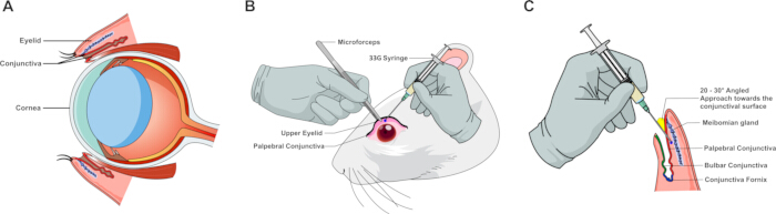
Figure 1: Anatomical landmarks and schematic illustration of the subconjunctival injection procedure in rodents. (A) Sagittal cross-section of the rodent eye depicting primary anatomical landmarks, including the eyelid, cornea, conjunctival sac, and targeted subconjunctival space. (B) Surgical demonstration of tissue stabilization and palpebral conjunctiva exposure using microforceps; the blue dot indicates the target needle insertion site for microinjection using a 33-gauge syringe. (C) High-magnification schematic illustrating the needle orientation and insertion trajectory relative to the palpebral conjunctival surface, conjunctival fornix, and underlying ocular structures. Please click here to view a larger version of this figure.
Screening of TGF-β isoforms in rats and dose-response titration of TGF-β2 in mice for axial elongation
Experiments were conducted in sequential, independent experimental cohorts comprising the dose-screening cohorts (Figure 2) and primary validation cohorts (Figure 3). Bilateral parameters were measured across cohorts, with individual animals (N) designated as the primary biological replicate. Inter-eye dependencies (OD versus OS within the same animal) were accounted for using two-way repeated-measures ANOVA or mixed-effects models, where appropriate. Continuous data are presented as mean ± SD. Differences between control and treatment groups were evaluated using two-tailed Student's t-tests, one-way ANOVA, or two-way repeated-measures ANOVA, where appropriate. A p-value < 0.05 was considered statistically significant.
To determine the most potent transforming growth factor-β (TGF-β) isoform and establish an optimal dosage regimen for inducing axial elongation, the effects of repeated subconjunctival injections in rat and mouse cohorts from baseline (Day 0) to Day 21 (Figure 2) were evaluated. Biometric recordings were performed using the two-day split regimen to prevent anesthesia-induced artifacts.
Three recombinant human TGF-β isoforms (TGF-β1, TGF-β2, and TGF-β3) were first screened in the rat cohort at a standardized concentration of 500 pg/eye (Figure 2A). One-way ANOVA, followed by post hoc multiple comparisons, revealed that all three isoforms significantly stimulated posterior ocular growth compared with the vehicle-treated control group over the 21-day period. While both TGF-β1 (0.444 ± 0.042 mm) and TGF-β3 (0.454 ± 0.049 mm) promoted significant axial elongation (p < 0.05 and p < 0.01 versus the control of 0.328 ± 0.071 mm, respectively), TGF-β2 induced the greatest axial length increase (0.486 ± 0.074 mm) and demonstrated the highest statistical significance (p < 0.001), establishing it as the most effective inducer of the myopic phenotype.
Based on the rat screening results, a dose-response titration of TGF-β2 in the mouse cohort was performed, testing three concentrations: 250, 375, and 500 pg/eye (Figure 2B). Subconjunctival TGF-β2 delivery resulted in an inverted U-shaped dose-response profile. The lowest dose (250 pg/eye) yielded the most significant acceleration in axial elongation compared to vehicle controls (0.177 ± 0.038 mm versus 0.044 ± 0.011 mm; p < 0.0001). At higher doses (375 and 500 pg/eye), the magnitude of axial length change progressively diminished, although it remained significantly higher than controls (0.123 ± 0.029 mm; p < 0.001 and 0.095 ± 0.024 mm; p < 0.01, respectively).
Together, these data establish a weekly subconjunctival regimen of 500 pg/eye for rats and 250 pg/eye for mice as the optimal parameters for subsequent structural and molecular investigations.
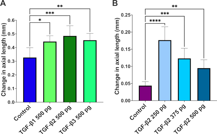
Figure 2: Dose-selection screening of transforming growth factor-β (TGF-β) isoforms and concentrations on axial length in rat and mouse eyes. (A) Change in axial length in rats (initial screening cohort), calculated as the difference between baseline (Day 0, before the first injection) and the final endpoint (Day 21), after three sequential subconjunctival injections of TGF-β1, TGF-β2, or TGF-β3 (500 pg/eye administered on Days 0, 7, and 14), compared with vehicle-treated controls. (B) Change in axial length in mice (dose-screening cohort), calculated as the difference between baseline (Day 0) and Day 21, after three sequential subconjunctival injections of TGF-β2 at three distinct concentrations (250, 375, or 500 pg/eye administered on Days 0, 7, and 14), compared with vehicle-treated controls. Data are presented as mean ± SD (N = 5 animals/group, representing 10 eyes per group). Statistical significance was evaluated by one-way ANOVA followed by post hoc multiple comparisons; *p < 0.05, **p < 0.01, ***p < 0.001, ****p < 0.0001 versus the vehicle control group. Please click here to view a larger version of this figure.
Repeated subconjunctival TGF-β2 injections induce ocular axial elongation and myopic shifts in rats and mice
To evaluate the biometric impact of repetitive subconjunctival TGF-β2 delivery (3 weekly injections), longitudinal changes in ocular axial length and refractive error from baseline to Week 3 in both rat and mouse models (Figure 3) were monitored. The two-day split protocol (refractive status captured on Day 20, followed by B-scan ultrasonography on Day 21) minimized potential anesthetic artifacts and ocular stress. Both species exhibited phenotypic shifts consistent with experimental myopia development following this multi-dose regimen.
In the rat cohort, animals receiving repeated subconjunctival injections of 500 pg/eye TGF-β2 showed a significantly greater increase in axial length over the 21-day period compared to vehicle controls (0.466 ± 0.045 mm versus 0.388 ± 0.032 mm; p < 0.01; Figure 3A). This elongation was accompanied by a significant myopic shift in refractive error (p < 0.05; Figure 3B). While vehicle-control eyes exhibited an expected slight hyperopic drift (+0.31 ± 0.93 D), TGF-β2–treated eyes transitioned into a myopic refractive state (−1.38 ± 0.91 D).
Consistent with the rat data, mice treated with 250 pg/eye TGF-β2 also demonstrated distinct structural and refractive changes. Axial elongation was significantly enhanced in TGF-β2–treated mice compared to vehicle controls (0.197 ± 0.012 mm versus 0.110 ± 0.014 mm; p < 0.0001; Figure 3C). Furthermore, this structural elongation resulted in a significant myopic shift (p < 0.05; Figure 3D), effectively reversing the natural hyperopic refraction seen in control mice (+2.25 ± 1.77 D) to a myopic profile (−1.58 ± 0.63 D).
To verify inter-ocular response symmetry and confirm that pooling bilateral ocular measurements does not introduce statistical bias, axial elongation dynamics were evaluated separately for left (OS, n = 5) and right (OD, n = 5) eyes within each group (Supplementary Figure 1). In both rats (Supplementary Figure 1A) and mice (Supplementary Figure 1B), no statistically significant difference was observed between OS and OD eyes within either the vehicle-control or TGF-β2–treated cohorts, confirming symmetrical response dynamics across eyes. Furthermore, both OS and OD eyes in TGF-β2–treated animals demonstrated consistent and robust axial elongation relative to their corresponding vehicle controls (p < 0.05 for rats; p < 0.0001 for mice), supporting the validity of accounting for bilateral measurements in the primary biometric analysis.
Collectively, these findings demonstrate that repeated subconjunctival administration of TGF-β2 effectively drives posterior ocular growth and a corresponding myopic refractive shift in both rodent models (Figure 3 and Supplementary Figure 1).
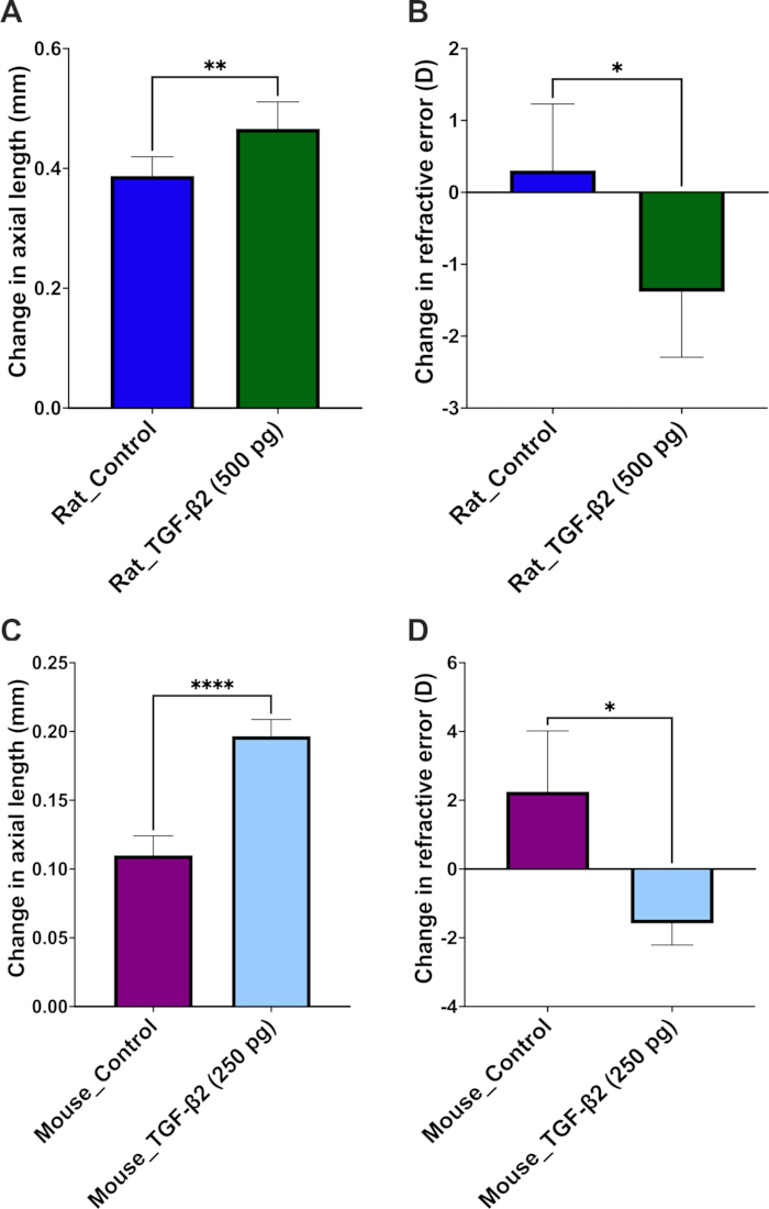
Figure 3: Primary validation of TGF-β2 effects on axial elongation and refractive error in independent rat and mouse cohorts. (A) Change in axial length in rats, calculated as the difference between baseline and the Week 3 final endpoint in eyes receiving subconjunctival TGF-β2 (500 pg/eye administered sequentially on Days 0, 7, and 14) or vehicle control. (B) Change in refractive error in the same rat eyes, delineating a significant myopic shift in the TGF-β2-treated group compared with the vehicle-control group. (C) Change in axial length in mice (independent primary validation cohort), calculated as the difference between baseline and the Week 3 final endpoint in eyes receiving subconjunctival TGF-β2 (250 pg/eye administered on Days 0, 7, and 14) or vehicle control. (D) Change in refractive error in the same mouse eyes, revealing a corresponding negative dioptric shift under optimal TGF-β2 induction. Data are presented as mean ± SD (N = 5 animals/group, representing 10 eyes per group). Statistical significance was rigorously evaluated using unpaired two-tailed t-tests; *p < 0.05, **p < 0.01, ****p < 0.0001 versus the corresponding vehicle control group. Please click here to view a larger version of this figure.
Subconjunctival TGF-β2 administration upregulates scleral MMP2 expression during experimental myopia progression
To assess downstream tissue remodeling along the retino-scleral axis, matrix metalloproteinase 2 (MMP2) expression in scleral cross-sections following subconjunctival TGF-β2 administration (Figure 4) was evaluated. Quantitative immunofluorescence analysis of posterior ocular sections in the rat model demonstrated a significant upregulation of MMP2 protein levels within the scleral stroma of TGF-β2–treated eyes (500 pg/eye) at Day 21 compared with vehicle controls (p < 0.0001; Figure 4A, B).
A prominent MMP2 fluorescent signal was localized throughout the scleral matrix underlying the retinal pigment epithelium (RPE) and choroidal layers, confirming localized MMP2-associated extracellular matrix (ECM) remodeling in the outer ocular coat during experimental myopia development. These structural changes in the sclera prompted further investigation into upstream molecular alterations within the adjacent retina.
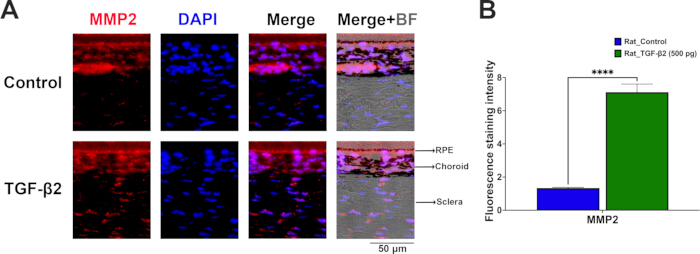
Figure 4: Subconjunctival TGF-β2 administration upregulates MMP2 expression in the rat sclera. (A) Representative immunofluorescence images of posterior ocular cross-sections showing scleral MMP2 expression (red) in vehicle-control and TGF-β2–treated (500 pg/eye) rats at Day 21. Cell nuclei were counterstained with DAPI (blue). Brightfield overlay (Merge+BF) delineates the anatomical boundaries of the RPE, choroid, and sclera. Scale bar = 50 µm. (B) Quantitative analysis of fluorescence staining intensity confirming a significant increase in scleral MMP2 expression in TGF-β2–treated rat eyes. Data are presented as mean ± SD (N = 3 independent biological replicates per group, 1 eye per animal). Statistical significance was evaluated using an unpaired two-tailed t-test; ****p < 0.0001 versus the vehicle control group. Abbreviations: RPE, retinal pigment epithelium; BF, brightfield. Please click here to view a larger version of this figure.
TGF-β2 induction upregulates MMP2 and inflammasome-related proteins in both rat and mouse retinas
To investigate the downstream molecular alterations induced by repetitive subconjunctival injections, immunofluorescence staining on posterior ocular sections from both Brown Norway rats and C57BL/6 mice at Day 21 (Figure 5A–D) was performed. All evaluations were performed on sagittal retinal sections strictly aligned to the optic nerve head to ensure consistent spatial orientation. Quantitative analysis revealed that weekly TGF-β2 administration significantly altered specific protein expression profiles across the retinal laminae in both species compared to vehicle controls (Figure 5E,F).
In the rat model, administration of 500 pg/eye TGF-β2 led to a significant increase in retinal TGF-β2 levels (p < 0.0001; Figure 5A,E). This upregulation was accompanied by elevated expression of matrix metalloproteinase 2 (MMP2) (p < 0.0001; Figure 5B,E), indicating active extracellular matrix remodeling. Furthermore, TGF-β2 treatment activated the retinal inflammatory cascade, evidenced by the upregulation of the inflammasome sensor NLRP3 (p < 0.0001; Figure 5C,E) and the pro-inflammatory cytokine IL-1β (p < 0.0001; Figure 5D,E).
Similarly, mice treated with 250 pg/eye TGF-β2 exhibited consistent upward trends in these biomarkers (Figure 5F). Retinal TGF-β2 (p < 0.01; Figure 5A,F) and MMP2 (p < 0.001; Figure 5B,F) expressions were significantly elevated. The inflammasome pathway was also activated in the mouse retina, with significant increases in NLRP3 expression (p < 0.01; Figure 5C,F) and a pronounced upregulation of IL-1β (p < 0.0001; Figure 5D,F) compared to near-undetectable baseline levels in the control group.
Together, these findings demonstrate that subconjunctival TGF-β2 administration promotes retinal MMP2-mediated matrix remodeling drivers and activates the NLRP3/IL-1β inflammasome pathway in the posterior retina of both rodent species.
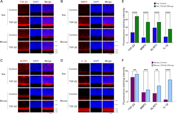
Figure 5: Repeated subconjunctival TGF-β2 administration upregulates MMP2 and inflammasome-related protein expression in the posterior retina of rats and mice. (A–D) Representative immunofluorescence images of retinal sagittal sections from control and TGF-β2–treated groups in rats (upper panels) and mice (lower panels), stained for (A) TGF-β2, (B) MMP2, (C) NLRP3, and (D) IL-1β (red), with nuclei counterstained by DAPI (blue). Scale bar = 50 µm. The representative fields selectively showcase the alterations within the outer retinal layers, highlighting prominent protein expression spanning from the outer plexiform layer (OPL) down to the basal retinal pigment epithelium (RPE) complex. (E) Quantification of fluorescence staining intensity for TGF-β2, MMP2, NLRP3, and IL-1β in rat retinas from the control group and the TGF-β2–treated group (500 pg). (F) Quantification of fluorescence staining intensity for TGF-β2, MMP2, NLRP3, and IL-1β in mouse retinas from the control group and the TGF-β2–treated group (250 pg). Data are presented as mean ± SD (N = 3 independent biological replicates per group, 1 eye per animal). Statistical significance was rigorously evaluated using unpaired two-tailed t-tests; **p < 0.01, ***p < 0.001, ****p < 0.0001 versus the respective vehicle-control group. Abbreviations: OPL, outer plexiform layer; ONL, outer nuclear layer; PIS, photoreceptor inner segments; POS, photoreceptor outer segments; RPE, retinal pigment epithelium. Please click here to view a larger version of this figure.
Supplementary Figure 1: Inter-ocular symmetry and evaluation of bilateral data pooling following subconjunctival TGF-β2 administration. (A) Comparative analysis of axial length changes between left eyes (OS, n = 5) and right eyes (OD, n = 5) in rats receiving vehicle control or bilateral subconjunctival TGF-β2 (500 pg/eye). (B) Corresponding inter-ocular comparison between OS (n = 5) and OD (n = 5) eyes in mice under vehicle control or bilateral TGF-β2 treatment (250 pg/eye). Statistical testing confirmed no significant difference between OS and OD eyes within the same treatment cohort, validating that bilateral measurement pooling does not introduce interocular bias. Simultaneously, both OS and OD eyes in TGF-β2–treated animals exhibited significant axial elongation relative to their respective vehicle controls. Data are presented as mean ± SD (N = 5 animals per group, representing 5 OS and 5 OD eyes per group). Statistical significance was evaluated using two-way repeated-measures ANOVA followed by Sidak’s post hoc test; *p < 0.05, ****p < 0.0001 versus the corresponding vehicle control. Abbreviations: OS, oculus sinister (left eye); OD, oculus dexter (right eye). Please click here to download this file.
Supplementary Figure 2: Safety evaluation of structural retinal integrity and electrophysiological function following subconjunctival TGF-β2 administration. (A) Representative hematoxylin and eosin (H&E)-stained sagittal retinal cross-sections from control and TGF-β2–treated mice (250 pg/eye) and rats (500 pg/eye) at Day 21, demonstrating preserved retinal layer architecture across species (N = 3 independent biological replicates per group). Scale bars = 20 µm. (B) Representative scotopic electroretinography (ERG) waveforms recorded from mice at Day 21, comparing vehicle control eyes (grey line) and TGF-β2–treated eyes (red line). Comparable a-wave and b-wave response dynamics confirm preserved retinal light responsiveness and electrophysiological safety (N = 3 mice per group, representing 6 eyes per group). Abbreviations: GCL, ganglion cell layer; INL, inner nuclear layer; ONL, outer nuclear layer. Please click here to download this file.
Discussion
A notable finding in the study is the non-linear, inverted U-shaped dose-response profile elicited by TGF-β2 in the mouse cohort, where the lowest administered dose (250 pg/eye) induced the most prominent axial elongation (Figure 2B). This biphasic phenomenon is characteristic of many potent cytokine signaling pathways, including the TGF-β superfamily29,30. Excessively high concentrations of exogenous TGF-β2 may lead to receptor saturation or trigger intracellular negative feedback loops—such as the upregulation of inhibitory SMAD family member 7 (Smad7) or receptor-mediated endocytosis—thereby attenuating downstream biological responses31,32. The immunofluorescence data further support this pharmacological window; while the 250 pg dose in mice (and 500 pg in rats) resulted in a moderate elevation in retinal TGF-β2 retention, it simultaneously triggered a robust downstream inflammatory cascade (Figure 5F). This suggests that the optimized low-dose threshold resides within a highly sensitive physiological window, maximizing targeted tissue efficacy while avoiding ligand-induced desensitization or localized suppression33. Therefore, these findings highlight the necessity of rigorous dose titration when establishing reproducible rodent models of chemical-induced myopia.
Compared with traditional form-deprivation myopia (FDM) or hyperopic defocus lens-induced myopia (LIM) models—which require customized hardware and are prone to lens displacement, grooming-induced detachment, and corneal complications—the subconjunctival protocol offers direct pharmacological control and minimal equipment costs13,34. Consistent with prior work showing that periocular delivery modulates scleral and retinal TGF-β signaling35, these data demonstrate posterior-segment biological responses following anterior subconjunctival TGF-β2 administration (Figure 4 and Figure 5). This efficient posterior targeting is attributed to the unique ocular scale and fluid dynamics of small rodents. Given the compact axial length of rodent eyes (~3 mm in mice and ~6 mm in rats), subconjunctival administration bathes a relatively large proportion of the episcleral plane. Driven by high transscleral permeability and the anatomical continuity of the sub-Tenon space, the injected formulation may diffuse across the sclera and choroid toward the posterior pole. Crucially, this route is associated with biological effects in posterior ocular tissues while avoiding the severe risks of globe perforation and optic nerve injury inherent to deep posterior sub-Tenon or intravitreal injections in small rodents. This approach avoids the mechanical and behavioral confounds of head-mounted lenses13,36, as well as the well-documented risks of retinal detachment and endophthalmitis associated with intravitreal injection37,38. Furthermore, partitioning the biometric measurements into two brief sessions improves upon conventional single-session paradigms. Prolonged anesthetic exposure frequently causes ocular disruptions—such as anesthetic cataracts in mice and spontaneous palpebral closure in rats—which compromise the accuracy of refractive and axial measurements39,40. By separating the refractive error (Days –1 and 20) and axial length (Days 0 and 21) assessments, the protocol is designed to minimize potential cumulative ocular stress and maintain consistent imaging conditions, which may help mitigate intra-experimental variability. To further eliminate potential confounding variables arising from systemic cross-contamination—such as systemic vascular absorption or lymphatic transport affecting non-target tissue—as well as contralateral sympathetic responses, a strictly between-subject design was implemented. By administering bilateral vehicle control or bilateral active rhTGF-β2 within individual animals, this paradigm prevents interocular contamination while ensuring robust pharmacological evaluation. Consistently, separate biometric analyses of the paired left and right eyes confirmed high inter-ocular symmetry, validating that bilateral data pooling did not introduce statistical or physiological bias across treatment cohorts (Supplementary Figure 1).
This rodent model effectively recapitulates the TGF-β2–driven inflammatory and matrix-remodeling axis previously established in studies by the authors in tree shrews and Brown Norway rats21,41. Previous investigations by the authors showed that exogenous TGF-β or TGF-β2 amplified intraocular inflammation—specifically via NF-κB and IL-1β activation—while upregulating MMP2 expression, depleting scleral collagen I, and driving axial elongation42,43. Conversely, dampening complement and inflammasome activity via complement decay-accelerating factor (CD55) suppressed both localized inflammation and myopia progression41,44. Similar chronic inflammatory signatures, marked by elevated MMP2 activation and subsequent collagen I loss, are also evident in classical form-deprivation models and in patients with pathological high myopia42,43,45,46. Importantly, rather than functioning as an isolated or universal primary inducer operating uniformly across all tissues, TGF-β (specifically the TGF-β2 isoform) acts as a context-dependent signaling bridge. It links early-stage retinal/RPE inflammatory signaling—specifically via NLRP3 inflammasome and complement activation—to downstream scleral extracellular matrix (ECM) execution21,41. Recognizing this spatiotemporal transition resolves apparent literature inconsistencies regarding TGF-β expression, as its dynamics shift between early local inflammatory amplification in the retina/RPE and late MMP-mediated matrix degradation in the sclera47,48. Specifically, while the structural execution of scleral ECM degradation ultimately dictates physical eye elongation (Figure 4), the molecular findings highlight the outer retina and RPE as the primary upstream signaling origin (Figure 5). Here, while the present study exclusively utilized quantitative immunofluorescence (IF) to map the in situ spatial distribution and laminar upregulation of these markers, the overall gene and protein dynamics (via RT-qPCR and Western blot, respectively) of this retinal phosphorylated nuclear factor kappa B (p-NF-κB)/IL-1β/tumor necrosis factor-alpha (TNF-α)/NLRP3 cascade under identical TGF-β2 induction paradigms have been independently established in the previous studies by the authors46,49. Subconjunctival TGF-β2 activates the NLRP3/IL-1β–MMP2 cascade within the retina/RPE, which propagates across the retino-choroidal-scleral axis to drive downstream scleral MMP2 expression and matrix remodeling. These parallels strongly support the central role of low-grade inflammation and MMP-mediated remodeling of the scleral extracellular matrix (ECM) in accelerated eye growth.
Despite these procedural advantages, several limitations of the present protocol warrant consideration. First, as a purely pharmacological model driven by exogenous cytokine delivery, this approach bypasses the optical defocus and retina-to-sclera light signal transduction pathways characteristic of traditional form-deprivation myopia (FDM) or lens-induced myopia (LIM) models16,50. Consequently, while highly suitable for targeting downstream biochemical cascades—specifically the inflammatory-MMP2 remodeling axis—it is complementary to, rather than a replacement for, models investigating upstream visual processing or optical manipulations51. Second, although subconjunctival injection avoids systemic toxicity and intravitreal risks, the requirement for repeated micro-injections (once weekly for 3 weeks) introduces transient localized mechanical perturbation and brief anesthesia exposure, which could subtly influence ocular surface homeostasis if not rigorously executed38,52. Achieving reproducible induction of experimental myopia via this route relies heavily on precise technical execution during subconjunctival delivery. The primary technical determinant of success is the correct shallow bevel insertion beneath the palpebral conjunctiva to establish a localized, well-circumscribed submucosal bleb (chemosis). Verification via immediate transillumination ensures that fluid remains within the tissue plane, preventing inadvertent scleral perforation, deep orbital extravasation, or microvascular laceration. To prevent post-injection fluid reflux or active leakage—a common failure mode that compromises effective pharmacological dosing—the micro-needle should be held in situ for 5 s post-delivery before slow, steady withdrawal under visual control. In cases where immediate reflux, insufficient bleb height, or inadvertent tissue hemorrhage occurs upon withdrawal, animals should be documented and excluded according to pre-specified criteria to avoid confounding variability. Adherence to these strict technical benchmarks guarantees high-fidelity, targeted posterior drug retention while minimizing ocular surface trauma. Furthermore, while the absolute refractive status (e.g., -1.58 D in mice and -1.38 D in rats) in rodent models may appear modest relative to their high baseline dioptric power, the net myopic shifts relative to vehicle controls are substantial (+3.83 D in mice and +1.69 D in rats; both p < 0.05) and strictly concordant with structural axial elongation and downstream scleral MMP2 upregulation across both species. Importantly, safety and physiological tolerability evaluations confirmed the non-cytotoxic nature of this low-dose subconjunctival administration paradigm. Histological cross-sections (H&E) revealed well-preserved retinal layer architecture across both mice and rats, while scotopic electroretinography (ERG) at Day 21 demonstrated intact a-wave and b-wave responses without implicit time delays or amplitude depression (Supplementary Figure 2), confirming that localized TGF-β2 delivery induces targeted axial elongation without causing structural retinopathy or electrophysiological impairment. Third, the endpoint evaluations at Week 3 capture acute-to-subacute inflammatory responses and matrix remodeling; long-term studies are needed to determine whether these phenotypic changes persist post-treatment or exhibit recovery dynamics following washout41,53. Finally, while direct pharmacokinetic and ocular tissue penetration analyses of exogenous TGF-β2 were beyond the scope of this model validation study, the robust and consistent biometric shifts (axial elongation and refractive error change), alongside the specific retinal/RPE molecular activations established in the previous studies by authors46,49, support effective biological action in posterior ocular structures. Future studies incorporating further protein expression profiling (e.g., Western blotting), RT-qPCR, or fluorescently tagged TGF-β2 tracking will further clarify its precise tissue distribution and clearance kinetics. Recognizing these boundaries will guide researchers in selecting this model for appropriate therapeutic screening and mechanistic validation.
By demonstrating that subconjunctival TGF-β2 activates the NLRP3/IL-1β–MMP2 axis within the outer retina and retinal pigment epithelium (RPE) of both Brown Norway rats and C57BL/6 mice, the protocol provides a pharmacologically defined framework that validates these inflammatory and matrix-remodeling pathways across rodent species. Consequently, the TGF-β2–induced model serves as a reliable platform for standardizing the induction of axial elongation, dissecting retino-scleral signaling cascades, and screening preclinical interventions targeting TGF-β2, complement components, the inflammasome, or MMP2.
Déclarations de divulgation
Les auteurs n'ont aucune divulgation ni conflit d'intérêts à déclarer en lien avec cette étude. Des outils linguistiques assistés par l'intelligence artificielle ont été utilisés uniquement pour la correction grammaticale et l'amélioration de la syntaxe anglaise lors de la préparation du manuscrit. Aucun outil d'intelligence artificielle générative n'a été utilisé pour l'acquisition des données, le traitement des images, l'analyse quantitative ou la conception des figures.
Remerciements
Les expériences et l'analyse des données ont été réalisées en partie grâce aux installations centrales de recherche médicale, Bureau de la recherche & ; Développement, à l'Université médicale de Chine, Taichung, Taïwan, République de Chine. Les auteurs remercient sincèrement le National Center for Biomodels (NCB, NIAR, Taïwan) et BioLASCO Taiwan Co., Ltd. pour leur soutien technique exceptionnel dans les services de reproduction contractuelle et de fourniture d'animaux. De plus, l'architecture schématique et les procédures microchirurgicales illustrées dans la Figure 1 ont été conçues et personnalisées à l'aide de ressources vectorielles haute résolution provenant de Mind the Graph (mindthegraph.com, licence Premium) et ont été assemblées et affinées professionnellement à l'aide d'Affinity Designer (Serif Europe Ltd.). Le financement de la recherche a été fourni en partie par le Conseil national des sciences et de la technologie, Taïwan, République de Chine (NSTC-112-2314-B-039-029 ; 115-2320-B-039-056), l'Hôpital universitaire médical de Chine, Taichung, Taïwan (DMR-114-091 ; DMR-115-117) et l'Université médicale de Chine, Taichung, Taïwan (CMU114-MF-78 ; CMU-114-ASIA-10). Les financeurs n'ont joué aucun rôle dans la conception de l'étude, la collecte et l'analyse des données, la décision de publier ou la rédaction du manuscrit. Le commanditaire ou l'organisme de financement n'a eu aucun rôle dans la conception ou la réalisation de cette recherche.
Matériaux
| Nom | Entreprise | Numéro de catalogue | Commentaires |
|---|---|---|---|
| Photoréfractomètre infrarouge automatisé | StriaTech GmbH (Tübingen, Allemagne) | ST-PR.01 | Avec des paramètres prédéfinis spécifiques aux espèces souris/rats |
| Solution saline équilibrée (BSS) | Alcon Laboratories, Inc. (Fort Worth, États-Unis) | N/A | Solution stérile d'irrigation intraoculaire |
| Albumine sérique bovine (BSA) | Sigma-Aldrich (St. Louis, États-Unis) | A3059 | Poudre lyophilisée, fraction V à 0,1 % |
| Rats Brown Norway (BN) | BioLASCO Taiwan Co., Ltd. et/ou le National Center for Biomodels | https://www.biolasco.com.tw/product/brown-norway-rats/ | |
| Souris C57BL/6 (B6) | BioLASCO Taiwan Co., Ltd. et/ou le National Center for Biomodels | https://www.ncb.niar.org.tw/p3_animal2_detail.asp?cid=1&nid=3&ppage=0&type=1 | |
| Solution de DAPI (1 mg/mL) | Thermo Scientific (Thermo Fisher Scientific, Waltham, États-Unis) | 62248 | Utilisée à une dilution de travail de 1:500 pour l'immunofluorescence. |
| Éthanol absolu (≥99,5 %) | J.T.Baker (Avantor Performance Materials, Radnor, États-Unis) | N/A | Réactif de qualité, 75 % pour rinçage, 20 %–100 % pour déshydratation |
| Éclaireur à fibre optique (150 W, double col de cygne) | Stoelting Co. (Wood Dale, États-Unis) | N/A | Source lumineuse haute intensité |
| Microforceps à pointe fine | Stephens Instruments (Lexington, États-Unis) | S5-1375 | Droits, cannelés, avec broche de guidage |
| Gel ophtalmique Ginpol | Winston Medical Supply Co., Ltd. (Tainan, Taïwan) | N/A | Carbomère 0,2 %, protection cornéenne |
| Glycérol (biologie moléculaire, ≥99,0 %) | Sigma-Aldrich (St. Louis, États-Unis) | G5516 | Réactif de qualité, matrice de travail à 50 % |
| Anticorps secondaire chèvre anti-lapin IgG (H+L), Alexa Fluor Plus 555 | Invitrogen (Thermo Fisher Scientific, Waltham, États-Unis) | A32732 | Utilisé à une dilution de travail de 1:800 pour l'immunofluorescence. |
| Système d'électrorétinographie portable multi-espèces (HMSERG) | OcuScience (Henderson, États-Unis) | Modèle 2000 (HMsERG-2000), système d'ERG à champ complet par flash pour rongeurs avec logiciel ERGView. | |
| Fixateur de Hartman′s (fixateur de Davidson) | Sigma-Aldrich (St. Louis, États-Unis) | H0290 | Cocktail de fixation rapide pour la géométrie oculaire |
| Mini-sonde à ultrasons haute fréquence | ASUSTeK Computer Inc. (Taipei, Taïwan) | LU710LH | Portative, fréquence de 16 MHz |
| Aiguille de micro-injection (33-G) | Hamilton Company (Reno, États-Unis) | 7803-05 | Style de pointe 4, angle de biseau peu prononcé |
| Seringue de micro-injection (10 µL) | Hamilton Company (Reno, États-Unis) | 80014 (#1701) | Seringue micro-litrique étanche |
| Solution ophtalmique Mydrin-P | Santen Pharmaceutical Co., Ltd. (Osaka, Japon) | N/A | Tropicamide 0,5 % / Phényléphrine 0,5 % |
| Sérum de chèvre normal | Jackson ImmunoResearch Laboratories, Inc. (West Grove, États-Unis) | 005-000-121 | Utilisé comme solution de blocage à 10 % pour l'immunofluorescence. |
| Logiciel d'acquisition du photoréfractomètre | StriaTech GmbH (Tübingen, Allemagne) | N/A | Traitement automatisé de l'équivalent sphérique moyen |
| Anticorps monoclonal lapin anti-IL1 bêta (HL1421) | GeneTex, Inc. (Irvine, États-Unis) | GTX636887 | Utilisé à une dilution de travail de 1:500 pour l'immunofluorescence. |
| Anticorps polyclonal lapin anti-MMP2 | GeneTex, Inc. (Irvine, États-Unis) | GTX104577 | Utilisé à une dilution de travail de 1:500 pour l'immunofluorescence. |
| Anticorps polyclonal lapin anti-NLRP3 | Abcam (Cambridge, Royaume-Uni) | ab214185 | Utilisé à une dilution de travail de 1:400 pour l'immunofluorescence. |
| Anticorps polyclonal lapin anti-TGF-bêta 2 | AbboMax, Inc. (San Jose, États-Unis) | 500-3784 | Utilisé à une dilution de travail de 1:250 pour l'immunofluorescence. |
| TGF-β1 humain recombinant | PeproTech (Thermo Fisher Scientific, Cranbury, États-Unis) | 100-21 | Poudre lyophilisée, haute pureté |
| TGF-β2 humain recombinant | PeproTech (Thermo Fisher Scientific, Cranbury, États-Unis) | 100-35 | Poudre lyophilisée, haute pureté |
| TGF-β3 humain recombinant | PeproTech (Thermo Fisher Scientific, Cranbury, États-Unis) | 100-36 | Poudre lyophilisée, haute pureté |
| Gel à ultrasons stérile Aquasonic 100 | Parker Laboratories, Inc. (Fairfield, États-Unis) | N/A | Matrice acoustique couplante standard |
| Logiciel d'imagerie par ultrasons | ASUSTeK Computer Inc. (Taipei, Taïwan) | N/A | Pour visualisation en direct en mode B-scan et mesures au pied à coulisse |
Références
- Holden BA, et al. Myopia: a growing global problem with sight-threatening complications. Community eye health. 2015;28(90):35.
- Holden BA, et al. Global prevalence of myopia and high myopia and temporal trends from 2000 through 2050. Ophthalmology. 2016;123(5):1036-42.
- McMonnies CW. An examination of the relation between intraocular pressure, fundal stretching, and myopic pathology. Clinical and Experimental Optometry. 2016;99(2):113-9.
- Fledelius HC. Myopia and significant visual impairment: global aspects [Presentation]. Presented at: 7th International Conference on Myopia; Taipei, Taiwan; 1998.
- Park SC, Liebmann JM, Ritch R. Glaucoma in myopia. Pathologic Myopia: Springer; 2013. p. 283-92.
- Gale J, Ikuno Y. Ii. B. Myopic Vitreopathy. Vitreous: In Health and Disease: Springer; 2014. p. 113-29.
- Saw SM, Gazzard G, Shih-Yen EC, Chua WH. Myopia and associated pathological complications. Ophthalmic and Physiological Optics. 2005;25(5):381-91.
- Tkatchenko TV, Shen Y, Tkatchenko AV. Mouse Experimental Myopia Has Features of Primate Myopia. Investigative Ophthalmology & Visual Science. 2010;51(3):1297-303.
- Morgan IG, Ashby RS, Nickla DL. Form deprivation and lens-induced myopia: are they different? Ophthalmic Physiol Opt. 2013;33(3):355-61.
- Barathi VA, Boopathi VG, Yap EP, Beuerman RW. Two models of experimental myopia in the mouse. Vision Res. 2008;48(7):904-16.
- Zhao HL, Wang RQ, Wu MQ, Jiang J. Dynamic changes of ocular biometric parameters: a modified form-deprivation myopia model of young guinea pigs. Int J Ophthalmol. 2011;4(5):484-8.
- Wu XH, et al. Unaltered retinal dopamine levels in a C57BL/6 mouse model of form-deprivation myopia. Invest Ophthalmol Vis Sci. 2015;56(2):967-77.
- Faulkner AE, Kim MK, Iuvone PM, Pardue MT. Head-mounted goggles for murine form deprivation myopia. J Neurosci Methods. 2007;161(1):96-100.
- Wang Y, Zhang Y, Tian N. Cause and Management of Suture-related Ocular Complications after Buried-suture Double-eyelid Blepharoplasty. J Plast Reconstr Aesthet Surg. 2021;74(12):3431-6.
- Pang K, Lennikov A, Yang M. Hypoxia adaptation in the cornea: Current animal models and underlying mechanisms. Animal Model Exp Med. 2021;4(4):300-10.
- Schaeffel F, Feldkaemper M. Animal models in myopia research. Clinical and Experimental Optometry. 2015;98(6):507-17.
- Harper AR, Summers JA. The dynamic sclera: extracellular matrix remodeling in normal ocular growth and myopia development. Exp Eye Res. 2015;133:100-11.
- Jobling AI, Nguyen M, Gentle A, McBrien NA. Isoform-specific changes in scleral transforming growth factor-beta expression and the regulation of collagen synthesis during myopia progression. J Biol Chem. 2004;279(18):18121-6.
- Vranka JA, Kelley MJ, Acott TS, Keller KE. Extracellular matrix in the trabecular meshwork: intraocular pressure regulation and dysregulation in glaucoma. Exp Eye Res. 2015;133:112-25.
- Tien P-T, et al. Diacerein inhibits myopia progression through lowering inflammation in retinal pigment epithelial cell. Mediators of Inflammation. 2021;2021(1):6660640.
- Ku H, et al. The role of transforming growth factor beta in myopia development. Molecular immunology. 2024;167:34-42.
- Yin X, Ge J. The role of scleral changes in the progression of myopia: a review and future directions. Clinical Ophthalmology. 2025:1699-707.
- Schippert R, Brand C, Schaeffel F, Feldkaemper MP. Changes in scleral MMP-2, TIMP-2 and TGFβ-2 mRNA expression after imposed myopic and hyperopic defocus in chickens. Experimental eye research. 2006;82(4):710-9.
- Xin J, et al. Crosstalk between myopia and inflammation: a mini review. International journal of medical sciences. 2024;21(9):1589.
- Zhang K, et al. Elevated Transforming Growth Factor-β2 in the Aqueous Humor: A Possible Explanation for High Rate of Capsular Contraction Syndrome in High Myopia. Journal of Ophthalmology. 2016;2016(1):5438676.
- Zhang M, et al. Complement and local ocular inflammatory response: Chronic immune factors in myopia. Science Progress. 2025;108(3):00368504251383046.
- Meng L, et al. Aqueous Humour Cytokines as Predictors of Axial Elongation in High Myopia: A 2-Year Longitudinal Cohort Study. Clinical & Experimental Ophthalmology. 2026;54(6):756-768.
- Miller DA, et al. Murine transforming growth factor-β2 cDNA sequence and expression in adult tissues and embryos. Molecular Endocrinology. 1989;3(7):1108-14.
- Langer JC, Henckaerts E, Orenstein J, Snoeck HW. Quantitative trait analysis reveals transforming growth factor-beta2 as a positive regulator of early hematopoietic progenitor and stem cell function. J Exp Med. 2004;199(1):5-14.
- Shinar DM, Rodan GA. Biphasic effects of transforming growth factor-beta on the production of osteoclast-like cells in mouse bone marrow cultures: the role of prostaglandins in the generation of these cells. Endocrinology. 1990;126(6):3153-8.
- Heldin CH, Moustakas A. Signaling Receptors for TGF-β Family Members. Cold Spring Harb Perspect Biol. 2016;8(8).
- Zhao B, Chen YG. Regulation of TGF-β Signal Transduction. Scientifica (Cairo). 2014;2014:874065.
- Akhurst RJ. Targeting TGF-β Signaling for Therapeutic Gain. Cold Spring Harb Perspect Biol. 2017;9(10).
- Pardue MT, Stone RA, Iuvone PM. Investigating mechanisms of myopia in mice. Exp Eye Res. 2013;114:96-105.
- Cordeiro MF, Gay JA, Khaw PT. Human Anti-Transforming Growth Factor-β2 Antibody: A New Glaucoma Anti-Scarring Agent. Investigative Ophthalmology & Visual Science. 1999;40(10):2225-34.
- Guo ZV, et al. Procedures for Behavioral Experiments in Head-Fixed Mice. PLOS ONE. 2014;9(2):e88678.
- Jager RD, Aiello LP, Patel SC, Cunningham ET, Jr. Risks of intravitreous injection: a comprehensive review. Retina. 2004;24(5):676-98.
- Lommatzsch AP, et al. Endophthalmitis als schwere Komplikation der intravitrealen Medikamentengabe. Klin Monbl Augenheilkd. 2013;230(11):1130-4.
- Li XT, Qin Y, Zhao JY, Zhang JS. Acute lens opacity induced by different kinds of anesthetic drugs in mice. Int J Ophthalmol. 2019;12(6):904-8.
- Tkatchenko TV, Tkatchenko AV. Ketamine-xylazine anesthesia causes hyperopic refractive shift in mice. J Neurosci Methods. 2010;193(1):67-71.
- Lin S-C, et al. The Function of Transforming Growth Factor 2 in Facilitating Inflammas ome Activation to Enhance the Development of Myopia via Complement Sys tem. Cells. 2025;14(16):1295.
- Wei CC, et al. Allergic Conjunctivitis-induced Retinal Inflammation Promotes Myopia Progression. EBioMedicine. 2018;28:274-86.
- Wei CC, et al. Corrigendum to "Allergic Conjunctivitis-induced Retinal Inflammation Promotes Myopia Progression" [EBioMedicine 28 (2018) 274-286]. EBioMedicine. 2019;41:717-8.
- Lin HJ, et al. Role of Chronic Inflammation in Myopia Progression: Clinical Evidence and Experimental Validation. EBioMedicine. 2016;10:269-81.
- Lin HJ, et al. Sclera-related gene polymorphisms in high myopia. Mol Vis. 2009;15:1655-63.
- Lin CF, et al. Harnessing Lactiplantibacillus plantarum EP21 and its membrane vesicles to inhibit myopia development. Gut Microbes. 2025;17(1):2534677.
- Chou Y-L, et al. Complement decay-accelerating factor inhibits inflammation-induced myopia development. Molecular Immunology. 2024;171:47-55.
- Ku H, et al. Myopia development in tree shrew is associated with chronic inflammatory reactions. Current Issues in Molecular Biology. 2022;44(9):4303-13.
- Chou Y-L, et al. Vitamin D3 Reshapes Gut Microbiota and Metabolite Profiles in a Rat Model of Inflammation-Induced Myopia. Biomolecules. 2026;16(7):939.
- Wallman J, Winawer J. Homeostasis of eye growth and the question of myopia. Neuron. 2004;43(4):447-68.
- Morgan IG, et al. IMI Risk Factors for Myopia. Investigative Ophthalmology & Visual Science. 2021;62(5).
- Wildsoet C, et al. IMI-interventions for controlling myopia onset and progression report. Investigative Ophthalmology and Visual Science. 2019;60(3):M106-M31.
- Rada JAS, Shelton S, Norton TT. The sclera and myopia. Experimental eye research. 2006;82(2):185-200.
Réimpressions et autorisations
Cet article a été publié
Vidéo bientôt disponible