The adoption of laparoscopic duodenum-preserving pancreatic head and neck resection, which is challenged by complex anatomy and high risk, has been optimized with preoperative three-dimensional visualization, intraoperative indocyanine green fluorescence, and augmented-reality navigation, thereby enabling dissection and vessel management and reducing complications, including pancreatic fistula.
Article de méthode
Laparoscopic Duodenum-Preserving Pancreatic Head and Neck Resection
Dans cet article
Résumé
Résumé
The selection of surgical approaches for benign and low-grade malignant tumors of the pancreatic head and neck, such as serous cystadenomas, neuroendocrine tumors, and intraductal papillary mucinous neoplasms (IPMNs), has long been controversial. Traditional pancreaticoduodenectomy (PD) is associated with substantial trauma and requires gastrointestinal reconstruction, which alters normal physiological structures. With a deeper understanding of pancreatic function and the advancement of minimally invasive techniques, duodenum-preserving pancreatic head resection (DPPHR) has gained increasing attention because it preserves the integrity of the duodenum and bile duct, better maintaining physiological conditions. However, this procedure imposes extremely high technical demands on surgeons, particularly when performed laparoscopically. Laparoscopic duodenum-preserving pancreatic head resection (LDPPHR) is frequently confronted with high risks of intraoperative bleeding and postoperative pancreatic fistula (POPF). With proficiency in LDPPHR attained by the present authors and its indications extended to lesions of the pancreatic head and neck, the surgical workflow was optimized to reduce the incidence of complications. Under precise preoperative three-dimensional visualization planning and meticulous dissection guided by intraoperative ICG fluorescence and AR navigation, the anterior and posterior pancreaticoduodenal arterial arcades were preserved, and the pancreatic cut surface was reconstructed using a modified Blumgart pancreaticojejunostomy, so that curative tumor resection is accomplished while organ function is maximally preserved and patient outcomes are improved. This report is presented as a demonstration of the technical feasibility of completing each step of the protocol; its safety and effectiveness require evaluation in larger series.
Introduction
Benign or low-grade malignant tumors of the pancreatic head, such as IPMNs and solid pseudopapillary neoplasms (SPNs), typically exhibit slow growth and favorable prognoses. For such lesions, PD, first proposed by Whipple in 1935, enables complete resection1,2; however, it involves extensive trauma, requiring removal of the distal stomach, duodenum, gallbladder, and part of the common bile duct, along with complex gastrointestinal reconstruction. This often leads to long-term complications, including digestive dysfunction, nutritional malabsorption, and new-onset diabetes mellitus.
Consequently, DPPHR emerged, first described by Beger in 19723, primarily for chronic pancreatitis complicated by inflammatory masses in the pancreatic head. Its core principle is to preserve the integrity of the duodenum and bile duct while resecting the diseased pancreatic head tissue, thereby avoiding the digestive dysfunction and postoperative diabetes risk associated with traditional Whipple procedures4,5,6. As an extension of LDPPHR, LDPPHNR requires the transection plane to be carried beyond the right lateral border of the superior mesenteric vein (SMV) so that the diseased pancreatic head and neck are removed en bloc. Whether an additional portion of the pancreatic body is resected is decided on the basis of the intraoperative frozen-section result, in order to secure a negative margin.
Its key procedural steps and indications for tumor resection are essentially the same as those of LDPPHR; however, a longer segment of the pancreas must be mobilized, and a pancreaticojejunostomy to the body–tail remnant is required, which increases operative risk to some extent. The shared core of LDPPHR and LDPPHNR lies in the need for a technically demanding pancreaticojejunostomy and the preservation of the pancreaticoduodenal arterial arcades, thereby ensuring an adequate blood supply to the duodenum and the common bile duct7,8. Therefore, the key to the successful performance of LDPPHNR lies in the safe separation of the vascular network between the pancreatic head and the duodenum and in the effective management of the pancreatic cut surface.
In this study, a standardized surgical protocol was adopted, emphasizing meticulous dissection and protection of the anterior and posterior pancreaticoduodenal arterial arcades, as well as a specialized suture technique for the pancreatic stump, to minimize postoperative complications and ensure complete tumor resection.
Protocole
The study was approved by the Ethics Review Committee of the Third Affiliated Hospital of Southern Medical University, and written informed consent was obtained from the patient (Approval number: 2024-PR-001). See the Table of Materials for details on all materials and equipment used in this protocol.
1. Patient selection
- Include patients in the study if any of the following criteria are present: i) a cystic or solid lesion that, on preoperative imaging, involves both the pancreatic head and the neck (lesion extends across the right lateral border of the SMV); (ii) a preoperative diagnosis of a benign or low-grade malignant tumor (e.g., intraductal papillary mucinous neoplasm, serous cystadenoma, neuroendocrine tumor, or solid pseudopapillary neoplasm).
- Exclude patients if there is radiological or cytological evidence of invasive malignancy; involvement of the main pancreatic duct such that an oncological pancreaticoduodenectomy would be required; uncorrectable coagulopathy; or an inability to tolerate pneumoperitoneum.
NOTE: One patient who underwent LDPPHNR at our institution was included in this study.
2. Preoperative evaluation
- aDetermine the origin and course of the gastroduodenal artery (GDA) and the anterior and posterior pancreaticoduodenal arterial arcades based on preoperative CT or MRI and preliminarily confirm the tumor nature as IPMN located in the pancreatic head and neck region (Figure1A-D).
- Perform fully automated artificial intelligence (AI)-based three-dimensional reconstruction of the liver and its internal vasculature (Figure 1E) and apply manual correction as an adjunct (Figure 1F). Based on the resulting three-dimensional model, define the course of the gastroduodenal artery (GDA) and of the pancreaticoduodenal arterial arcades in three-dimensional space, and formulate a preliminary surgical plan (Figure 1G–J).
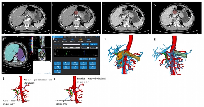
Figure 1: Preoperative CT and three-dimensional visualization of surgical planning. (A,B) Arterial phase; the mass is indicated by red circles. (C,D) Portal venous phase; the mass is indicated by red circles. (E) AI-assisted three-dimensional reconstruction. (F) Interface for manual adjustment of the AI segmentation result. (G) Three-dimensional visualization model of the pancreas and surrounding vessels. (H) Three-dimensional surgical plan for transection of the pancreatic head and neck (the remnant pancreas is shown in light green). (I,J) The anterior and posterior pancreaticoduodenal arterial arcades intended to be preserved. Abbreviations: CT = computed tomography; AI = artificial intelligence. Please click here to view a larger version of this figure.
3. Anesthesia and preoperative preparation
- Administer general anesthesia using endotracheal intubation. Simultaneously, perform internal jugular vein puncture under ultrasound guidance and place a 7Fr venous catheter. Insert a 21 G arterial needle into the radial artery for arterial pressure monitoring.
- Place a 16Fr nasogastric tube and a 16Fr urinary catheter. Administer antibiotics (cefuroxime sodium 1.5 g) 30 min before surgery for infection prophylaxis.
- Position the patient supine, fix the lower limbs, and abduct them 50°–60°. Position the surgeon on the patient's right side, the first assistant on the left, and the camera holder between the patient's legs. Reposition the patient promptly according to intraoperative requirements.
- Administer an oral laxative on the day before surgery and perform an enema on the morning of the procedure. Thirty minutes before surgery, administer ICG intravenously at a dose of 0.25 mg (reconstitute 25 mg of ICG with 10 mL of normal saline to give a concentration of 2.5 mg/mL, and withdraw 0.1 mL of this solution).
4. Surgical procedure
- Open the gastrocolic ligament to expose the anterior surface of the pancreas. Explore the tumor location and confirm that the tumor is located in the pancreatic head and neck region (Figure 2A).
- Open the suprapancreatic space. Under augmented reality (AR) navigation—for which the laparoscopic video and the preoperative three-dimensional model are imported into the referenced platform and registered manually—dissect the GDA and mark it with a vessel loop. During the subsequent steps, real-time AR navigation may be used as required by the surgeon (Figure 2B–D).
- Open the infrapancreatic space. Dissect the superior mesenteric vein (SMV) and dissect upward along the SMV. Divide corresponding venous branches and establish the retropancreatic tunnel (Figure 2E,F).
- Transect the pancreatic head and neck from the pancreatic body and tail. Resect the pancreatic duct on the normal pancreatic side and send it for intraoperative frozen section examination (Figure 2G,H).
- Turn the pancreatic head upward. Carefully dissect and protect the anterior inferior pancreaticoduodenal artery (AIPDA), posterior inferior pancreaticoduodenal artery (PIPDA), and anterior inferior pancreaticoduodenal vein (AIPDV) (Figure 2I,J).
- Separate the pancreatic parenchyma along the medial border of the duodenum and the anterior superior pancreaticoduodenal artery (ASPDA) by turning the pancreas leftward (Figure 2K,L).
- Identify and protect the common bile duct (CBD) under ICG fluorescence navigation, and completely resect the tumor (Figure 2M,N).
- If the intraoperative frozen section examination indicates a positive pancreatic duct margin, perform extended resection (Figure 2O).
- Transect the proximal jejunum 15 cm distal to the ligament of Treitz (Figure 2P).
- Perform a modified Blumgart pancreaticojejunostomy between the pancreatic stump and the jejunum as follows.
- Using a double-armed 4-0 polypropylene (Prolene) suture, insert one needle into the anterior surface of the pancreas approximately 1 cm from the cut edge, pass it through the full thickness of the parenchyma and out through the posterior surface of the pancreas (Figure 2Q), then through the seromuscular layer of the lower jejunal wall (Figure 2R). Reverse the needle, re-enter from the posterior surface of the pancreas, traverse the parenchyma, and exit through the anterior surface (Figure 2S), thereby completing a U-shaped mattress suture.
- Perform the duct-to-mucosa anastomosis next. Place the distal end of the pancreatic duct stent into the jejunal lumen and direct it distally, and perform an interrupted duct-to-mucosa anastomosis between the pancreatic duct and the jejunum using 5-0 absorbable Vicryl sutures (Figure 2T); pass the middle suture through the pancreatic duct stent to secure it in position (Figure 2U).
- Finally, use the second needle of the double-armed 4-0 polypropylene suture to take a bite of the jejunal seromuscular layer and tie it to the U-shaped suture (Figure 2V), so that the jejunal wall is invaginated to wrap the pancreatic stump (Figure 2W).
- Perform a side-to-side jejunojejunostomy between the jejunum 40 cm distal to the pancreaticojejunostomy and the proximal jejunum (Figure 2X).
- At the end of the procedure, place drainage tubes: anterior and posterior to the pancreaticojejunostomy, and the foramen of Winslow (Figure 2Y). Send the specimen for routine postoperative pathological examination (Figure 2Z).
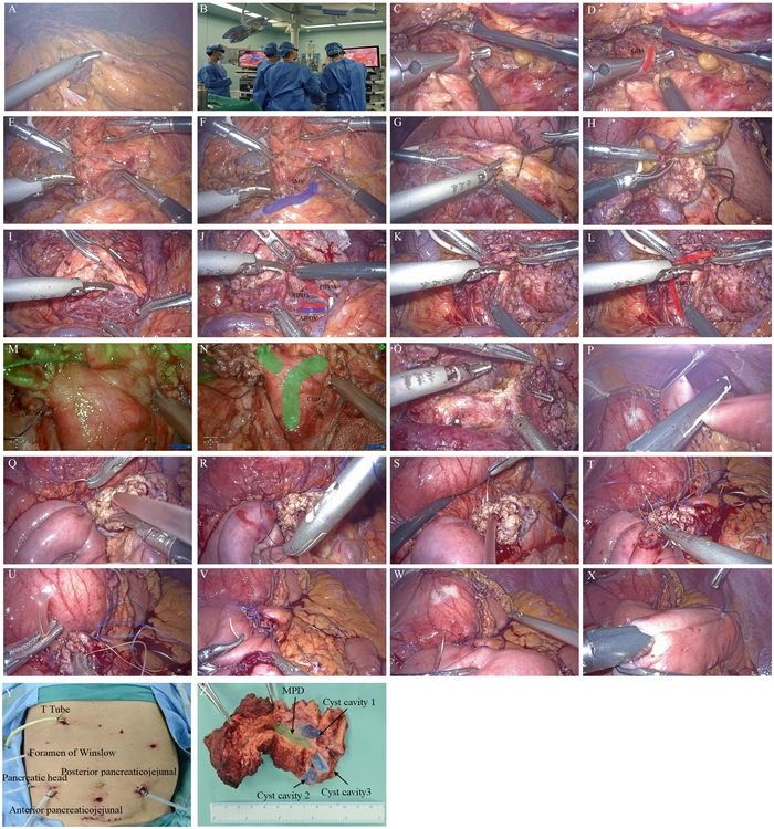
Figure 2: Detailed surgical steps. (A) Open the gastrocolic ligament; (B) intraoperative AR navigation; (C,D) dissection and suspension of the gastroduodenal artery; (E,F) dissection of the superior mesenteric vein; (G) transection of the pancreas; (H) resection of the pancreatic duct on the normal side for examination; (I,J) upward turning of the pancreas along the anterior inferior pancreaticoduodenal artery, posterior inferior pancreaticoduodenal artery, and anterior inferior pancreaticoduodenal vein; (K,L) leftward turning of the pancreas along the anterior superior pancreaticoduodenal artery; (M,N) Indocyanine Green visualization of the common bile duct and main pancreatic duct; (O) extended resection margin; (P) transection of the proximal jejunum; (Q–S) The first needle of the double-armed 4-0 Prolene suture is used to place a U-shaped mattress suture. (T,U) Mucosa-to-mucosa pancreaticojejunostomy. (V) The second needle of the double-armed 4-0 Prolene suture is used to take a bite of the jejunal wall. (W) The jejunal wall is invaginated to wrap the pancreatic stump. (X) Side-to-side jejunojejunostomy. (Y) Placement of the drainage tubes. (Z) Gross specimen. Abbreviation: MPD = main pancreatic duct. Please click here to view a larger version of this figure.
Résultats
Routine pathological examination and immunohistochemical staining of the resected specimen confirmed the diagnosis of IPMN of the pancreatic head and neck (Figure 3A,B). The operative time was 6 h, and the estimated intraoperative blood loss was 200 mL. No postoperative complications occurred, and the postoperative hospital stay was 2 weeks. Follow-up computed tomography showed no obvious fluid collection at the operative site (Figure 3C,D), and the amylase and bilirubin concentrations in the drainage fluid from each drain, measured on postoperative days 3 and 7, were within normal limits.
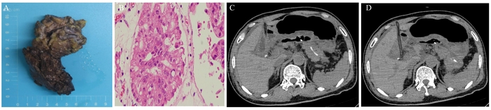
Figure 3. Postoperative findings. (A) Gross specimen. (B) Hematoxylin and eosin staining (original magnification ×40). (C,D) Postoperative follow-up abdominal computed tomography. Please click here to view a larger version of this figure.
Discussion
With the growing concept of precision surgery, organ-preserving pancreatic surgeries, such as LDPPHNR, have improved patients' quality of life and survival. However, it is undeniable that the clinical implementation of this technique demands high technical proficiency and involves a lengthy learning curve. Currently, it is routinely performed only in larger medical centers. Life-threatening complications such as pancreatic fistula, biliary fistula and biliary stricture, duodenal ischemia and necrosis, and postoperative hemorrhage are not uncommon.
In 2015, Beger et al.9 demonstrated that DPPHR was associated with significantly lower rates of ISGPF grade B and C pancreatic fistulas compared to PD. Statistical data indicate that the incidence of grade B and C pancreatic fistulas in LDPPHR is lower than that in open DPPHR9,10. In patients with adenomas, neuroendocrine tumors, solid pseudopapillary neoplasms, and similar diseases, the pancreatic texture is often soft, and the pancreatic duct is relatively small, which makes pancreaticojejunostomy technically challenging and leads to a high incidence of postoperative pancreatic fistula. The modified Blumgart pancreaticojejunostomy method employed in this study has been demonstrated to yield satisfactory surgical outcomes11.
With the widespread application of ICG fluorescence navigation in pancreatic surgery, the incidence of biliary fistula and biliary stricture has decreased markedly. Huang et al.12 reported that the incidence of intraoperative bile duct injury in the ICG group was lower than that in the non-ICG group (10% vs. 60%, P = 0.009), which indicates that ICG fluorescence navigation is feasible for use in LDPPHR and plays an important role in the exposure and protection of the pancreatic segment of the common bile duct (CBD). Reports describing the use of ICG fluorescence navigation in LDPPHR nevertheless remain limited13⁻15. These findings also point to a direction for future research, namely, the application of ICG fluorescence navigation to LDPPHNR.
Duodenal stenosis or ischemia most commonly results from injury to the periduodenal vascular arcades. Mild ischemia may lead to duodenal stenosis and delayed gastric emptying, whereas severe ischemia can cause duodenal necrosis and postoperative hemorrhage. Therefore, protection of the periduodenal vascular arcades is crucial in LDPPHNR16. Early postoperative hemorrhage is usually attributable to inadequate hemostasis, whereas late hemorrhage is often caused by erosion of vessels due to pancreatic or biliary fistula.
Accordingly, the authors propose that several points should be emphasized when performing LDPPHNR: First, a thorough understanding of the local anatomical relationships should be achieved preoperatively through CT/MRCP and precise three-dimensional visualization. Second, intraoperative manipulation should be performed patiently and meticulously, especially during separation of the pancreatic head from the duodenum, combining blunt and sharp dissection to avoid violent tearing. The integration of ICG fluorescence navigation and AR navigation technology can further enhance surgical precision. Third, careful management of the pancreatic stump should be ensured with reliable suturing. Fourth, close postoperative monitoring of drainage fluid characteristics and amylase levels should be conducted to facilitate the timely detection and management of related complications.
Two further points must be emphasized in this article. First, the indications for this procedure are extremely rare. LDPPHNR is suitable only for lesions that fulfill all of the following conditions simultaneously: the lesion is benign or of low-grade malignancy; it involves or abuts the pancreatic neck; and it spares the duodenum, the intrapancreatic segment of the common bile duct, and both the anterior and the posterior pancreaticoduodenal arterial arcades. For most patients with a lesion in this region, a standard DPPHR or a PD is the more appropriate operation; in our practice, only a very small minority of patients referred with a pancreatic head lesion meet all the requirements described above. Second, the reproducibility of the procedure is limited. Because it combines advanced laparoscopic pancreatic resection with three-dimensional reconstruction and AR navigation, the procedure is currently performed routinely in only a small number of specialist centers; its learning curve is long, and the two steps that carry the greatest risk—separation of the pancreatic head from the duodenum and construction of the pancreaticojejunostomy—are precisely the steps that are the most difficult to standardize. Readers should therefore regard this article as a technical demonstration performed by a team with experience in both advanced laparoscopic pancreatic surgery and image-guided navigation and should not assume that the outcome reported here can be reproduced in units lacking comparable experience, operative volume, and equipment.
Déclarations de divulgation
The authors have no conflicts of interest to declare.
Remerciements
This work is funded by the Medical Scientific Research Foundation of Guangdong Province of China (Grant No. B2026435), the General Program of Natural Science Foundation of Guangdong Province (Grant Nos. 2023A1515012906 and 2024A1515011531), and the Medical Research Special Fund of Shenzhen (Grant No. D2402006).
Matériaux
| Nom | Entreprise | Numéro de catalogue | Commentaires |
|---|---|---|---|
| Disposable negative-pressure drainage tube | Jiangsu Yangzijiang Medical Technology Co., Ltd. | 419280; 9.4/320 | |
| 16Fr nasogastric tube | |||
| 16Fr urinary catheter | |||
| 21 G arterial needle | |||
| 3DV Works | Shenzhen Nov3D Intelligent Technology Co., Ltd | AS-070-1965 | |
| 4-0 Prolene (polypropylene) suture | Ethicon (Johnson & Johnson MedTech), Somerville, NJ, USA | W8557 | |
| 5-0 Vicryl suture | Ethicon (Johnson & Johnson MedTech), Somerville, NJ, USA | SXMD1B402 | |
| 7Fr venous catheter | |||
| Cefuroxime sodium | |||
| Disposable multiple-fire clip applier and ligating clips | EndoSystem (Wuhan) Medical Technology Co., Ltd. | ELC02ML15 | |
| Disposable powered endoscopic linear cutting stapler and reloads | Ningbo Verykind Medical Device Co., Ltd. | TCR60Y | |
| Enema supplies | |||
| Gstation | Shenzhen Nov3D Intelligent Technology Co., Ltd | MS490 | |
| Indocyanine green / ICG | |||
| Normal saline | |||
| Oral laxative | |||
| Pancreatic duct stent | |||
| Peng's multifunctional surgical dissector (electrocoagulation and cutting device, PMOD) | Zhejiang Shuyou Instrument & Equipment Co., Ltd. | SY-ISY-IIIB-2 | |
| Storz 3D Laparoscope | KARL STORZ | 26003 BA | |
| TuGe 3D 4K Fluorescence Laparoscope | Nanjing TUGE Healthcare Co., Ltd | TCS-M420C | |
| Vessel loop |
Références
- Basar O, Brugge WR. My treatment approach: pancreatic cysts. Mayo Clin Proc. 2017;92(10):1519-31.
- Tan Y, et al. Laparoscopic vs. open pancreaticoduodenectomy: a comparative study in elderly people. Updates Surg. 2020;72(3):701-7.
- Beger HG, Krautzberger W, Bittner R, Büchler M, et al. Duodenum-preserving resection of the head of the pancreas in patients with severe chronic pancreatitis. Surgery. 1985;97(4):467-73.
- Zhou J, et al. Laparoscopic duodenum-preserving pancreatic head resection: a case report. Medicine (Baltimore). 2016;95(32):e4442.
- Beger HG, Friess H, Uhl W. Whipple resection for benign tumors and premalignant neoplasms of the pancreatic head: surgery-associated risk for complications and late metabolic morbidity matters. Ann Surg Open. 2023;4(3):e325.
- Justo Alonso I, et al. Whipple's cephalic pancreaticoduodenectomy versus pyloric-preserving pancreaticoduodenectomy: retrospective study. Cir Esp (Engl Ed). 2024;102(5):265-74.
- Jiang Y, et al. Robot-assisted duodenum-preserving pancreatic head resection with pancreaticogastrostomy for benign or premalignant pancreatic head lesions: a single-centre experience. Int J Med Robot. 2018;14(4):e1903.
- Cheng J. ASO author reflections: innovative techniques for laparoscopic duodenum-preserving pancreatic head resection. Ann Surg Oncol. 2021;28(6):3217-8.
- Beger HG, Mayer B, Rau BM. Parenchyma-sparing, limited pancreatic head resection for benign tumors and low-risk periampullary cancer—a systematic review. J Gastrointest Surg. 2016;20(1):206-17.
- Chen S, et al. Indocyanine green-enhanced fluorescence in laparoscopic duodenum-preserving pancreatic head resection: technique with video. Ann Surg Oncol. 2020;27(10):3926-7.
- Sun P, Yu Y, Li J, Cui X. A novel anastomosis technique for laparoscopic pancreaticoduodenectomy: case series of our center's experience. Front Surg. 2021;8:583671.
- Huang J, et al. Real-time fluorescence imaging with indocyanine green during laparoscopic duodenum-preserving pancreatic head resection. Pancreatology. 2024;24(1):130-6.
- Quaresima S, et al. Routine near infra-red indocyanine green fluorescent cholangiography versus intraoperative cholangiography during laparoscopic cholecystectomy: a case-matched comparison. Surg Endosc. 2020;34(5):1959-67.
- Xia Z, et al. Short-term clinical outcomes of laparoscopic duodenum-preserving pancreatic head resection for the management of pancreatic-head cystic neoplasms. BMC Surg. 2023;23(1):104.
- Gao Y, Huang J, Zheng Y, Han J. Effect of comfort nursing on postoperative quality of life, negative emotions and nursing satisfaction in patients undergoing laparoscopic surgery. Am J Transl Res. 2021;13(12):13825-34.
- Hong D et al. How to perform total laparoscopic duodenum-preserving pancreatic head resection safely and efficiently with innovative techniques. Ann Surg Oncol. 2021;28(6):3209-16.
Réimpressions et autorisations
Cet article a été publié
Vidéo bientôt disponible