This video demonstrates the procedure of co-culturing amoebae with mutant bacterial strains to evaluate their intracellular survival and identify virulence markers through differences in colony counts.
הרשמה ל-JoVE נדרשת לצפייה בתוכן זה. התחברו או התחילו בגרסת ניסיון בחינם.
מאמר שיטה
Detecting Bacterial Virulence Using Amoeba as a Host System
159 צפיות
⸱
1 ביולי 2026
במאמר זה
תקציר
פרוטוקול
- Co-culture of amoebae-Mycobacterium abscessus Tn mutant: first screen
NOTE: This takes 3–4 months for the screening of 6,000 mutants.- Spread 5 x 10⁴ amoebae(Acanthamoeba castellanii) /well to a 96-well plate in 100 µL of AC (Acanthamoeba castellanii) buffer. Incubate for 1 h at 32 °C without shaking. Allow the floating amoeba trophozoïtes time to adhere to the wells. While incubating, thaw bacteria on ice for 1 h.
- Add 5 µL of the thawed bacteria cultures with an electronic multichannel pipette. Infect for 1.5 h. Screen around 6,000 individualized clones from the 96-well plate Tn mutants stock.
NOTE: The MOI (multiplicity of infection) is unknown at this step of the protocol; however, all the 96-well plates containing Tn mutants were cultivated in exactly the same way. This first screen aims to quickly identify intracellular deficient mutants, without considering the variations in inoculum density. - After 1.5 h of infection, invert the 96-well plate on sterile gauze placed in a sterilized metal tray to remove the AC medium under a fume hood. Refill with 200 µL of AC medium. Repeat this wash twice more.
- Add 200 µL of fresh medium supplemented with 100 µg/mL amikacin to eliminate remaining extracellular mycobacteria.
- Incubate for 2 h before proceeding with 3 additional washings. After washing, maintain the cultures with 50 µg/mL amikacin in 200 µL of AC buffer.
- Add 5 x 10⁷ heat-inactivated Escherichia coli bacteria at the end of the infection and every 24 h to prevent encystment of amoebae.
- After 48 h of co-culture, lyse the cells by adding 10 µL of 10% SDS (Sodium dodecyl sulfate) to each well and incubate for 30 min at 32 °C. Proceed with the serial dilution and add on Columbia agar plates containing 5% sheep blood (COS plates) to evaluate the number of intracellular mycobacteria. Seal the plate with the parafilm and incubate at 37 °C.
- When colonies appear on the agar plates after 3-4 days, photograph the plate and identify the mutants impaired for intracellular growth by observing the deposits with the lowest number of bacterial colonies.
NOTE: Do not mind about the shape, height or density of the colony (Figure 1A). 136/6000 mutants were identified.
הגישה מוגבלת. התחברו או התחילו תקופת ניסיון כדי לצפות בתוכן זה.
תוצאות
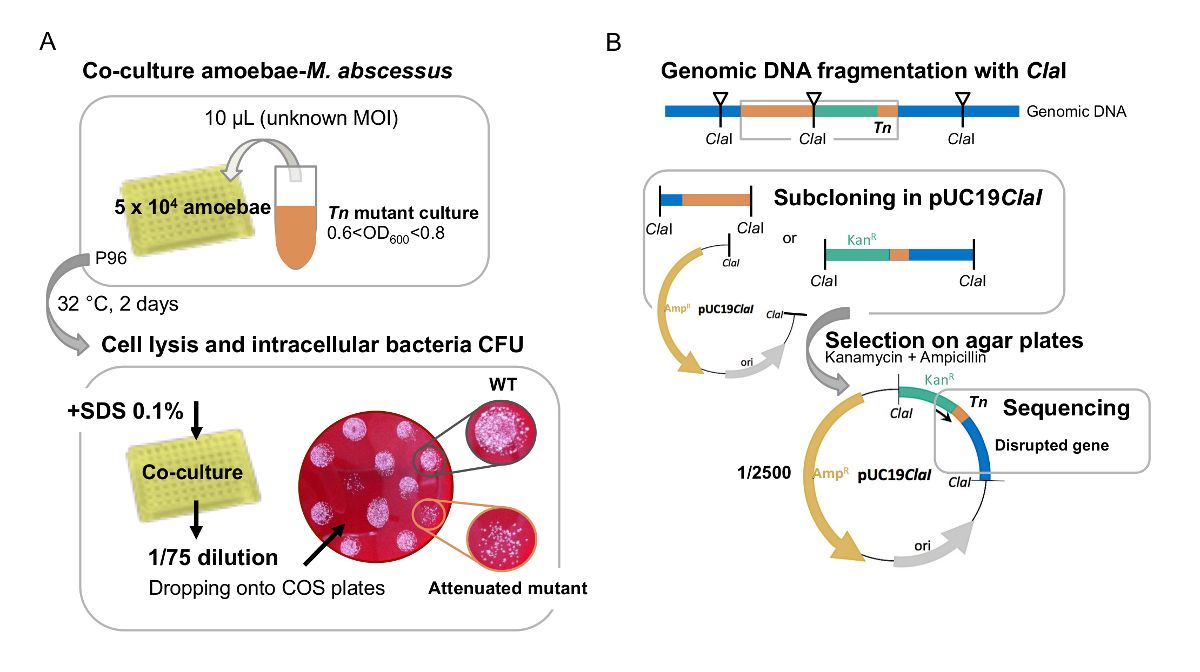
Figure 1: A. First visual screen of M. abscessus Tn mutants in amoeba. (A) large-scale screen of Tn mutants on amoebae is performed in...
הגישה מוגבלת. התחברו או התחילו תקופת ניסיון כדי לצפות בתוכן זה.
חומרים
| שם | חברה | מספר קטלוג | הערות |
|---|---|---|---|
| 24-well plates | Thermofisher | 11874235 | |
| 96-well plates | Thermofisher | 10687551 | |
| QuBit fluorometer | Thermofisher | Q33226 | |
| Acanthamoeba castellanii | ATCC | 30010 | strain |
| Amikacin | Mylan | 150927-A | powder |
| CaCl2 | Sigma-Aldrich | C1016 | >93% granular anhydrous |
| Columbia agar | Biomerieux | 43041 | 90 mm |
| D-Glucose | Sigma-Aldrich | G8270 | powder |
| DMEM | Thermofisher | 11500596 | medium |
| DNase and RNase free water | Invitrogen | 10977-035 | solution |
| Escherichia coli | Clinical isolate | personal stock | bacteria |
| Fetal Calf Serum | Gibco | 10270 | serum |
| Müller-Hinton agar | Biorad | 3563901 | powder |
| SDS 20% | Biorad | 1610418 | solution |
