This video demonstrates the purification of an oncolytic virus from infected mammalian cell cultures using nuclease treatment, sequential centrifugation, membrane filtration, and sucrose cushion centrifugation.
このコンテンツを表示するには、JoVEへの購読が必要です。 または、無料トライアルをお申し込みください。
方法論記事
Oncolytic Virus Purification from Infected Mammalian Cells
124 回視聴
⸱
2026年7月31日
この記事について
要約
プロトコル
1. Oncolytic herpes simplex virus (oHSV) purification
- Thaw (in a 37 °C warm water bath) the cells, followed by water bath-sonication for 1 min for a total of 3 cycles to ensure proper lysis of the cells to release the virus in the supernatant.
NOTE: Perform sonication for 1 min using 40 kHz, 120 V power. If a tunable sonicator is not available, use an ultrasonic water bath sonicator. Take a 50 μL aliquot for titration, which will help to identify which of the following steps is responsible for potential virus loss during the purification procedure. - Treat the cell lysate with Benzonase Nuclease (175 units/mL) + 2 mM magnesium chloride (MgCl2) (1 M stock = 2 µL/mL), vortex, and incubate for 30 min at 37 °C. Place the tube on ice and perform the following steps at 4 °C.
- Pellet the cell debris by low-speed centrifugation.
- Spin the cell lysate at 300 g for 10 min.
- Collect the supernatant in a new 50 mL conical centrifuge tube (re-suspend the cell pellet in 0.5 mL of virus buffer (VB), designate it as pellet-1, and store at 4 °C for use in step 1.3.5; see Figure 1).
- Spin the supernatant (obtained in step 1.3.2) again at 500 g for 10 min.
- Collect the supernatant in a new 50 mL conical centrifuge tube (re-suspend the cell pellet in 0.5 mL of VB, designate it as pellet-2, and store at 4 °C for use in step 1.3.5; see Figure 1).
- Combine the re-suspended pellet-1 (from 1.3.2) and pellet-2 (from 1.3.4) into a new 1.7 mL centrifuge tube, vortex/sonicate (water bath) 2x, and spin at 400 g for 10 min. Collect the supernatant and combine it with the supernatant obtained in step 1.3.4; see Figure 1.
NOTE: Perform sonication for 1 min using 40 kHz, 120 V power to prevent viral aggregation before the filtration procedure in step 1.4. Take a 50 μL aliquot of the combined supernatant for titration.
- Filter the combined supernatant using the following 3-step filtration method.
- Draw 21 mL of the supernatant using a 10 mL syringe (5-7 mL each time for easy passage through the filter) and pass it through a sterile 5 μm polyvinylidene difluoride (PVDF) membrane filter placed on a new 50 mL conical centrifuge tube (labeled as TUBE 1).
- Add 1 mL of VB to a 50 mL conical centrifuge tube emptied in 1.4.1 (to collect the remaining trace amount of virus supernatant), vortex, and pass through the same 5 μm PVDF filter placed on TUBE 1 (bringing the total to 22 mL of filtrate). Proceed to step 1.4.2.
- Draw 22 mL of the filtrate from TUBE 1 (as in 1.4.1) and pass it through a sterile 0.8 μm mixed cellulose ester (MCE) membrane filter placed on a new 50 mL conical centrifuge tube (labeled as TUBE 2).
- Add 1 mL of VB to TUBE 1 emptied in step 1.4.2, vortex, and pass it through the same 0.8 μm MCE filter placed on TUBE 2 (bringing the total to 23 mL of filtrate). Proceed to step 1.4.3.
- Draw 23 mL of filtrate from TUBE 2 (as in 1.4.1) and pass it through a sterile 0.45 µm PVDF filter placed on a new 50 mL conical centrifuge tube (labeled as TUBE 3).
- Add 1 mL of VB to TUBE 2 emptied in step 1.4.3, vortex, and pass it through the same 0.45 μm PVDF filter placed on TUBE 3 (bringing the total to 24 mL of filtrate).
NOTE: Take a 50 μL aliquot of the filtrate for titration.
- Add 1 mL of VB to TUBE 2 emptied in step 1.4.3, vortex, and pass it through the same 0.45 μm PVDF filter placed on TUBE 3 (bringing the total to 24 mL of filtrate).
- Draw 21 mL of the supernatant using a 10 mL syringe (5-7 mL each time for easy passage through the filter) and pass it through a sterile 5 μm polyvinylidene difluoride (PVDF) membrane filter placed on a new 50 mL conical centrifuge tube (labeled as TUBE 1).
- High-speed centrifugation using the sucrose-gradient method
NOTE: A fixed-angle F13-14x50cy rotor was used in this protocol. Both the rotor and the centrifuge must be at 4 °C before proceeding with the following steps.- Add 10 mL of an ice-cold, sterile-filtered 25% sucrose solution (prepared by dissolving 25 g of sucrose powder in 100 mL of Hank's Balanced Salt Solution) in a new 50 mL conical centrifuge tube.
- Slowly (3 mL/min) add 24 mL of the virus filtrate (obtained from step 1.4.3.1) on top of the sucrose layer. Take care to maintain separate layers of the virus filtrate and the sucrose solution.
NOTE: Up to 30 mL of the virus layer can be added over 10 mL of the sucrose solution. - Centrifuge the tube for 90 min at 22,620 g at 4 °C.
- Remove the supernatant and the sucrose layer from the 50 mL conical centrifuge tube.
NOTE: The virus pellet should be whitish. Take a 50 μL aliquot of the supernatant and a 50 μL aliquot of the sucrose layer for titration
アクセスが制限されています。このコンテンツを表示するにはログインするか、トライアルを開始してください。
結果
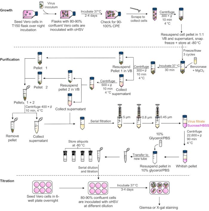
Figure 1: A schematic presentation of major steps involved in oHSV growth, purification, and the plaque assay. Abbreviations: oHSV = oncolytic herpes simplex virus; CPE = cytopathic effect; VB = Virus Buffer; HBSS = Hank's Balanced Salt S...
アクセスが制限されています。このコンテンツを表示するにはログインするか、トライアルを開始してください。
材料
| 名前 | 会社 | カタログ番号 | コメント |
|---|---|---|---|
| 1.7 mL Centrifuge tubes | Sigma | CLS3620 | |
| 15 mL Polypropylene centrifuge tubes | Falcon | 352097 | |
| 50 mL Polypropylene centrifuge tubes | Falcon | 352098 | |
| Benzonase Nuclease | Sigma | E8263-25KU | |
| Glycerol | Sigma | G5516 | |
| Magnesium chloride | Fisher Chemical | M33-500 | |
| Sorvall ST 16R Centrifuge | ThermoFisher Scientific | 75004381 | |
| Sorvall ST 21R Centrifuge | ThermoFisher Scientific | 75002446 | |
| Sucrose | Fisher Scientific | BP220-1 | |
| Syringe Filter, 0.45 PVDF | MilliporeSigma | SLHV033RS | |
| Syringe Filter, 0.8 MCE | MilliporeSigma | SLAA033SS | |
| Syringe filter, 5 µm PVDF | MilliporeSigma | SLSV025LS | |
| Ultrasonic water bath | Branson | CPX-952-116R |
