This video demonstrates the large-scale amplification of high-capacity adenoviral vectors in suspension-adapted human embryonic kidney cells expressing Cre recombinase for use in gene delivery applications requiring high-titer, helper-free vector preparations.
このコンテンツを表示するには、JoVEへの購読が必要です。 または、無料トライアルをお申し込みください。
方法論記事
Large-Scale Production of High-Capacity Adenoviral Vectors
153 回視聴
⸱
2026年7月31日
この記事について
要約
プロトコル
1. Large Scale Amplification of High-capacity adenoviral vectors (HCAdV) in 116 cells Growing in Suspension
- Add 900 ml of prewarmed (37 °C) fresh MEM (minimum essential medium) supplemented with 10% FBS (fetal bovine serum) and hygromycin B (100 µg/ml) into a 3-L spinner culture flask (Figure 1B).
- Remove medium from at least 10 individual 150-mm tissue culture dishes with 116 cells grown at a confluency of 90-100% and flush off cells with 10 ml of fresh pre-warmed (37 °C) MEM supplemented with FBS (10%) and hygromycin B (100 µg/ml) using a serological pipette. Pipette up and down several times to get a homogeneous cell suspension. Transfer cells directly into the spinner flask already containing 900 ml from step 1.1.
NOTE: Do not use Trypsin/EDTA (ethylene diamine tetraacetic acid) as it may negatively affect growth of suspension cells. After removal of medium immediately transfer cells into the spinner flask. Do not handle more than two tissue culture dishes at a time. The longer the waiting time after adding fresh media the more difficult it will be to detach the cells from the tissue culture dish. - To ensure optimal cell growth, incubate spinner flask on a magnetic stirrer in a tissue culture incubator for 24 hr at 37 °C and 5% CO2. Adjust the magnetic stirrer to 70 rpm to avoid attachment of cells to the glass surfaces.
- Monitor cell growth in spinner culture flask to examine whether cells grown in the spinner culture flask are viable and whether they grow to sufficient amounts. Each time prior to adding fresh medium transfer 2 ml from the bioreactor to a 60-mm tissue culture dish. Observe cell morphology under a microscope.
NOTE: Cells forming clumps floating in the culture medium indicate optimal growth and viability (see also Figure 2C). After 24 hr they form colonies on the surface of the tissue culture dish. 30-50% confluence indicates optimal density. - 24 hr after setting up the spinner culture flask add 500 ml of fresh media (MEM, 10% FBS) supplemented with hygromycin B (100 µg/ml). 24 hr later repeat this step.
- 72 hr after setting up the spinner culture, add 1,000 ml of fresh MEM media (10% FBS) with hygromycin B (100 µg/ml), resulting in a total volume of 3 L of cell suspension.
- 24 hr after reaching a volume of three litres, harvest 116 suspension cells by centrifugation for 10 min at 500 x g at RT (room temperature). Discard the supernatant. Keep the emptied spinner culture flask under the tissue culture hood.
- Resuspend cell pellets in fresh MEM supplemented with 5% FBS by pipetting up and down about 8-10x to get a homogeneous cell suspension. It is used for the infection of the cells. Use a total volume of 150 ml.
- Option a: For infection of 116 suspension cells with lysate from P3 to a 250-ml storage bottle equipped with a sterile magnetic stir bar or a 250-ml small-scale spinner flask, and co-infect cells with the virus within the lysate from P3 and 2 transducing units (TU) of helper virus (HV) per cell. Assuming a density of 3 x 10⁵ cells per ml, the total cell number is 9 x 10⁸ cells. Thus, add 1.8 x 10⁹ TU of the HV.
- Option b: For infection of 116 suspension cells with purified viral stock, transfer cells from step 1.8 to a 250-ml storage bottle equipped with a sterile magnetic stir bar or a 250-ml small-scale spinner flask and co-infect cells with the 100 viral particles (VP) per cell of formerly purified HCAdV. Thus, add 9 x 10¹⁰ VP according to the physical titer of the purified HCAdV and 1.8 x 10⁹ TU of the HV.
- Stir the cell-virus mixture from step 1.8.1 or 1.8.2 in on a magnetic stirrer in the tissue culture incubator at 37 °C and 5% CO2 for 2 hr at 60 rpm. Make sure that the storage bottle is not completely closed to allow air circulation.
- 2 hr post-infection transfer the total volume of the cell-virus mixture back into the 3-L spinner culture flask and add 1,850 ml of fresh pre-warmed (37 °C) MEM media supplemented with 5% FBS to a total volume of 2 L and incubate in the tissue culture incubator at 37 °C and 5% CO2 for 48 hr at 70 rpm.
- Harvest cells by centrifugation for 10 min at 890 x g at RT in 500-ml centrifuge tubes. Remove the medium and resuspend pellet cells in a total volume of 28 ml of DPBS (Dulbecco’s phosphate-buffered saline). Pipette up and down to obtain a homogeneous cell suspension. Freeze the cell-virus suspension in liquid nitrogen or at -80 °C making sure that the suspension is completely frozen. Store the cell-virus suspension at -80 °C until starting the virus purification.
アクセスが制限されています。このコンテンツを表示するにはログインするか、トライアルを開始してください。
結果
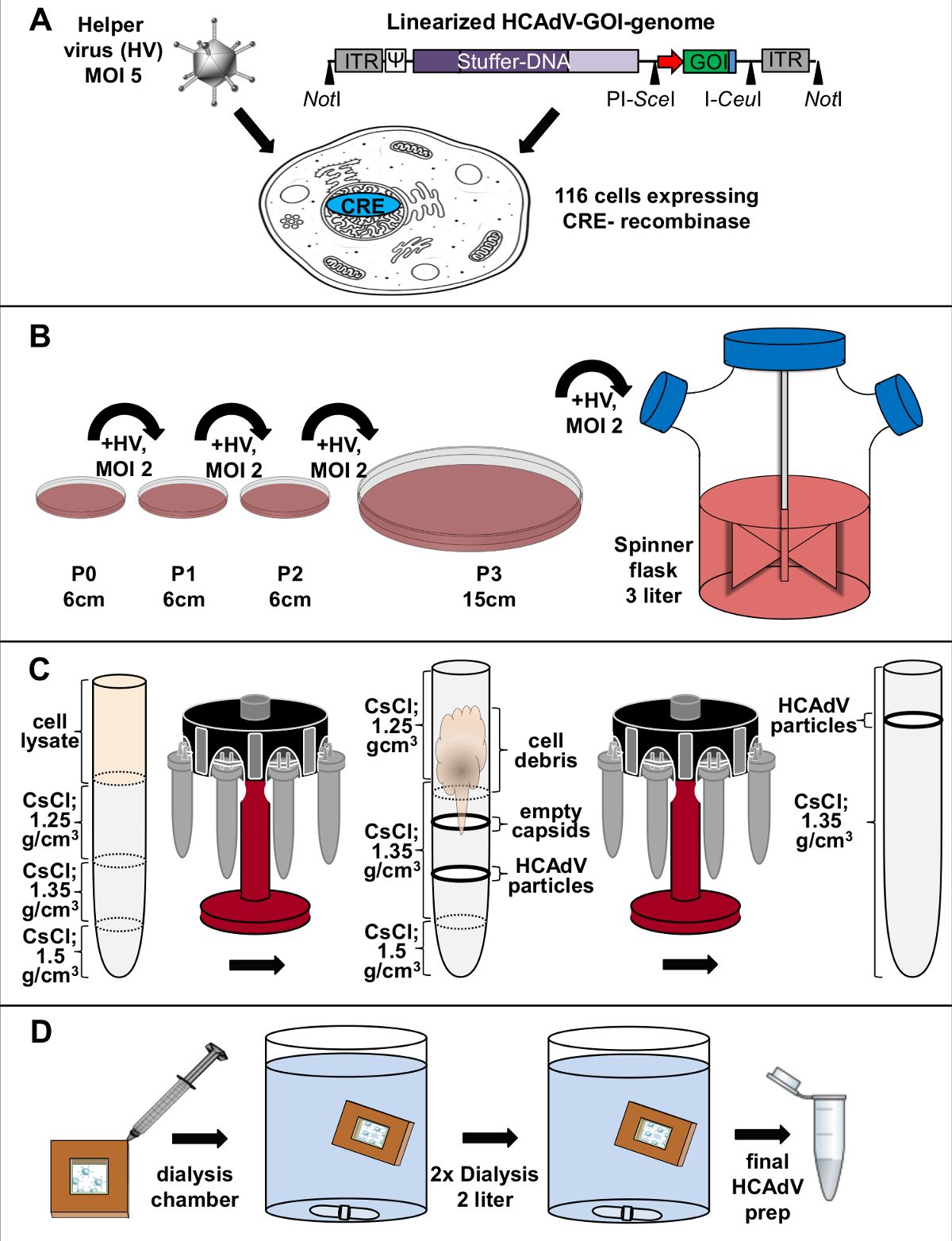
Figure 1. Schematic diagram for high-capacity adenoviral vector amplification and purification. (A) HCAdV DNA transfection and helper virus (HV) infection: Transfection of linearized HCAdV genome carrying the gene of inter...
アクセスが制限されています。このコンテンツを表示するにはログインするか、トライアルを開始してください。
材料
| 名前 | 会社 | カタログ番号 | コメント |
|---|---|---|---|
| Hygromycin B | PAN Biotech | P02-015 | Selection of CRE expressing 116 cells |
| DMEM | PAN Biotech | P04-03590 | Hek293T cell culture medium |
| Minimal Essential Medium (MEM) Eagle | PAN Biotech | P04-08500 | 116 cell culture medium |
| Dulbecco’s phosphate buffer saline (DPBS) | PAN Biotech | P04-36500 | Washing of cells, resuspension of cells |
| 250-ml storage bottle | Sigma | CLS430281-24EA | Infection of 116 cells grown in suspension |
| 500-ml PP CentrifugeTubes | Sigma | CLS431123-36EA | Sedimentation of cells from suspension culture |
| Spinner flask | Bellco | 1965-61030 | Growth of 116 cells in suspension |
