This video demonstrates the intravitreal injection of an adeno-associated viral vector into a mouse model with a compromised inner limiting membrane. The resulting fluorescent protein expression reveals the extent of retinal barrier disruption based on viral penetration into deeper retinal layers.
方法論記事
Intravitreal Injection of Adeno-Associated Virus to Assess Retinal Barrier Disruption in Mice
42 回視聴
⸱
2026年10月29日
この記事について
要約
プロトコル
All procedures involving animal models have been reviewed by the local institutional animal care committee and the JoVE veterinary review board.
1. Intravitreal Injection of Adeno-Associated Virus (AAV)
- Anesthetize C57BL6J mice with ketamine (50 mg/kg) and xylazine (10 mg/kg) and verify the loss of righting reflex, the withdrawal reflex, and tail pinch response, indicating a proper anesthetization. Dilate pupils by corneal application of neosynephrine 5% and mydriaticum 0.5% eye drops. Use vet ointment on the eyes to prevent dryness while under anesthesia.
- Pass an ultrafine 30 G disposable needle through the equator and next to the limbus, into the vitreous cavity. Inject 1 µL stock containing 1 to 4 x 10¹¹ viral particles of ShH10-GFP or AAV5-GFP with direct observation of the needle in the center of the vitreous cavity (Figure 1). Use one eye as a control for Inner Limiting Membrane experiments. Inject both eyes for BRB (Blood-Retinal Barrier) experiments. Apply an anti-inflammatory and antibacterial topical treatment to the injected eyes.
- Dilate pupils with neosynephrine and mydriaticum eye drops for imaging. Perform fundus examinations with an eye fundus camera to follow GFP (Green Fluorescent Protein) expression 1 week, 2 weeks, 1 month, and 2 months after intravitreal injection (Figure 1). Sacrifice mice by CO2 inhalation 1 to 2 months post-injection.
結果
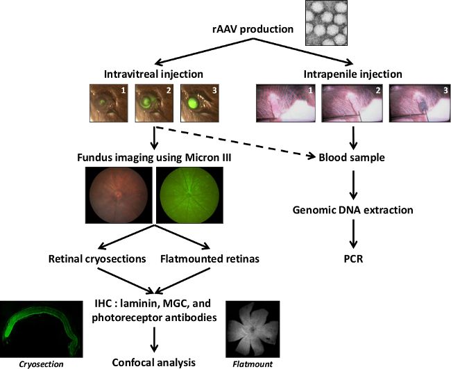
Figure 1: Schematic representation of the general protocol. After AAV production, particles are injected into the vitreous or into the penile vein of anesthetized adult mice. Fundus images with the Micron III camera are obtained to monitor GFP expression. GFP expression is analyzed in retinal cryosections and flat-mounted retinas using immunohistochemistry and confocal microscopy. The AAV passage through the BRB is assessed by PCR analysis on blood samples coming from intravitreally or intrapenally injected mice, in order to detect the presence of the GFP sequence, an indicator of the AAV particles presence into the bloodstream.
材料
| 名前 | 会社 | カタログ番号 | コメント |
|---|---|---|---|
| C57BL6J mice strain | JANVIER LABS | mice | |
| Ketamine 500 | Virbac France | anesthetic | |
| Xylazine Rompun 2% | Bayer Healthcare | anesthetic | |
| Neosynephrine 5% Faure | Europhta | dilatant | |
| Mydriaticum 0.5% | Thea | dilatant | |
| Sterdex | Novartis | anti-inflammatory | |
| Micron III | Phoenix Research Labs | Microscopy system based on 3-CCD color camera, frame grabber, and off-the-shelf software enables researchers to image mouse retinas. | |
| Insulin Syringes | Terumo | SS30M3109 | |
| Syringe 10 µl Hamilton | Dutscher | 74487 | Seringue 1701 |
| Needle RN G33, 25 mm, PST 2 | Fisher Scientific | 11530332 | Intravitreal Injection |
| UltraMicroPump UMP3 | World Precision Instruments | UMP3 | Versatile injector uses microsyringes to deliver picoliter volumes |
| UltraMicroPump (UMP3) (one) with SYS-Micro4 Controller | UMP3-1 | Digital controller |
