心臓外科手術における標準の経食道心エコー技術を使用して渦の形成時間、左心室充填効率の指標を測定するプロトコルについて述べる.異なる心臓疾患患者のいくつかのグループの渦の形成時間を分析するこの手法を適用します。
このコンテンツを表示するには、JoVEへの購読が必要です。 または、無料トライアルをお申し込みください。
この記事について
サマリー
要約
トランス僧帽弁血流を生成する三次元回転体流体、強化の効率性左心室 (LV) を充填する渦輪と呼ばれる連続的な線形ジェットと比較します。渦リング開発は最もしばしば渦形成時間 (VFT) 硬質チューブから流体吐出に基づく無次元パラメーターで定量化されます。私たちのグループは、心臓手術中に充填性 LV に影響を与える要因に関心があります。このレポートで標準の 2 次元 (2 D) およびドップラーを使用する方法を示します VFT を計算に必要な経食道心エコー (t シャツ) 非侵襲的変数を派生させる。トランス僧帽弁初期 LV 充填と心房収縮期血流速度波形半ば食道 4 室ティー ビューで測定の速度時間積分から心房の占有率 (β) を計算します。LV の流出経路の径の製品が半ば食道長軸ティー ビューで測定し、パルス波を用いた深い transgastric ビューで流出経路を通る血の流れの速度時間積分決定ストローク量 (SV) を計算します。ドップラー。最後に、僧帽弁の直径 (D) は、メジャーとマイナーの半径の長さをそれぞれ直交半ば食道 bicommissural および長軸画像平面で測定の平均値として決定されます。VFT、4 × (1-β) × SV として計算されます/(πD3)。VFT 異なる心臓の異常の患者の複数のグループを分析するこのテクニックを使いました。この技術とその潜在的な限界の我々 のアプリケーションと日付を我々 の結果を確認し.T シャツを使用して VFT の非侵襲的測定、心臓手術麻酔下の患者で簡単です。充填性をリアルタイムに心臓麻酔科医と外科医病理学の条件および LV の外科的介入の影響を評価するには、テクニックが使用されることができます。
概要
流体力学は、左心室 (LV) 充填の重要なまだしばしば過小評価の決定要因です。三次元回転体流体がオリフィス1,2,3を通過するたびに、流体の渦輪と呼ばれるが生成されます。この渦輪と連続的な線形ジェット4比較流体輸送の効率が向上します。初期 LV 充填中に僧帽弁を通って血液の動き5,6,7、8を形成する渦輪を引き起こすし、容易に流体の運動量を維持することで室内に伝播し、運動エネルギーの9。これらのアクションは、充填効率4,10、11,12,13LV を強化します。リングだけでなく LV apex14,15,16,17血流うっ滞を抑制する、また僧帽弁前7,の下に優先的に流れ、指示18、根尖部の血栓形成のリスクを減らす、LV 流出の充填を容易にする効果はそれぞれ19を追跡します。コントラスト心エコー法17、ドップラー ベクトル フロー マッピング6,20,21, 磁気共鳴画像7、および粒子画像流速測定法9,22 ,23,24は外観と正常および病的条件下でトランス僧帽弁渦輪の挙動を示すために使用されています。左心房左心室圧勾配拡張期僧帽弁環状遠足、弛緩期, 率と LV 緩和の範囲で得られる最小 LV 圧力の程度、期間、サイズ、フロー強度の位置の 4 つの主要な決定要因トランス僧帽弁輪2,12,では、26,27,28,29が25、。
渦リング開発は無次元パラメーター (渦の形成時間で定量化され、ほとんどの場合VFT) は、硬質チューブから3VFT が時間平均流速の積とオリフィスの直径で割った放出の期間として定義されている流体吐出に基づいています。渦輪の最適なサイズは、VFT は 4の in vitro末尾のジェットとエネルギッシュな制限が大きいサイズ3,4を達成するからそれを防ぐために行われます。僧帽弁 VFT は、臨床的に経胸壁心エコー図8,30,31を使用して近似されています。簡単にすることができますトランス僧帽弁血流速度と僧帽弁の直径 (D) の分析に基づいて、その VFT8を示す = 4 × (1-β) × EF × α3、どこ β 心房収縮率、EF = = 左心室駆出分画、α = EDV1/3/D、どこ EDV =末期容積。駆出率、拍出量 (SV) の比率、EDV、VFT を簡素化するこの方程式を許可する = 4 × (1-β) × SV/(πD3)。VFT は無次元 (ボリューム/ボリューム)、ので、このインデックスは体重や体の表面積の8の調整することがなくさまざまなサイズの患者間の直接比較をできます。3.3 と健常者の8、および結果に 5.5 の間の最適な VFT 範囲流体力学モデル3,32で得られたものと一致しています。VFT は ≤ 2.0 落ち込んで左室収縮機能、患者も理論的予測8でサポートされている調査結果を示した。VFT の削減は、罹患率と30心不全患者の死亡率を個別に予測しました。高 LV の後負荷33アルツハイマー病34、拡張機能の異常19、および義足35ネイティブ僧帽弁弁置換は、VFT 低下に示されています。VFT の測定は急性心筋梗塞36,37血流うっ滞や血栓症患者を識別するために有用なことがあります。
当社グループは、充填性心臓手術38,39,40,41中 LV に影響を与える要因に関心があります。非侵襲的 VFT の計算に必要な変数を派生させる標準的な二次元とドップラー経食道心エコー (TEE) を使用します。本報告では、この方法を詳しく説明して日付に私達の結果を確認します。
アクセスが制限されています。このコンテンツを表示するにはログインするか、トライアルを開始してください。
プロトコル
クレメント ・ J サブロッキ退役軍人医療センターの制度検討委員会は承認プロトコルです。書面によるインフォームド コンセントは、侵襲的な心臓モニタリングと t シャツは日常的に当院での心臓手術を受けるすべての患者に使用するために放棄されました。ティー、これらを繰り返し胸骨正中切開や緊急手術、心房または心室の頻脈性不整脈とのそれらを受けての相対的または絶対的な禁忌症は、参加から除外しました。
1. 麻酔
- 手術前に鎮静のため (1 〜 3 mg) ミダゾラム静脈内投与とフェンタニル (50 に 150 μ g) 各患者を提供します。
- 静脈と橈骨動脈カテーテルを挿入するために局所麻酔 (皮下 1% リドカイン) を使用します。針を刺したと局所麻酔の品質をテストします。
- 患者が鼻カニューレ (2 から 4 L/分) を使用して酸素を受け取ることを確認します。
- 右または左の適切な臨床徴候に基づいて超音波ガイドで内頸静脈を無菌条件下で局所麻酔 (皮下 1% リドカイン) を用いた中心静脈や肺動脈カテーテルを配置します。
- フェンタニル (5 マイクログラム/kg)、プロポ フォール (1 に 2 mg/kg)、ロクロニウム (0.1 mg/kg) を使用して麻酔を誘導します。イソフルラン (終末呼気濃度 1% の) フェンタニル (1 に 2 μ g/kg/h)、空気・酸素混合物の吸入麻酔を使用して、神経モニタリングを用いた効果に滴定ロクロニウム (0.05 mg/kg) を維持します。
- 経口胃管を用いて胃を吸引します。
- 患者の咽頭に超音波ゼリーを置きます。前方顎を持ち上げ、TEE プローブを hypopharygeus 筋肉の抵抗を克服するために穏やかな圧力を食道に進出。
2 経食道心エコー
- 次のアメリカ心エコー検査の社会/各患者の心臓血管麻酔ガイドライン42会包括的なティー検査を実行します。
- パルス波ドップラー サンプル ボリューム面 (図 1) をイメージング半ば食道 4 室 t レコード トランス僧帽弁血流速度に僧帽弁尖の先端の間に配置します。
- 初期 LV 充填およびトランス僧帽弁血流速の心房収縮期血流速波形を識別し、対応するピーク速度と速度の時間積分を測定 (VTIEと vti 社は、それぞれ) を使用して、心エコー検査機器の統合ソフトウェア パッケージ (図 1)。
- 合計 LV 充填に心房の比として心房収縮率 (β) を計算します。
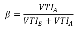
- 半ば収縮期 (図 2 a) に半ば食道大動脈弁長軸ティー ビューで大動脈弁直下左室流出路の最大径を測定します。
- Π/4 の製品および直径の正方形として円形ジオメトリを想定して左室流出路の面積を計算 (2.5 上記の手順を参照してください)。
- 深い transgastric 長軸ティー ビューを取得し、直径は測定される、(2.5 上記の手順を参照してください); 同じレベルで (図 2 b) 血液流れ速度封筒を記録する遠位左心室流出路のパルス波ドップラー サンプル ボリュームを配置心エコー装置のソフトウェア パッケージを使用して、VTI を取得するこの波形部を統合します。
- 乗算結果速度時間積分 (VTI) LV 流出トラック血流波形 (図 2 b) 流出域の追跡 (手順 2.6 参照) ストローク量 (SV) を取得します。
- 半ば食道 bicommissural、LV 長軸 t シャツ画像面、それぞれ42のビデオ クリップを記録。必ず各レコーディングにいくつか心臓のサイクルを含めるしてください。
- 僧帽弁の弁尖の最大開口部を選択する心電図 T 波の後 (手順上記 2.9 参照) のビデオ クリップのスローモーション画像を調べます。
- 僧帽弁リーフレット (図 3Aと3B) の間の距離を測定エコー機器の「キャリパー」関数を使用しますします。
- メジャー (transcommissural 前横方向-後方の内側部) とマイナー (前後) の長さの平均として僧帽弁直径 (D) を計算します。
- VFT の数式を使用して計算します。
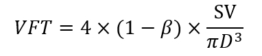
- 最後の有効期限で 3 通すべての定量的超音波測定を実行します。
3. 実験計画
- 前に、冠動脈の下で通常術前左心室駆出率 10 患者の心肺のバイパス (CPB) 後 15、30、60 分、30 分定常状態中に VFT、左心室拡張機能、および血行動態の指標を決定します。CPB VFT39一過性低下すること仮説をテストするために手術。
- 大動脈弁狭窄による左心室圧負荷肥大は重症大動脈弁狭窄および 8 の別のグループに観測を比較 (8 症例で大動脈弁置換の 1 つのグループ) で調べれば VFT を減少させること仮説をテストします。通常 LV 患者肉厚40冠動脈手術を受けています。定常状態の 30 分 CPB 前に、中 VFT、左心室拡張機能、循環動態と末期の後部壁の厚さを測定します。
- 大動脈弁狭窄症、中等度大動脈弁閉鎖不全大動脈弁狭窄症は逆流弁を持っていない患者が 8 対 8 の患者における効率充填トランス僧帽弁の LV を LV に入る異常な拡張期血流に影響の仮説します。38. メジャー VFT およびその他のパラメーター (ステップ 3.2) 前述のよう。
- 年齢を高度な仮説のテストは LV 充填性 7 ぐらい (82 ± 2 年) 7 若い (55 ± 6 歳)41受ける冠動脈手術症例と比較しての VFT を用いて定量化の削減に関連付けられます。両方のグループに通常術前左室駆出率があることを確認します。(ステップ 3.2) 上記 VFT と他のパラメーターを測定します。
4. 統計
- 平均 ± 標準偏差としてデータを表示します。
- 分散分析 (ANOVA) スチューデントのtのボンフェローニの修正後を使用して、データを評価・ テストします。
- 線形回帰分析を使用して、VFT と末期の後部壁の厚さと VFT と年齢との間の関係を調べます。
- 無仮説とき p < 0.05。
アクセスが制限されています。このコンテンツを表示するにはログインするか、トライアルを開始してください。
結果
現在の技術では、血流と標準 t シャツ画像平面で二次元的記録からそれぞれの行列式を取得することにより様々 な病態下での心臓手術中に VFT を確実に測定することができました。パルス波ドップラー サンプル ボリューム トランス僧帽弁血流流速の心房の占有率 (β; を計算する必要を取得する - 食道 4 部屋ビューで僧帽弁尖の先端に置かれました。図 1)。ストローク ボリューム (LV 流出トラック血流波形の流出経路の面積を乗算の速度時間積分) 継続の同等化を使用して決定し、半ば食道 LV 長軸像 (LV 流出トラック径を測定しました。図 2 a) 流出路を介して血流が深い transgastric 短い軸平面 (図 2 b) をイメージングで決定されたに対し。最後に、僧帽弁平均直径をメジャーの平均として求めた、短軸径測定半ば食道 bicommissural と LV の長い軸平面 (
アクセスが制限されています。このコンテンツを表示するにはログインするか、トライアルを開始してください。
ディスカッション
現在の結果は、VFT がここで説明されているティー技術を使用して心臓の手術中に確実に測定することができますを示しています。VFT の前の記述は意識した科目の経胸壁心エコーを使用、胸が開いているときにこのアプローチを活かすことができません。術中 TEE を使って中に LV 充填ダイナミクスの変化が虚血再灌流障害や外科的介入の結果として頻出心臓手術麻酔をかけられた患者の VFT を決定します。VFT 測定が充填性過渡 CPB 誘導障害緩和パターン拡張不全、大動脈弁膜症、老化によって生成される LV の変更を反映することが示唆されました。心臓手術中に VFT の計算現在手法僧帽弁と左室流出路寸法と血流 (の精密な測定を保証するために循環定常状態時に高品質 t シャツ画像とビデオ クリップが必要です。図 1、図 2、および図 3)。すべての患者は心臓や心臓の幾何学の病理学的変化の非主軸回転のため最適な画像ウィンドウを持ちます。これら潜在的な制限があるにもかかわらず経験...
アクセスが制限されています。このコンテンツを表示するにはログインするか、トライアルを開始してください。
開示事項
著者ない競合する金銭的な利益またはこの作業により他の利害の対立があります。
謝辞
この資料は、リソースと、クレメント ・ J サブロッキ退役軍人医療センター、ウィスコンシン州ミルウォーキーでの施設の使用サポート作業の結果です。
アクセスが制限されています。このコンテンツを表示するにはログインするか、トライアルを開始してください。
材料
| 名前 | 会社 | カタログ番号 | コメント |
|---|---|---|---|
| 心エコー検査装置 | Philips Ultrasound, Bothall, WA | iE33 | |
| 経食道心エコー検査プローブ | Philips Ultrasound, Bothall, WA | X7-2t | |
| Statistical Software | AnalystSoft, Walnut, CA | StatPlus:mac Pro |
参考文献
- Collier, E., Hertzberg, J., Shandas, R. Regression analysis for vortex ring characteristics during left ventricular filling. Biomedical Sciences Instrumentation. 38 (2), 307-311 (2002).
- Kheradvar, A., Gharib, M. Influence of ventricular pressure drop on mitral annulus dynamics through the process of vortex ring formation. Annals of Biomedical Engineering. 35 (12), 2050-2064 (2007).
- Gharib, M., Rambod, E., Shariff, K. A universal time scale for vortex ring formation. Journal of Fluid Mechanics. 360 (1), 121-140 (1998).
- Krueger, P. S., Gharib, M. The significance of vortex ring formation to the impulse and thrust of a starting jet. Physics of Fluids. 15 (5), 1271-1281 (2003).
- Reul, H., Talukder, N., Muller, W. Fluid mechanics of the natural mitral valve. Journal of Biomechanics. 14 (5), 361-372 (1981).
- Kim, W. Y., et al. Two-dimensional mitral flow velocity profiles in pig models using epicardial Doppler echocardiography. Journal of the American College of Cardiology. 24 (2), 532-545 (1994).
- Kilner, P. J., et al. Asymmetic redirection of flow through the heart. Nature. 404 (6779), 759-761 (2000).
- Gharib, M., Rambod, E., Kheradvar, A., Sahn, D. J., Dabiri, J. O. Optimal vortex formation as an index of cardiac health. Proceedings of the National Academy of Sciences USA. 103 (16), 6305-6308 (2006).
- Rodriguez Munoz, D., et al. Intracardiac flow visualization: current status and future directions. European Heart Journal Cardiovascular Imaging. 14 (11), 1029-1038 (2013).
- Martinez-Legazpi, P., et al. Contribution of the diastolic vortex ring to left ventricular filling. Journal of the American College of Cardiology. 64 (16), 1711-1721 (2014).
- Dabiri, J. O., Gharib, M. The role of optimal vortex formation in biological fluid transport. Proceedings of the Royal Society B. 272 (1572), 1557-1560 (2003).
- Kheradvar, A., Gharib, M. On mitral valve dynamics and its connection to early diastolic flow. Annals of Biomedical Engineering. 37 (1), 1-13 (2009).
- Linden, P. F., Turner, J. S. The formation of "optimal" vortex rings, and the efficiency of propulsion devices. Journal of Fluid Mechanics. 427 (1), 61-72 (2001).
- Domenichini, F., Pedrizzetti, G., Baccani, B. Three-dimensional filling flow into a model left ventricle. Journal of Fluid Mechanics. 539 (1), 179-198 (2005).
- Sengupta, P. P., et al. Left ventricular isovolumic flow sequence during sinus and paced rhythms: new insights from use of high-resolution Doppler and ultrasonic digital particle imaging velocimetry. Journal of the American College of Cardiology. 49 (8), 899-908 (2007).
- Rodriguez Munoz, D., et al. Flow mapping inside a left ventricular aneurysm: a potential tool to demonstrate thrombogenicity. Echocardiography. 31 (1), E10-E12 (2014).
- Son, J. W., et al. Abnormal left ventricular vortex flow patterns in association with left ventricular apical thrombus formation in patients with anterior myocardial infarction: a quantitative analysis by contrast echocardiography. Circulation Journal. 76 (11), 2640-2646 (2012).
- Kheradvar, A., Falahatpisheh, A. The effects of dynamic saddle annulus and leaflet length on trans-mitral flow pattern and leaflet stress of a bileaflet bioprosthetic mitral valve. The Journal of Heart Valve Disease. 21 (2), 225-233 (2012).
- Kheradvar, A., Assadi, R., Falahatpisheh, A., Sengupta, P. P. Assessment of trans-mitral vortex formation in patients with diastolic dysfunction. Journal of the American Society of Echocardiography. 25 (2), 220-227 (2012).
- Chen, R., et al. Assessment of left ventricular hemodynamics and function of patients with uremia by vortex formation using vector flow mapping. Echocardiography. 29 (9), 1081-1090 (2012).
- Hendabadi, S., et al. Topology of blood transport in the human left ventricle by novel processing of Doppler echocardiography. Annals of Biomedical Engineering. 41 (12), 2603-2616 (2013).
- Sengupta, P. P., Pedrizetti, G., Narula, J. Multiplaner visualization of blood flow using echocardiographic particle imaging velocimetry. Journal of the American College of Cardiology Cardiovascular Imaging. 5 (5), 566-569 (2012).
- Sengupta, P. P., et al. Emerging trends in CV flow visualization. Journal of the American College of Cardiology Cardiovascular Imaging. 5 (3), 305-316 (2012).
- Hong, G. R., Kim, M., Pedrizzetti, G., Vannan, M. A. Current clinical application of intracardiac flow analysis using echocardiography. Journal of Cardiovascular Ultrasound. 21 (4), 155-162 (2013).
- Kheradvar, A., Milano, M., Gharib, M. Correlation between vortex ring formation and mitral annulus dynamics during ventricular rapid filling. American Society for Artificial Internal Organs Journal. 53 (1), 8-16 (2007).
- Hong, G. R., et al. Characterization and quantification of vortex flow in the human left ventricle by contrast echocardiography using vector particle image velocimetry. Journal of the American College of Cardiology Cardiovascular Imaging. 1 (6), 705-717 (2008).
- Zhang, H., et al. The evolution of intraventricular vortex during ejection studied by using vector flow mapping. Echocardiography. 30 (1), 27-36 (2013).
- Nogami, Y., et al. Abnormal early diastolic intraventricular flow 'kinetic energy index' assessed by vector flow mapping in patients with elevated filling pressure. European Heart Journal Cardiovascular Imaging. 14 (3), 253-260 (2013).
- Zhang, H., et al. The left ventricular intracavity vortex during the isovolumic contraction period as detected by vector flow mapping. Echocardiography. 29 (5), 579-587 (2012).
- Poh, K. K., et al. Left ventricular filling dynamics in heart failure: echocardiographic measurement and utilities of vortex formation time. European Heart Journal Cardiovascular Imaging. 13 (5), 385-393 (2012).
- Belohlavek, M. Vortex formation time: an emerging echocardiographic index of left ventricular filling efficiency? European Heart Journal Cardiovascular Imaging. 13 (5), 367-369 (2012).
- Dabiri, J. O., Gharib, M. Starting flow through nozzles with temporally variable exit diameter. Journal of Fluid Mechanics. 538 (1), 111-136 (2005).
- Jiamsripong, P., et al. Impact of acute moderate elevation in left ventricular afterload on diastolic trans-mitral flow efficiency: analysis by vortex formation time. Journal of the American Society of Echocardiography. 22 (4), 427-431 (2009).
- Belohlavek, M., et al. Patients with Alzheimer disease have altered trans-mitral flow: echocardiographic analysis of the vortex formation time. Journal of Ultrasound in Medicine. 28 (11), 1493-1500 (2009).
- Pedrizzetti, G., Domenichini, F., Tonti, G. On the left ventricular vortex reversal after mitral valve replacement. Annals of Biomedical Engineering. 38 (3), 769-773 (2010).
- Martinez-Legazpi, P., et al. Stasis mapping using ultrasound: a prospective study in acute myocardial infarction. Journal of the American College of Cardiology Cardiovascular Imaging. 11 (3), 514-515 (2018).
- Harfi, T. T., et al. The E-wave propagation index (EPI): a novel echocardiographic parameter for prediction of left ventricular thrombus. Derivation from computational fluid dynamic modeling and validation on human subjects. International Journal of Cardiology. 227 (1), 662-667 (2017).
- Pagel, P. S., Boettcher, B. T., De Vry, D. J., Freed, J. K., Iqbal, Z. Moderate aortic valvular insufficiency invalidates vortex formation time as an index of left ventricular filling efficiency in patients with severe degenerative calcific aortic stenosis undergoing aortic valve replacement. Journal of Cardiothoracic and Vascular Anesthesia. 30 (5), 1260-1265 (2016).
- Pagel, P. S., Gandhi, S. D., Iqbal, Z., Hudetz, J. A. Cardiopulmonary bypass transiently inhibits intraventricular vortex ring formation in patients undergoing coronary artery bypass graft surgery. Journal of Cardiothoracic and Vascular Anesthesia. 26 (3), 376-380 (2012).
- Pagel, P. S., Hudetz, J. A. Chronic pressure-overload hypertrophy attenuates vortex formation time in patients with severe aortic stenosis and preserved left ventricular systolic function undergoing aortic valve replacement. Journal of Cardiothoracic and Vascular Anesthesia. 27 (4), 660-664 (2013).
- Pagel, P. S., Dye, L., Boettcher, B. T., Freed, J. K. Advanced age attenuates left ventricular filling efficiency quantified using vortex formation time: a study of octogenarians with normal left ventricular systolic function undergoing coronary artery surgery. Journal of Cardiothoracic and Vascular Anesthesia. 32 (4), 1775-1779 (2018).
- Shanewise, J. S., et al. ASE/SCA guidelines for performing a comprehensive intraoperative multiplane transesophageal echocardiography examination: recommendations of the American Society of Echocardiography Council for Intraoperative Echocardiography and the Society of Cardiovascular Anesthesiologists Task Force for Certification in Perioperative Transesophageal Echocardiography. Journal of the American Society of Echocardiography. 12 (10), 884-900 (1999).
- Gaspar, T., et al. Three-dimensional imaging of the left ventricular outflow tract: impact on aortic valve area estimation by the continuity equation. Journal of the American Society of Echocardiography. 25 (7), 749-757 (2012).
- Karamnov, S., Burbano-Vera, N., Huang, C. C., Fox, J. A., Shernan, S. A. Echocardiographic assessment of mitral stenosis orifice area: a comparison of a novel three-dimensional method versus conventional techniques. Anesthesia and Analgesia. 125 (3), 774-780 (2017).
- Pagel, P. S., Kampine, J. P., Schmeling, W. T., Warltier, D. C. Comparison of end-systolic pressure-length relations and preload recruitable stroke work as indices of myocardial contractility in the conscious and anesthetized, chronically instrumented dog. Anesthesiology. 73 (2), 278-290 (1990).
- Pagel, P. S., Kampine, J. P., Schmeling, W. T., Warltier, D. C. Alteration of left ventricular diastolic function by desflurane, isoflurane, and halothane in the chronically instrumented dog with autonomic nervous system blockade. Anesthesiology. 74 (6), 1103-1114 (1991).
- De Hert, S. G., Rodrigus, I. E., Haenen, L. R., De Mulder, P. A., Gillebert, T. C. Recovery of systolic and diastolic left ventricular function early after cardiopulmonary bypass. Anesthesiology. 85 (5), 1063-1075 (1996).
- Gorcsan, J., Diana, P., Lee, J., Katz, W. E., Hattler, B. G. Reversible diastolic dysfunction after successful coronary artery bypass surgery. Assessment by transesophageal Doppler echocardiography. Chest. 106 (5), 1364-1369 (1994).
- Djaiani, G. N., et al. Mitral flow propagation velocity identifies patients with abnormal diastolic function during coronary artery bypass graft surgery. Anesthesia and Analgesia. 95 (3), 524-530 (2002).
- Casthely, P. A., et al. Left ventricular diastolic function after coronary artery bypass grafting: a correlative study with three different myocardial protection techniques. Journal of Thoracic and Cardiovascular Surgery. 114 (2), 254-260 (1997).
- Tulner, S. A., et al. Perioperative assessment of left ventricular function by pressure-volume loops using the conductance catheter method. Anesthesia and Analgesia. 97 (4), 950-957 (2003).
- Firstenberg, M. S., et al. Relationship between early diastolic intraventricular pressure gradients, an index of elastic recoil, and improvements in systolic and diastolic function. Circulation. 104 (12 Suppl 1), I330-I335 (2001).
- Cooke, J., Hertzberg, J., Boardman, M., Shandas, R. Characterizing vortex ring behavior during ventricular filling with Doppler echocardiography: an in vitro study. Annals of Biomedical Engineering. 32 (2), 245-256 (2004).
- Grossman, W., Jones, D., McLaurin, L. P. Wall stress and patterns of hypertrophy in the human left ventricle. Journal of Clinical Investigation. 56 (1), 56-64 (1975).
- Hess, O. M., et al. Diastolic function and myocardial structure in patients with myocardial hypertrophy. Special reference to normalized viscoelastic data. Circulation. 63 (2), 360-371 (1981).
- Hess, O. M., et al. Diastolic stiffness and myocardial structure in aortic valve disease before and after valve replacement. Circulation. 69 (5), 855-865 (1984).
- Sandstede, J. J. W., et al. Cardiac systolic rotation and contraction before and after valve replacement for aortic stenosis: a myocardial tagging study using MR imaging. American Journal of Roentgenology. 178 (4), 953-958 (2002).
- Stuber, M., et al. Alterations in the local myocardial motion pattern in patients suffering from pressure overload due to aortic stenosis. Circulation. 100 (4), 361-368 (1999).
- Nagel, E., et al. Cardiac rotation and relaxation in patients with aortic valve stenosis. European Heart Journal. 21 (7), 582-589 (2000).
- Rakowski, H., et al. Canadian consensus recommendations for the measurement and reporting of diastolic dysfunction by echocardiography: from the Investigators of Consensus on Diastolic Dysfunction by Echocardiography. Journal of the American Society of Echocardiography. 9 (5), 736-760 (1996).
- Homeyer, P., Oxorn, D. C. Aortic regurgitation: echocardiographic diagnosis. Anesthesia and Analgesia. 122 (1), 37-42 (2016).
- Landzberg, J. S., et al. Etiology of the Austin Flint murmur. Journal of the American College of Cardiology. 20 (2), 408-413 (1992).
- Flint, A. On cardiac murmurs. American Journal of Medical Sciences. 91 (1), 27(1886).
- Botvinick, E. H., Schiller, N. B., Wickramasekaran, R., Klausner, S. C., Gertz, E. Echocardiographic demonstration of early mitral valve closure in severe aortic insufficiency. Its clinical implications. Circulation. 51 (5), 836-847 (1975).
- Mann, T., McLaurin, L., Grossman, W., Craige, E. Assessing the hemodynamic severity of acute aortic regurgitation due to infective endocarditis. New England Journal of Medicine. 293 (3), 108-113 (1975).
- Borlaug, B. A., et al. Longitudinal changes in left ventricular stiffness: a community-based study. Circulation Heart Failure. 6 (5), 944-952 (2013).
- Wong, J., et al. Age-related changes in intraventricular kinetic energy: a physiological or pathological adaptation? American Journal of Physiology Heart Circulatory Physiology. 310 (6), H747-H755 (2016).
- Carrick-Ranson, G., et al. Effect of healthy aging on left ventricular relaxation and diastolic suction. American Journal of Physiology Heart Circulatory Physiology. 303 (3), H315-H322 (2012).
- Iskandrian, A. S., Hakki, A. H. Age-related changes in left ventricular diastolic performance. American Heart Journal. 112 (1), 75-78 (1986).
- Schulman, S. P., et al. Age-related decline in left ventricular filling at rest and exercise. American Journal of Physiology. 263 (6 Pt 2), H1932-H1938 (1992).
- Stork, M., et al. Age-related hemodynamic changes during diastole: a combined M-mode and Doppler echo study. Internal Journal of Cardiovascular Imaging. 6 (1), 23-30 (1991).
- Sanders, D., Dudley, M., Groban, L. Diastolic dysfunction, cardiovascular aging, and the anesthesiologist. Anesthesiology Clinics. 27 (3), 497-517 (2009).
アクセスが制限されています。このコンテンツを表示するにはログインするか、トライアルを開始してください。
再版と許可
