眼状レンズが網膜に光を集中させる機能は、組織の透明度、生体力学、屈折率に依存します。本レビューでは、レンズの三次元構造を強調し、組織の恒常性を維持する複雑なメカニズムについて論じます。
このコンテンツを表示するには、JoVEへの購読が必要です。 または、無料トライアルをお申し込みください。
この記事について
サマリー
要約
水晶体は眼の前房にある透明な楕円形の器官で、光を網膜に集めます。上皮細胞と線維細胞から成るこの3次元組織は、光の経路をクリアにするために高度に組織化されており、高い屈折率と透明性を確立・維持するために複数の戦略を用いています。人間では、レンズは異なる距離からの光を集束させるために形状を変える過程を「調整」と呼び、これらの弾性的・生体力学的特性は組織、細胞、分子のメカニズムに依存しています。白内障とは、通常透明な水晶体の不透明度のことであり、世界で失明の主な原因として依然として存在しています。老眼は、加齢に伴うレンズの硬さの変化によるもので、調節性の低下や老眼鏡の必要性と関連しています。本レビューでは、哺乳類のレンズの発達と生涯成長、結晶体および細胞骨格タンパク質、細胞間コミュニケーションと微小循環、レンズの生体力学、そしてこの独特な組織を研究するための課題と新しい方法を探ります。
概要
眼状レンズは、一見すると上皮細胞と線維細胞の2つの細胞タイプからなる比較的単純な組織です。水晶体の大部分は繊維細胞で構成されており、前半球1を覆う上皮細胞の単層層があります(図1A)。組織全体は、主にコラーゲンIVとラミニンでできた薄い基底膜であるレンズカプセルに包まれており、これは以前のレビュー2で説明されています。血液供給や神経を欠く水晶体は、レンズの被膜と毛様体をつなぐ弾性帯状繊維(ゾヌール)によって眼の前房に吊り下げられています。前方または中央の水晶体上皮細胞は立方体で、石畳のパターン3,4,5に配置されています(図1A、淡い青色の細胞、上部)。これらの細胞は静止しており、増殖しません。レンズ赤道のすぐ前方で、上皮細胞は発芽帯で分裂します(図1A、オレンジ色の細胞、右上)。これらの赤道細胞は赤道に向かって移動し、六角形の細胞形7,8,9,10の子午線列に高度に秩序立てます(図1A)、緑色のセル、右上)。子午線の列細胞は頂端を水晶体の支点(モディオルス)に固定し、伸長して新たな二次繊維細胞へと分化し始めます(図1A、左下)。二次線維の先端は上皮細胞の頂端側と相互作用して前端に移動し、異例の頂端-頂端接合部3を形成します(図1A、赤線)。新たに形成された線維の後部基部先端は、レンズカプセルに沿って後極に向かって伸びています(図1A、左下)。伸長した線維細胞の先端が前極または後極に達すると、細胞は前上皮細胞や後被膜から離れ、組織の反対側から突き出た細胞の先端と合流して縫合糸11、12、13を形成します(図1A、左上)。レンズファイバーセルは断面が細長い六角形で、長辺が2つ、短辺が4つ、三細胞接合部が6つあります(図1A、右下)14。隣接するファイバーセル間の細胞間空間が最小限であることは光の散乱を減少させます14。周辺部に皮質線維細胞を継続的に追加し、前の世代の線維に重ねることで、レンズの生涯にわたる成長を促進します14,15。水晶体の中心部は核と呼ばれ、この領域は周辺部や皮質に比べて剛性が増加しており、これは以前のレビュー16で説明されています。
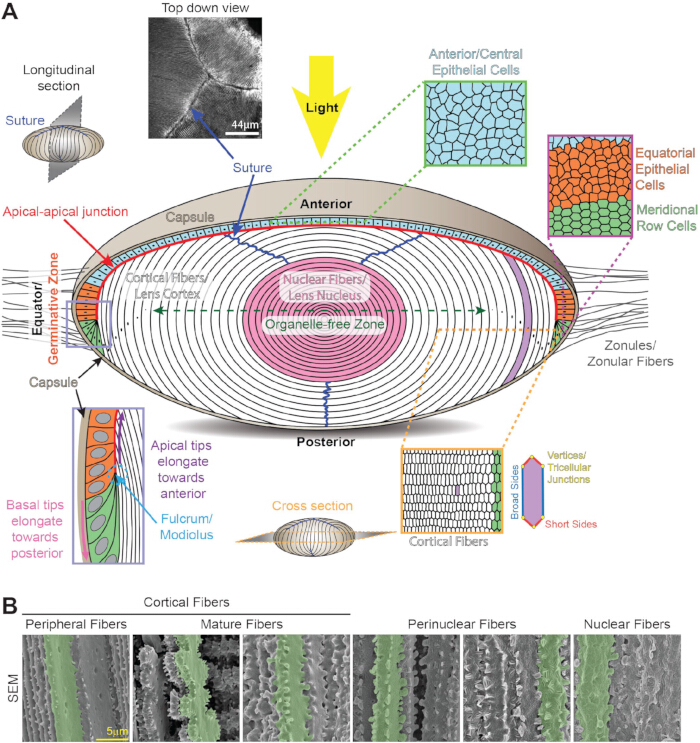
図1:水晶体の解剖学と構造。(A) 水晶体は前半球を覆う上皮細胞(淡い青、オレンジ、淡い緑)の単層と、白とピンクの水晶体繊維の塊で構成されています。薄いコラーゲン膜であるレンズカプセル(タン)が組織を包みます。水晶体は、レンズ赤道付近の嚢内に挿入された帯状線維(ゾヌール)によって眼の前方部屋に吊り下げられています。前部・中央上皮細胞(淡い青色、上部)は直方体状で断面は石畳です。赤道上皮細胞(オレンジ)は発芽帯にランダムに詰め込まれた細胞(オレンジ)から、組織化された六角形細胞列(緑)へと変化します。経絡列細胞はレンズ周辺で新たな皮質二次線維細胞(白色)層へと伸長します。新たに形成された二次繊維の先端は支点/モディオルス(ペリウィンクルボックス、左下)に固定されています。細長い二次線維の先端は水晶体上皮細胞の頂端側に沿って前極へ移動し、異例の頂端-頂端接合部(赤線)を形成します。伸長した線維の基部先端は後嚢に沿って後極に向かって移動します。二次線維が完全に伸長すると、先端が前上皮または後被殻から剥がれて水晶体縫合線を形成します(濃い青色の線と左上の画像)。レンズファイバーセルは断面が細長い六角形(右下)で、2つの長辺/広い辺、4つの短辺、6つの三細胞接合部を持っています。線維細胞の成熟過程では、すべての細胞小器官が除去され、細胞小器官のない領域(濃い緑色の二頭矢印)が形成され、光の散乱を最小限に抑えます。最も古い水晶体繊維細胞は組織の中心に圧縮され、水晶体核(ピンク色)を形成します。アニメはスケールで描かれているわけではありません。(B)レンズ内の異なる深さにあるファイバーセルの走査型電子顕微鏡画像は、分化と成熟過程で変化する細胞形態を示します。単一セルは強調のために緑色でハイライトされています。皮質末梢線維は長辺に球状の指状交差があり、短辺には小さな突起があります。繊維細胞の成熟期には、小さな突起や大きな突起が飾られた大きなパドルが成熟細胞の短辺を飾ります。核周線維は膜全体に舌と溝の指状の交差があり、短辺には大きな突起があります。核繊維は短辺に大きな突起があり、球状膜の形態を示します。スケールバー、5μm。16,328から修正。この図の拡大版はこちらをクリックしてご覧ください。
レンズの複雑さは、高度に秩序立てられた三次元(3D)細胞配置、すべての細胞小器官を分解・除去する特殊な繊維成熟、そして組織の透明性と高い屈折率を確立・維持するための分子および細胞のメカニズムにあります。ほとんどのレンズ線維細胞に細胞小器官が存在しないことは、生物の生涯を通じて作られたすべてのレンズ細胞の健康と寿命を維持する困難を増しています。レンズは加齢や加齢に伴う変化の研究に優れた組織です。なぜなら、中心細胞は胚発生初期に形成され、生涯を通じて組織の周辺で新しい細胞を生成し続けるため、年代的に古い細胞と老年期に作られた細胞の研究を可能にします。レンズの細かいピント合わせ機能により、異なる距離の物体から網膜上に鮮明な画像を形成でき、レンズの形状変化や調整能力に依存します。水晶体は結合性や筋肉でない組織であり、通常の機能には生体力学的特性が必要です。
視覚障害の主な原因は17歳の加齢です。加齢に伴う変化により、白内障と老眼という2つの主要な水晶体病理が生じます。白内障やレンズの不透明度は世界の失明の主な原因であり、紫外線、喫煙、糖尿病、酸化ストレスなどの遺伝的・環境的要因によって引き起こされ、タンパク質の凝集や細胞の欠陥を引き起こします。これは以前のレビューで説明されています。18,20,21,22,23,24,25.老眼とは、加齢に伴う適応の低下であり、近くの物体に焦点を合わせた際にレンズの形状変化能力が低下すること、そして拡大解釈して老眼鏡の必要性が低下することによって引き起こされます。世界保健機関(WHO)は、世界で少なくとも10億人が未解決の視覚障害を持つと推定しており、そのうち白内障が6,520万人、老視が8億2,600万人です。高齢化が進む中で、レンズの生理学や生物学をより深く理解する必要性が急務です。
本レビューでは、哺乳類の水晶体の機能と恒常性に重要な組織構造、細胞配置、分子メカニズムを扱い、他の脊椎動物のレンズに関する情報も含めます。目的は、レンズの生物学と生理学の一般的な概要を提供し、さらに詳細なレビューを参照することです。トピックには胚性水晶体の発生、出生後の水晶体成長、3次元細胞および組織の配列、組織の生体力学的特性、微小循環が含まれます。以下では、結晶体、細胞骨格ネットワーク、細胞間通信といった主要なレンズタンパク質について説明します。レビューは、この独特な組織を研究するための現在の課題と新しい方法をまとめて締めくくります。
アクセスが制限されています。このコンテンツを表示するにはログインするか、トライアルを開始してください。
レビューと展望
胚性水晶体の発達と出生後のレンズ成長
脊椎動物では、水晶体は表面の外胚葉細胞から発達し、将来の水晶体は視小胞6,29,30の上方の表面外胚葉の肥厚化から始まります(図2A)。視カップの形成と同時に、肥厚した外胚葉または水晶体板が水晶体くぼの6,29,30に陥ります(図2B)。アクチンフィラメント(F-アクチン)は、後部のレンズ窩を発達中の光学カップの前表面に結合させ、これらの発達中の組織の動きや形状の変化を調整しています。31,32,33,34。また、レンズの凹入には水晶体上皮細胞の頂端収縮が必要です。35,36,37,38,39,40 (図2C)レンズの穴の開口部は狭くなり続け、穴が閉じるまで続き、レンズ柄細胞はアポトーシス(41,42,43)を経験します。水晶体小胞は将来の角膜外胚葉から分離され、水晶体小胞を形成します。これは上皮細胞の中空球体(6,29,30)(図2D)です。水晶体胞内の後上皮細胞は前上皮に向かって伸長し、一次線維細胞に分化して空洞を満たし胚性水晶体を形成します(図2E)。前半球を覆う上皮細胞の単層は維持されますが、完全に形成された胚性水晶体には後部上皮細胞は存在しません(図2F)。一次線維細胞は水晶体核の最中心部を形成し、細胞周辺や皮質から生じる新しい次晶体線維に囲まれています。これらは生涯6,29,30です。ほとんどの脊椎動物の水晶体は陥落によって形成されますが、ゼブラフィッシュでは板状が厚くなり、その後層状化してレンズ44を形成します。
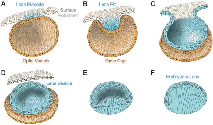
図2:哺乳類の眼における胚性水晶体の発生。 表面の外胚葉細胞は白色、レンズ細胞は淡い青色、視カップ細胞は砂色です。 (A) 胚性水晶体板(淡い青色)は表面の外胚葉から発達します。表面の外胚葉およびレンズ板の後方には視小胞があります。 (B) 水晶体板の陥入がレンズの凹部を形成し、視小胞の陥入が視カップを形成します。前視カップは虹彩と毛様体に発達し、後部視カップは網膜と網膜色素上皮を形成します。 (C) レンズプレートの開口部が狭くなり、開口部が閉じるまで続きます。 (D) 水晶体小胞は表面の外胚葉から分離し、レンズ上皮細胞で覆われた中空の球体です。 (E) 水晶体小胞内の後方水晶体上皮細胞が伸長し、一次水晶体線維へと分化し始めます。 (F) 一次水晶体線維が水晶体小胞の空洞を満たし胚性水晶体を形成します。アニメはスケールで描かれているわけではありません。 411をモデルにしています。 この図の拡大版はこちらをクリックしてご覧ください。
胚発生期には、水晶体はバスケット状の血管網(6,45,46)に囲まれています。これらの血管は網膜の硝子動脈から始まり、水晶体くぼの後方でネットワークを形成し始め、最終的には胚性水晶体を養うために、前房と房水生成が完全に発達するまで、排卵レンズに栄養を与えるために網膜の網目を作り出します。6,45。これらの血管は、種によっては出生前または目開ける前に明確な光路を確保するために後退し、消失します。6、45、46、47。
Pax6は初期の眼の発達に必要であり、48、49、50、51、52に必要であり、レンズの発達53、54、55、56には必須かつ十分です。Pax6のリン酸化およびSUM酸化は、脳、中枢神経系、眼の発達過程でこの転写因子の機能を調節します 57,58,59,60。胚性眼および水晶体の発生に必要な他の重要なシグナル伝達経路はPax6シグナル伝達と交差しており、強化子[Meis、骨形態形成タンパク質(Bmp)、線維芽細胞成長因子(FGF)]および下流タンパク質[Foxe3およびレチノイン酸(RA)]などが含まれており、これは以前のレビュー61で述べられています。Meis1およびMeis2はPax6遺伝子の増強領域に結合し、脊椎動物62のレンズ誘導に必要です。遺伝学的研究では、Bmp7がPax6発現を調節することで水晶板の発生に必要であることが示されています。63,64。Foxe3はPax6シグナルの下流に位置し、水晶体65,66の発達、特に水晶体小胞が形成される際にレンズ柄を表面外胚葉からきれいに分離させるために必要です。Foxe3の喪失は先天性眼の異常(Peters Anomaly67)を引き起こします。FGF受容体(FGFR)シグナル伝達はBmp7と協力してPax6発現を調節し、その結果、形成中の水晶体68のFoxe3レベルに影響を与えます。抑制性FGFRの発現は、水晶体プラコード形成の遅延、水晶体柄の不完全な分離、そして小さな水晶体小胞68を引き起こす。RAシグナル伝達の妨害は眼形成欠損を引き起こし、Pax6が破壊されるとRAの発現が減少し、2つのシグナル伝達経路間にクロストークが存在することを示唆する70。
胚性水晶体が完全に形成された後、赤道上皮細胞の増殖と分化による連続的なレンズ成長は、主に哺乳類レンズにおけるFGFによる成長因子シグナル伝達によって駆動されると考えられており、これは以前のレビュー71で説明されています。FGFは房水よりも硝子体に多く存在し、レンズ赤道付近で成長と分化信号の勾配が形成されます。このレンズは4つのFGFRsのうち3つ、FGFR1、FGFR2、FGFR3を発現し、赤道74,75のレンズ後方で発現が増加します。FGFsの過剰発現は、通常は静止している前上皮細胞の異常な伸長と分化を誘導し、線維様細胞へと移行させます。FGFシグナル伝達に加え、阻害剤アッセイではBmp経路が線維細胞の伸長に関与していることが示されました79,80、異常な形質転換因子β(TGFβ)受容体が正常な線維細胞成熟を妨害します81。
レンズの透過率と屈折率
レンズファイバー細胞は、成熟時にすべての細胞小器官を除去し、光経路中の光散乱対象を排除し、オルガネルフリーゾーン(OFZ)82(図1A)を形成するという特殊なプロセスを受けます。すべての細胞小器官の協調的な喪失は、繊維細胞が成熟プログラムの特定の段階に達すると急速に起こります。83,84,85,86,87,88。異常な細胞小器官の劣化は白内障を引き起こし、いくつかのノックアウト(KO)ゼブラフィッシュおよびマウスモデルは、クロマチンおよびDNA分解の遺伝子89,90,91,92,93、サイクリン依存性キナーゼ94,95、熱ショック因子4の遺伝子91,96,97,98、PLATホスホリパーゼ99の遺伝子を特定していますこれらはOFZの形成にとって重要です。細胞膜および細胞骨格タンパク質100,101,102,103,104、結晶体105、106、107、がん遺伝子108,109の異常発現や変異も、細胞小器官の除去やレンズの透明性に影響を与えることがあります。しかし、線維細胞における細胞小器官の分解を引き起こす正確な信号は依然として明確ではありません。最近の研究では、レンズの発達とカタラクトジェネシスにおけるRNA結合タンパク質の重要性が明らかになりました(これは以前のレビュー110で述べられています)。Tdrd7111、caprin2112、Rbm24113、Celf1 114,115,116などのRNA結合タンパク質の破壊は、異常な水晶体発生、細胞小器官の分解障害、白内障を引き起こします。
体内のほとんどの細胞タイプと比べて、哺乳類のレンズファイバー細胞は非常に高いタンパク質濃度(200〜450 mg/ml)を持っています。タンパク質はレンズ全体の重量の30%〜35%を占めるため、レンズの水分含有量は比較的低く、約65%〜70%です。レンズタンパク質の質量比で90%が結晶123であり、哺乳類レンズには3つのファミリー(α、β、γ)があり、これは以前のレビュー124,125で記述されています。高タンパク質含有量は通常、光の波長の半分を超える集合体のリスクを高め、光散乱を引き起こす126ですが、結晶質は短距離秩序で組織され、高い屈折率と透明性を促進するために117,127です。タンパク質凝集は、以下で説明するα結晶のシャペロン様機能によって弱まります。レンズ全体の勾配屈折率(GRIN)は光の集束を促進し、以前のレビューで述べられているように128,129、そしてレンズ中心部の最大屈折率は核繊維セルの圧縮130,131,132と相関しています。魚レンズでは、核の高い屈折率はタンパク質が組織中心への輸送によって起こることがデータに示唆されています133。結晶タンパク質の細胞間および繊維の殻間における均一な分布は、細胞繊維134,135に沿った膜融合による高分子輸送経路によって促進されると仮説されています。しかし、マウスレンズ136,137,138では膜融合が明確に観察されておらず、複数種のレンズでのインピーダンス研究では膜融合139,140,141,142は検出されていません。ギャップ接合部通信の妨害を受けたマウスモデルのデータは、細胞間通信や線維細胞の伸長も高分子輸送経路に寄与していることを示唆しています143。
結晶の分子量は~20〜30 kDaで、レンズ内で構造的および屈折的な役割を果たします。α結晶体はαAとαBのサブユニットで構成されており、これらは55%〜60%の相同性を共有しています144,145,146。αBはほとんどの細胞で広く発現していますが、αA結晶体は主に水晶体で発現し、脾臓や胸腺など他の組織でも低レベルで検出されています 147,148.最近の研究では、網膜色素上皮細胞や網膜神経節細胞がストレスや疾患中にαA結晶を発現できることが示されており、神経保護効果をもたらすと考えられています。α結晶は通常、30–35個の混合αAとαBサブユニットからなる大きなヘテロマルチマーに結合します123。マウス、ウシ、ヒトの水晶体上皮では、前方細胞ではαBのみが検出されますが、赤道細胞ではαAとαBが1:3の比率で存在します。この比率は二次繊維細胞の分化と伸長時に3:1に反転します。123,152,153。アルファ結晶体は小さな熱ショックタンパク質ファミリーに属し、シャペロン様機能154を持ち、異常または折りたたまれていないタンパク質に結合・隔離することで、さらなるタンパク質の凝集を防ぎます。小さなα結晶ペプチドは、以前のレビュー155で記述されたように、白内障を含むタンパク質凝集誘発疾患の予防に探求されています。しかし、マウスモデルにおけるα結晶のシャペロン様活性の増加または減少は、いずれのシナリオも白内障の重症度増加をもたらすことを示し、α結晶の適切なシャペロン様活性がレンズの透明性に不可欠であることを示唆しています(156)。ベータ結晶およびγ結晶は独自のβ/γ結晶タンパク質ファミリーに属し、線維細胞特異的です。哺乳類では、β結晶体には二量体、四量体、または高次のオリゴマーとして存在する7種類のアイソフォームがあり、以前のレビュー125で述べたように、γ結晶体にはモノマーであるアイソフォームが7種類存在します。構造機能に加え、β結晶およびγ結晶はカルシウムイオンに結合し、レンズ繊維中のカルシウムイオンの利用可能性と予備量を調節します。カルシウムイオンは157、158、159、160、161です。
レンズ細胞骨格および膜タンパク質
レンズ上皮細胞および線維細胞は、F-アクチン、ケラチンまたはビメンチン中間フィラメント、特殊なビーズ状中間フィラメント、微小管のネットワークを持ち、これは以前のレビューで述べられています。162、163、164、165、166。眼および水晶体の発達における機能(31,32,33,34)に加え、水晶体上皮細胞および線維細胞内の多様なFアクチンネットワークが水晶体の透明性に重要であり、167,168,169,170,171,172。最近の研究では、Fアクチンネットワークの変化がレンズの生体力学に影響を与えることが示されています。173、174、175、176。ビメンチンまたはケラチンからなる中間フィラメントは、レンズ線維細胞分化の初期段階または胚発生期にのみ存在します。それぞれ177、178、179、180、181;対照的に、すべてのレンズ繊維は、フィレンシン(別名CP95/CP115、Bsfp1で符号化)およびCP49(別名ファキニン、Bsfp2で符号化)182,183,184のヘテロダイマーからなるビーズ状中間フィラメントを持っています。これらのヘテロダイマーのビーズ状の外観は、フィラメントコア185,186を装飾するCP49とフィレンシンからなるオリゴマーと、これらの特殊な中間フィラメント187,188,189,190,191にα結晶体が結合していることによるものです。フィレンシンまたはCP49のいずれかが失われると、このネットワークは完全に失われます。192,193,194,195。ビメンチン中間フィラメントまたはビーズ状中間フィラメントの破壊は、人間および動物モデルにおいて白内障を引き起こす 192,193,196,197,198,199,200,201,202,203,204これらの細胞骨格ネットワークがレンズの透明性においていかに重要かを示しました。最近の研究では、微小管がレンズ線維細胞の伸長205,206および小器官フリーゾーン177の形成に重要であることが示唆されています。
アクチン細胞骨格と相互作用するアディエレン接合部は、レンズ形成207および線維細胞分化に重要です。E-カデリンとN-カデリンは共にレンズ上皮細胞208,209,210,211で発現し、E-カデリンは線維細胞分化の開始時に212でオフにされます。N-カデリンはレンズ線維細胞で高発現し、伸長する線維細胞8,209の基底六角足の中点で濃縮されています。一次線維細胞形成後のN-カデリンの喪失は、前上皮細胞の頂端に沿った分化線維の頂端の異常移動により二次線維細胞伸長の障害を引き起こします。レンズ内のエフリン双方向シグナル伝達の妨害は、アディエレン接合部4,7,214,215,216に欠陥を引き起こします。
その他の主要なレンズ膜タンパク質にはアクアポリン、コネクシン;MP20は、以前のレビュー217で説明された通りです。ガレクチン3;ガレクチン関連繊維間タンパク質(GRIFIN);インテグリンは、前回のレビュー218で説明されていました。そして酸性でシステインが豊富なタンパク質(SPARC)を分泌します。アクアポリンとコネキシンについては次のセクションで説明します。MP20(別名MP17またはLim2)は、細胞間接着機能85,219を持つレンズ特異的膜タンパク質で、線維細胞内に豊富に存在します。MP20の変異は白内障222,223,224,225,226,227,228,229,230を引き起こす。最近の研究では、MP20ともう一つの重要な細胞間接着分子であるガレクチン3(231,232,233)との相互作用が示されています。グリフィンはガレクチンのレンズ特異的アイソフォームであり、細胞繊維に局在し、Pax6、結晶体、カデリン234、235、236、237と相互作用します。インテグリンは細胞外マトリックス(ECM)と細胞骨格を結ぶ重要なリンクであり、正常な水晶体の発達に必要です。レンズ内で発現するインテグレインアイソフォームは、ラミニン、コラーゲン、またはフィブロネクチンなどのカプセル成分の受容体として機能します。8,238,239,240,241,242,243,244,245。各マトリックス成分は複数のインテグリンと相互作用し、水晶体の発達と成長中のインテグリンの時空間的発現を通じて多様なシグナル伝達をもたらします218。特に、白内障手術後の残存水晶体上皮細胞における上皮から間葉への移行におけるTGFβ、インテグリン、ECM成分の変化、すなわち前述のレビュー218で述べたように後被膜不透明化の理解に関心があります。SPARCは細胞間接着および増殖分子であり、水晶体上皮に局在し、分化線維には存在しません。SPARCの喪失は白内障246,247,248を引き起こし、最近の研究ではSPARCがシャペロン様機能を持つ可能性249、インテグリンおよびECMタンパク質250,251,252、微小循環経路253に影響を与えることが示されています。
無血管組織の微小循環経路
血液供給が不足しているため、完全に形成された水晶体は栄養素を供給し老廃物を除去するために独自の微小循環経路を生成します。これは以前のレビュー254,255,256,257で説明されています。イオン電流は前後の極からレンズに入り、赤道258で出ます。内向きの電流は、能動輸送によって生成される濃度勾配に沿って、各極の晶体細胞間の細胞外空間にナトリウムが移動することに依存し、外向きの電流は細胞膜の隙間接合チャネルによって促進され、これらは水晶体赤道付近に集中し、ナトリウムイオンが水晶体赤道259から排出されるようにします。赤道上皮細胞および末梢線維細胞は、細胞外環境260,261,262のナトリウムイオンと細胞外のナトリウムイオン2個を活性的に交換するNa+/K+ ATPaseポンプの高い発現を持っています。レンズは高カリウム濃度(135mM)と低ナトリウム濃度(16mM)を維持し、ナトリウムが極から再入りし、濃度勾配259,263を下る条件を作り出します。Na+/K+ ATPaseに加え、イオン共輸送体Na-K-2Cl共輸送体1(NKCC1)も、前述のレビュー264で述べたように、メカノ感受性チャネルによる活性化を通じてレンズイオン濃度の調節にも寄与します。
レンズ内のギャップ接合は、3つのコネキシン、すなわちコネキシン43(Cx43またはα1)、Cx46(またはα3)、およびCx50(またはα8)265,266で構成されています。Cx43は主に水晶体上皮細胞267で発現し、Cx43 KOレンズでは線維細胞形成の開始が正常である一方で、上皮細胞と線維細胞の界面は細胞外空間と細胞内液孔を拡大しており、Cx43が浸透圧バランス268に機能していることを示唆している。Cx46はレンズ線維細胞に集中し、Cx50は上皮細胞と線維細胞の両方に存在します269。Cx23はマウスの水晶体の繊維細胞内でも発現しますが、ヒトレンズでは発現しません(270,271,272,273)。Cx23の優性変異はマウスに線維細胞の欠損を引き起こすが、Cx23 KOレンズには顕著な欠損はない(274)。6つのコネキシンサブユニットがコネクソンまたはヘミチャネルを形成し、ある細胞のコネクソンは隣接するコネクソンとドッキングしてギャップジョインクションを形成し、隣接する細胞間に約10オングストロームの孔を形成し、小分子、イオン、水275,276の交換を可能にします。コネキシン半チャネルは、前述のレビューで述べたように細胞内細胞質と細胞外環境間の物質の通過を可能にし、ヘミチャネルは微小循環279、栄養素や抗酸化物質の機械感受性輸送280,281,282,283、酸化ストレス応答284,285に重要です、286、287、288。ギャップ接合部は繊維細胞膜上の大きなプラークに集まります。レンズ内のプラークは直径ミクロンサイズで、隣接するファイバーセル289の長辺に沿って整列しています。マウスのレンズでCx46の喪失は、レンズの流出経路の閉塞により核白内障が密集します。マイクロ循環290,291。繊維細胞膜上の大きなCx46プラークの組み立ておよび/または安定性には、Fアクチンおよびビーズ状中間フィラメントネットワーク289が必要です。Cx50 KOは、異常な上皮細胞増殖による小眼症、軽度の核白内障、そして小さな水晶体を引き起こす292,293。Cx50の喪失は、レンズファイバー細胞139に残るCx46ギャップ接合プラークのゲートに影響を与えます。Cx50を内因性Cx50遺伝子座下で発現するノックインCx46(4コピー)に置き換えることで、Cx50の喪失による白内障を救い、線維間のギャップ接合結合を増加させますが、これらの透明ノックインレンズは依然として小さいままです(142,294です)。したがって、Cx46はCx50のレンズ成長関数に代わることはできません。ノックイン戦略は、γB結晶変異タンパク質による核白内障の救済にも用いられており、ギャップ接合部の結合増加がレンズの恒常性向上および白内障予防の戦略となり得ることを示唆している295。
ナトリウムイオンによって生成されるイオンフラックスは、アクアポリンチャネル259を通じてレンズセルへの水の流れを伴います。アクアポリンチャネルは上皮細胞のAqp1で構成され、線維細胞では成熟度に応じてAqp0(MIP)とAqp5が水分チャネルを形成しており、これは以前のレビューで記載されている2億6429万6297人です。ギャップ接合チャネルを通る水の移動はレンズに静水圧勾配298,299を生み出し、この勾配は過渡受容体ポテンシャルバニロイド1(TRPV1)およびTRPV4の機械感受チャネルを通じてレンズ浸透圧を維持し、それぞれレンズ静水圧299,300,301,302を増加・減少させます.レンズのゾノラー張力が低下すると静水圧が低下し、TRPV1およびその後NKCC1が活性化され、レンズ内のイオン濃度が上昇し、水の流れを誘導して通常の静水圧300,301を回復させます。逆に、帯状張力とレンズ体積の増加はTRPV4を活性化し、続いてNa+/K+ ATPaseを活性化させ、レンズイオン濃度を低下させ、静水圧300,301,302を低下させます。
必須栄養素とアミノ酸は微小循環経路303を通じて水晶体内に運ばれ、この輸送は特にOFZ繊維にとって重要です。促進性グルコーストランスポーター(GLUT)は水晶体、上皮細胞および線維細胞に存在します。GLUT1は両方の細胞型で発現し、GLUT3は上皮細胞304,305,306,307で発現します。GLUT1 KOマウスは白内障を発症し、レンズ透明性308にはグルコース取り込みが必要であることを示唆しています。グルタチオン(GSH)は水晶体透明度309,310の重要な抗酸化物質であり、水晶体内で作られ、房水311,312,313,314,315から水晶体内に運搬されます。システイン、グルタミン酸、グリシンはGSH合成に不可欠なアミノ酸であり、これらのアミノ酸はレンズ314,315の皮質および核領域に蓄積しています。加齢に伴うGSH合成の減少やGSH使用の増加は、特に核316,317,318,319,320,321において水晶体内のGSH利用可能性の低下を引き起こすことがあります。グルタチオン過酸化酵素(GPX)はGSHを補酵素として利用してヒドロペルオキシドを中和し、最近の研究では、有毒な脂質過酸化物によるフェロプトーシスがGPX4322,323の破壊により白内障を引き起こすことを示しています。
3Dレンズアーキテクチャとバイオメカニクス
水晶体繊維が六角形の細胞列に組織的に形成されるパターン化は、赤道上皮細胞が子午線列にパターン化されることに由来します(図1A)。ランダムに詰め込まれた赤道上皮細胞が六角形の子午線列細胞に組織化されるメカニズムはアクチン細胞骨格を中心に回っています。受容体チロシンキナーゼEphA2のシグナル伝達は、六角形の髄線細胞の頂点にF-アクチンを蓄積させ、細胞組織を促進するために必要です。ミオシンIIAはFアクチンに結合する収縮性タンパク質で、六角形細胞形状8,325,326の促進にも重要です。この高度に秩序化された繊維細胞の詰め込みはレンズの透明性に長らく必要だと理論されてきましたが、EphA2の喪失による誤包装、ミオシンIIA325,326の変異、または老年期に形成された130の異常細胞などは、六角形細胞充填は透明性に必須ではないことを示唆しています。
前極から後極に伸びる繊維細胞は、動物種やレンズの大きさによって数ミリメートルの長さになることがあります。通常、繊維セルは断面積327で4〜7μmしかありませんが、これらの細長く細い細胞は、前回のレビュー328で説明されたように、隣接するファイバーセル間で3次元ジッパーを形成する動的な細胞間指指によって結合されています。レンズ線維細胞の指指形成は、細胞の分化と成熟に伴い変化する複雑なパターンを持ちます。例えば、1) 短辺に小さな突起、長辺にボールソケット突起を持つ新たに形成された末梢線維、2) 細胞小器官を失い、短辺に小さな連結した突起を持つ大きなパドルを持つ成熟した線維細胞 3) 短辺に大きな連結突起と膜全体にわたる舌溝の指状交差を持つ核周線維、4) 短辺に大きな突起があり、膜全体に球状膜形態を持つ核繊維 137,328,329,330,331,332,333,334,335 336,337,338,339 (図1B)。最近の研究は、Fアクチンネットワークに依存する水晶体繊維細胞間の指指が組織の剛性に重要であることを直接的に示しており、長年の仮説を支持しています。接着接合部の成分であるARVCFの喪失は、異常な線維細胞の指指、細胞外空間の拡大、早期発症の皮質白内障340を引き起こす。レンズ核の剛性とサイズの欠損は、舌溝の指相変化や球状膜への遷移遅延と相関しています。131。
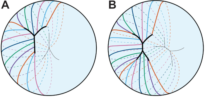
図3:レンズ縫合のパターン化。 各色の線は単一のファイバーセルを表します。 (A) 胚性ヒトレンズ内の線維細胞がY縫合糸を形成します。Y字縫合線は前極と後極に鏡像で形成されています。 (B) 人間のレンズが発達・成長を続けるにつれて、縫合線はより枝分かれになります。ここでは簡潔にするために4枝の縫合線が描かれています。縫合パターンは2つのポールの間でずれています。アニメはスケールで描かれているわけではありません。 45をモデルにしています。 この図の拡大版はこちらをクリックしてご覧ください。
レンズの全体的な形状は種によって異なり、線維細胞の曲率や縫合糸の種類によってレンズ形状341,342が決定されると理論化されています。縫合線は、前極と後極で伸長した線維細胞の先端が接合することで形成され、2つの極(11,12,13,343)の間に縫合線が鏡像として配置されています(図3)。マウスのレンズでは縫合糸はY字型であり、霊長類や人間のレンズでは、縫合線はY字型から始まり、11歳、12歳、45歳の年齢でより枝分かれします。興味深いことに、マウスレンズのY縫合は機械的荷重が除去された後のレンズの弾力性や回復力を制限し、エフリンシグナル伝達の障害を伴うKOレンズの分岐縫合は回復力の増加と相関しています344。レンズ縫合の正常な形成にはギャップ接合部345が必要であり、マウスレンズの加齢に伴う前方白内障は不完全縫合形成130の結果です。
レンズの生体力学的特性は、以前のレビューで述べられているように、分子、細胞、組織のさまざまな要因によって影響を受ける可能性があります。加齢に伴うレンズの硬さの増加が老眼346の主な要因と考えられています。人間では、遠くの物体を見る際に毛様体筋の弛緩によりカプセルに付着した帯状繊維が伸び、レンズ1が平坦化します。近くで焦点を合わせると、毛様体筋が収縮してゾヌールが弛緩し、レンズが丸めて近くの物体からの光を集束させます。この適応の過程には、レンズの生体力学的特性、毛様体筋の収縮、そして帯状の緊張が必要です。加齢に伴い、毛様体筋の収縮力や帯状の柔軟性、緊張にほとんど変化がないことが研究で示されています(347,348,349,350)。いくつかの研究では、レンズのサイズや核の大きさ、剛性の増加が26,351,352,353,354歳までにレンズ全体の硬性に寄与することが示唆されています。しかし、マウスのレンズ硬さの年齢に関する研究では、レンズと核の大きさと全水晶体の硬性130,132との相関はほとんどなく、他の要因が加齢に伴う水晶体の硬性に影響を与えることが示唆されています。最近の研究では、細胞骨格ネットワークの変化が水晶体の剛性に影響を与えることが示されています。F-アクチン安定化タンパク質であるトロポミオシン3.5の喪失は、線維細胞膜におけるF-アクチンネットワークの組成と配置に影響を与え、レンズの剛性を低下させます173。同様に、複数の近親交配野生型マウス系統における自発変異は、CP49192,355個、より柔らかいレンズ356,357個の自発的なKOを引き起こします。レンズ線維細胞膜はコレステロール含有量が異常に高く、358,359,360,361,362,363であり、膜の弾性を調節できます。膜コレステロール含有量の変化およびα結晶体が細胞繊維膜に結合する過程は、年齢とともにレンズの剛性を高めると理論されています。365,366,367,368,369,370,371,372。
レンズの研究における課題と新しい方法
レンズの3D構造は、細胞間の配列を理解し、組織構造を再現する適切なin vitroモデルの作成に課題をもたらします。シグナル伝達や変異タンパク質の局在化に関する貴重な知見は、不死化または一次培養細胞を用いて明らかにされていますが、分離されたレンズ上皮細胞は、ネイティブのレンズ上皮細胞と異なる直方体形状や遺伝子発現や細胞骨格構造を持たず、373を有しています。前回のレビューで述べたように、in vitroレントイドは374およびレンズレットモデル375で、それぞれミニレンズのような繊維状細胞と細胞の球状の細胞を作り出します。これらのモデルは迅速な薬物スクリーニングに有用な透明なレンズ状構造を作り出しますが、いずれも正常な線維細胞の組織やレンズの3D構造を再現するものではありません。レンズ組織切片中のタンパク質の局在化や免疫染色のための多くのプロトコルが開発されており、この方法はタンパク質が特定の細胞区画で発現しているかどうかを理解する最も効率的な方法です。しかし、組織学や免疫染色のための組織切片は、特に高齢成獣のサンプルではレンズ137の固定が不完全であるため困難です。また、2次元断面で3次元構造を研究することで、細胞の形状、細胞間の相互作用、タンパク質の局在化を理解する上で重要な詳細が失われます。電子顕微鏡(EM)は繊維細胞形状の研究において依然としてゴールドスタンダードですが、EMサンプル中のタンパク質の局在化は依然として困難であり、3億7,377万7,378回です。上皮細胞単層のフラットマウントによる最近の研究 4,7,324,373、単一繊維セル染色 85,173,174,379、全レンズイメージング 4,7,324,344,380,381細胞間癒着、レンズの生体力学、レンズ線維内の変化する細胞骨格ネットワークに関する重要な情報を明らかにしました。
レンズ上皮細胞と線維細胞は機能、トランスクリプトーム、プロテオームが異なり、過去の比較研究では、前半球を覆う単一層の上皮細胞が存在するため、多くのサンプルをプールせずに収集できるRNAやタンパク質の量が限られていました。RNA抽出技術、リアルタイムPCR、自動毛細血管電気泳動の進歩により、マウスの目1対から採取した水晶体上皮細胞サンプルの遺伝子およびタンパク質解析が可能になりました。マウスの目1対から採取したもの130,132,373,382、または水晶体上皮、皮質線維、核線維の微細解剖領域から採取されたもの383.単細胞トランスクリプトミクスにより、ゼブラフィッシュ胚および幼虫・ヒナ胚384,385のレンズ細胞の詳細なプロファイル解析が可能です。これらの新しい手法は、正常な老化や病理の過程で各晶体細胞区画を維持する分子メカニズムを明らかにする扉を開きます。
分野が進展するにつれ、マウスの遺伝的背景はレンズ病理386,387および生体力学355,356,357において重要な役割を果たすことを忘れてはなりません。したがって、実験における同兄弟の対照群はデータ解析において非常に重要です。ミュータントおよびKOマウスモデルは、加齢に関連した白内障やレンズ硬性の増加を研究するシステムを提供します。130,356,357,388,389,390,391,392,393,394,395,396,397マウスの寿命が比較的短いため、老化研究は若い動物からデータを外挿するのではなく、高齢動物に対して行うべきです。
アクセスが制限されています。このコンテンツを表示するにはログインするか、トライアルを開始してください。
結論
トランスクリプトミクス、プロテオミクス、画像診断、生体力学的検査技術の進歩により、レンズの生物学、生理学、病理学への理解が深まりました。水晶体、上皮細胞、線維細胞の異なる集団間の違いに関する知識の拡大は、加齢に伴う変化やタンパク質および細胞の寿命のメカニズムを理解する上で重要です。白内障や老眼の予防や遅延、白内障手術後のレンズ再生の可能性に関する医薬品介入の開発への関心が高まっています。白内障や老眼の予防または遅延のために水晶体への薬物投与には、レンズカプセルを通じた投与量を増やすために特定の化学的特性を持つ点眼薬が必要であり、レンズ内の異なる深さで浸透し蓄積する薬物の量を慎重に研究する必要があります。398,399,400,401,402.爬虫類...
アクセスが制限されています。このコンテンツを表示するにはログインするか、トライアルを開始してください。
開示事項
著者たちは何も明かすことはありません。
謝辞
この研究は国立眼科研究所の助成金R01 EY032056(CCへ)によって資金提供されました。著者たちは、アイザイア・J・イニス氏とグリフィン・M・フラワーズ氏の有益なコメントに感謝します。
アクセスが制限されています。このコンテンツを表示するにはログインするか、トライアルを開始してください。
参考文献
- Freddo, T. F., Chaum, E. Anatomy of the Eye and Orbit: The Clinical Essentials, 1e. , Lippincott Williams & Wilkins, a Wolters Kluwer business. (2018).
- Danysh, B. P., Duncan, M. K. The lens capsule. Exp Eye Res. 88 (2), 151-164 (2009).
- Zampighi, G. A., Eskandari, S., Kreman, M. Epithelial organization of the mammalian lens. Exp Eye Res. 71 (4), 415-435 (2000).
- Cheng, C., Gong, X. Diverse roles of Eph/ephrin signaling in the mouse lens. PLoS One. 6 (11), e28147(2011).
- Parreno, J., Cheng, C., Nowak, R. B., Fowler, V. M. The effects of mechanical strain on mouse eye lens capsule and cellular microstructure. Mol Biol Cell. 29 (16), 1963-1974 (2018).
- Lovicu, F. J., Robinson, M. L. Development of the Ocular Lens. , Cambridge University Press. (2011).
- Cheng, C., Ansari, M. M., Cooper, J. A., Gong, X. EphA2 and Src regulate equatorial cell morphogenesis during lens development. Development. 140 (20), 4237-4245 (2013).
- Bassnett, S., Missey, H., Vucemilo, I. Molecular architecture of the lens fiber cell basal membrane complex. J Cell Sci. 112 (Pt 13), 2155-2165 (1999).
- Bassnett, S., Sikic, H. The lens growth process. Prog Retin Eye Res. 60, 181-200 (2017).
- Sikic, H., Shi, Y., Lubura, S., Bassnett, S. A full lifespan model of vertebrate lens growth. R Soc Open Sci. 4 (1), 160695(2017).
- Kuszak, J. R. The development of lens sutures. Progress in Retinal and Eye Research. 14 (2), 567-591 (1995).
- Kuszak, J. R., Mazurkiewicz, M., Zoltoski, R. Computer modeling of secondary fiber development and growth: I. Nonprimate lenses. Mol Vis. 12, 251-270 (2006).
- Kuszak, J. R., Zoltoski, R. K., Tiedemann, C. E. Development of lens sutures. Int J Dev Biol. 48 (8-9), 889-902 (2004).
- Kuszak, J. R. The ultrastructure of epithelial and fiber cells in the crystalline lens. Int Rev Cytol. 163, 305-350 (1995).
- Kuszak, J. R., Macsai, M. S., Rae, J. L. Stereo scanning electron microscopy of the crystalline lens. Scan Electron Microsc. (Pt 3), 1415-1426 (1983).
- Cheng, C. Tissue, cellular, and molecular level determinants for eye lens stiffness and elasticity. Front Ophthalmol (Lausanne). 4, 1456474(2024).
- World Health Organization. World report on vision. , (2019).
- Michael, R., Bron, A. J. The ageing lens and cataract: a model of normal and pathological ageing. Philos Trans R Soc Lond B Biol Sci. 366 (1568), 1278-1292 (2011).
- Asbell, P. A., et al. Age-related cataract. Lancet. 365 (9459), 599-609 (2005).
- Hejtmancik, J. F. Oxidative Stress in Genetic Cataract Formation. Antioxidants (Basel). 13 (11), (2024).
- Shiels, A., Hejtmancik, J. F. Biology of Inherited Cataracts and Opportunities for Treatment. Annu Rev Vis Sci. 5, 123-149 (2019).
- Truscott, R. J. W., Friedrich, M. G. Molecular Processes Implicated in Human Age-related Nuclear Cataract. Invest Ophthalmol Vis Sci. 60 (15), 5007-5021 (2019).
- Fan, X., Monnier, V. M. Protein posttranslational modification (PTM) by glycation: Role in lens aging and age-related cataractogenesis. Exp Eye Res. 210, 108705(2021).
- Budnar, P., Tangirala, R., Bakthisaran, R., Rao, C. M. Protein Aggregation and Cataract: Role of Age-related Modifications and Mutations in alpha Crystallins. Biochemistry (Mosc). 87 (3), 225-241 (2022).
- Shiels, A., Hejtmancik, J. F. Inherited cataracts: Genetic mechanisms and pathways new and old. Exp Eye Res. 209, 108662(2021).
- Heys, K. R., Cram, S. L., Truscott, R. J. Massive increase in the stiffness of the human lens nucleus with age: the basis for presbyopia? Mol Vis. 10, 956-963 (2004).
- Glasser, A., Campbell, M. C. Biometric, optical and physical changes in the isolated human crystalline lens with age in relation to presbyopia. Vision Res. 39 (11), 1991-2015 (1999).
- Weeber, H. A., et al. Dynamic mechanical properties of human lenses. Exp Eye Res. 80 (3), 425-434 (2005).
- Piatigorsky, J. Lens differentiation in vertebrates. A review of cellular and molecular features. Differentiation. 19 (3), 134-153 (1981).
- Gunhaga, L. The lens: a classical model of embryonic induction providing new insights into cell determination in early development. Philos Trans R Soc Lond B Biol Sci. 366 (1568), 1193-1203 (2011).
- Chauhan, B. K., et al. Cdc42 and IRSp53 dependent contractile filopodia tether presumptive lens and retina to coordinate epithelial invagination. Development. 136 (21), 3657-3667 (2009).
- McAvoy, J. W. Cytoplasmic processes interconnect lens placode and optic vesicle during eye morphogenesis. Exp Eye Res. 31 (5), 527-534 (1980).
- Mann, I. The development of the human eye. , 3d, Grune & Stratton. (1964).
- Eiraku, M., et al. Self organizing optic cup morphogenesis in three dimensional culture. Nature. 472 (7341), 51-56 (2011).
- Lang, R. A., Herman, K., Reynolds, A. B., Hildebrand, J. D., Plageman, T. F. Jr p120 catenin dependent junctional recruitment of Shroom3 is required for apical constriction during lens pit morphogenesis. Development. 141 (16), 3177-3187 (2014).
- Plageman, T. F. Jr, et al. A Trio RhoA Shroom3 pathway is required for apical constriction and epithelial invagination. Development. 138 (23), 5177-5188 (2011).
- Borges, R. M., Lamers, M. L., Forti, F. L., Santos, M. F., Yan, C. Y. Rho signaling pathway and apical constriction in the early lens placode. Genesis. 49 (5), 368-379 (2011).
- Quintin, S., Gally, C., Labouesse, M. Epithelial morphogenesis in embryos: asymmetries, motors and brakes. Trends Genet. 24 (5), 221-230 (2008).
- Muccioli, M., Qaisi, D., Herman, K., Plageman, T. F. Jr Lens placode planar cell polarity is dependent on Cdc42 mediated junctional contraction inhibition. Dev Biol. 412 (1), 32-43 (2016).
- Houssin, N. S., Martin, J. B., Coppola, V., Yoon, S. O., Plageman, T. F. Jr Formation and contraction of multicellular actomyosin cables facilitate lens placode invagination. Dev Biol. 462 (1), 36-49 (2020).
- Glucksmann, A. Cell deaths in normal vertebrate ontogeny. Biol Rev Camb Philos Soc. 26 (1), 59-86 (1951).
- Ozeki, H., Ogura, Y., Hirabayashi, Y., Shimada, S. Suppression of lens stalk cell apoptosis by hyaluronic acid leads to faulty separation of the lens vesicle. Exp Eye Res. 72 (1), 63-70 (2001).
- Yan, Q., Liu, J. P., Li, D. W. Apoptosis in lens development and pathology. Differentiation. 74 (5), 195-211 (2006).
- Easter, S. S. Jr, Nicola, G. N. The development of vision in the zebrafish (Danio rerio). Dev Biol. 180 (2), 646-663 (1996).
- Mann, I. The development of the human eye. , The University Press. (1928).
- Darche, M., et al. Three dimensional characterization of developing and adult ocular vasculature in mice using in toto clearing. Communications Biology. 5 (1), 1135(2022).
- Wang, Z., Liu, C. H., Huang, S., Chen, J. Assessment and Characterization of Hyaloid Vessels in Mice. J Vis Exp. (147), e59222(2019).
- Hill, R. E., et al. Mouse small eye results from mutations in a paired like homeobox containing gene. Nature. 354 (6354), 522-525 (1991).
- Jordan, T., et al. The human PAX6 gene is mutated in two patients with aniridia. Nat Genet. 1 (5), 328-332 (1992).
- Hogan, B. L., et al. Small eyes (Sey): a homozygous lethal mutation on chromosome 2 which affects the differentiation of both lens and nasal placodes in the mouse. J Embryol Exp Morphol. 97, 95-110 (1986).
- Hanson, I. M., et al. Mutations at the PAX6 locus are found in heterogeneous anterior segment malformations including Peters' anomaly. Nat Genet. 6 (2), 168-173 (1994).
- Grindley, J. C., Davidson, D. R., Hill, R. E. The role of Pax 6 in eye and nasal development. Development. 121 (5), 1433-1442 (1995).
- Fujiwara, M., Uchida, T., Osumi Yamashita, N., Eto, K. Uchida rat (rSey): a new mutant rat with craniofacial abnormalities resembling those of the mouse Sey mutant. Differentiation. 57 (1), 31-38 (1994).
- Collinson, J. M., Hill, R. E., West, J. D. Different roles for Pax6 in the optic vesicle and facial epithelium mediate early morphogenesis of the murine eye. Development. 127 (5), 945-956 (2000).
- Quinn, J. C., West, J. D., Hill, R. E. Multiple functions for Pax6 in mouse eye and nasal development. Genes Dev. 10 (4), 435-446 (1996).
- Ashery Padan, R., Marquardt, T., Zhou, X., Gruss, P. Pax6 activity in the lens primordium is required for lens formation and for correct placement of a single retina in the eye. Genes Dev. 14 (21), 2701-2711 (2000).
- Kim, E. A., et al. Phosphorylation and transactivation of Pax6 by homeodomain interacting protein kinase 2. J Biol Chem. 281 (11), 7489-7497 (2006).
- Mikkola, I., Bruun, J. A., Bjorkoy, G., Holm, T., Johansen, T. Phosphorylation of the transactivation domain of Pax6 by extracellular signal regulated kinase and p38 mitogen activated protein kinase. J Biol Chem. 274 (21), 15115-15126 (1999).
- Yan, Q., et al. Sumoylation activates the transcriptional activity of Pax 6, an important transcription factor for eye and brain development. Proc Natl Acad Sci U S A. 107 (49), 21034-21039 (2010).
- Yan, Q., et al. Protein phosphatase 1 modulates the function of Pax 6, a transcription factor controlling brain and eye development. J Biol Chem. 282 (19), 13954-13965 (2007).
- Cvekl, A., Zhang, X. Signaling and Gene Regulatory Networks in Mammalian Lens Development. Trends Genet. 33 (10), 677-702 (2017).
- Zhang, X., Friedman, A., Heaney, S., Purcell, P., Maas, R. L. Meis homeoproteins directly regulate Pax6 during vertebrate lens morphogenesis. Genes Dev. 16 (16), 2097-2107 (2002).
- Dudley, A. T., Lyons, K. M., Robertson, E. J. A requirement for bone morphogenetic protein 7 during development of the mammalian kidney and eye. Genes Dev. 9 (22), 2795-2807 (1995).
- Wawersik, S., et al. BMP7 acts in murine lens placode development. Dev Biol. 207 (1), 176-188 (1999).
- Blixt, A., et al. A forkhead gene, FoxE3, is essential for lens epithelial proliferation and closure of the lens vesicle. Genes Dev. 14 (2), 245-254 (2000).
- Brownell, I., Dirksen, M., Jamrich, M. Forkhead Foxe3 maps to the dysgenetic lens locus and is critical in lens development and differentiation. Genesis. 27 (2), 81-93 (2000).
- Dimanlig, P. V., Faber, S. C., Auerbach, W., Makarenkova, H. P., Lang, R. A. The upstream ectoderm enhancer in Pax6 has an important role in lens induction. Development. 128 (22), 4415-4424 (2001).
- Faber, S. C., et al. Fgf receptor signaling plays a role in lens induction. Development. 128 (22), 4425-4438 (2001).
- Bavik, C., Ward, S. J., Chambon, P. Developmental abnormalities in cultured mouse embryos deprived of retinoic by inhibition of yolk sac retinol binding protein synthesis. Proc Natl Acad Sci U S A. 93 (7), 3110-3114 (1996).
- Enwright, J. F. 3rd, Grainger, R. M. Altered retinoid signaling in the heads of small eye mouse embryos. Dev Biol. 221 (1), 10-22 (2000).
- Lovicu, F. J., McAvoy, J. W. Growth factor regulation of lens development. Dev Biol. 280 (1), 1-14 (2005).
- Schulz, M. W., Chamberlain, C. G., de Iongh, R. U., McAvoy, J. W. Acidic and basic FGF in ocular media and lens: implications for lens polarity and growth patterns. Development. 118 (1), 117-126 (1993).
- Lovicu, F. J., Chamberlain, C. G., McAvoy, J. W. Differential effects of aqueous and vitreous on fiber differentiation and extracellular matrix accumulation in lens epithelial explants. Invest Ophthalmol Vis Sci. 36 (7), 1459-1469 (1995).
- de Iongh, R. U., Lovicu, F. J., Chamberlain, C. G., McAvoy, J. W. Differential expression of fibroblast growth factor receptors during rat lens morphogenesis and growth. Invest Ophthalmol Vis Sci. 38 (9), 1688-1699 (1997).
- de Iongh, R. U., Lovicu, F. J., Hanneken, A., Baird, A., McAvoy, J. W. FGF receptor 1 (flg) expression is correlated with fibre differentiation during rat lens morphogenesis and growth. Dev Dyn. 206 (4), 412-426 (1996).
- Lovicu, F. J., Overbeek, P. A. Overlapping effects of different members of the FGF family on lens fiber differentiation in transgenic mice. Development. 125 (17), 3365-3377 (1998).
- Robinson, M. L., et al. Extracellular FGF 1 acts as a lens differentiation factor in transgenic mice. Development. 121 (2), 505-514 (1995).
- Robinson, M. L., et al. Disregulation of ocular morphogenesis by lens specific expression of FGF 3/int 2 in transgenic mice. Dev Biol. 198 (1), 13-31 (1998).
- Faber, S. C., Robinson, M. L., Makarenkova, H. P., Lang, R. A. Bmp signaling is required for development of primary lens fiber cells. Development. 129 (15), 3727-3737 (2002).
- Belecky Adams, T., et al. Pax 6, Prox 1, and Chx10 homeobox gene expression correlates with phenotypic fate of retinal precursor cells. Invest Ophthalmol Vis Sci. 38 (7), 1293-1303 (1997).
- de Iongh, R. U., et al. Requirement for TGFbeta receptor signaling during terminal lens fiber differentiation. Development. 128 (20), 3995-4010 (2001).
- Bassnett, S. Lens organelle degradation. Exp Eye Res. 74 (1), 1-6 (2002).
- Bassnett, S. The fate of the Golgi apparatus and the endoplasmic reticulum during lens fiber cell differentiation. Invest Ophthalmol Vis Sci. 36 (9), 1793-1803 (1995).
- Bassnett, S., Beebe, D. C. Coincident loss of mitochondria and nuclei during lens fiber cell differentiation. Dev Dyn. 194 (2), 85-93 (1992).
- Bassnett, S., Shi, Y., Vrensen, G. F. Biological glass: structural determinants of eye lens transparency. Philos Trans R Soc Lond B Biol Sci. 366 (1568), 1250-1264 (2011).
- Counis, M. F., et al. Analysis of nuclear degradation during lens cell differentiation. Cell Death Differ. 5 (4), 251-261 (1998).
- Vrensen, G. F., Graw, J., De Wolf, A. Nuclear breakdown during terminal differentiation of primary lens fibres in mice: a transmission electron microscopic study. Exp Eye Res. 52 (6), 647-659 (1991).
- Costello, M. J., et al. Autophagy and mitophagy participate in ocular lens organelle degradation. Exp Eye Res. 116, 141-150 (2013).
- De Maria, A., Bassnett, S. DNase IIbeta distribution and activity in the mouse lens. Invest Ophthalmol Vis Sci. 48 (12), 5638-5646 (2007).
- He, S., et al. Chromatin remodeling enzyme Brg1 is required for mouse lens fiber cell terminal differentiation and its denucleation. Epigenetics Chromatin. 3 (1), 21(2010).
- Cui, X., et al. HSF4 regulates DLAD expression and promotes lens de nucleation. Biochim Biophys Acta. 1832 (8), 1167-1172 (2013).
- Nakahara, M., et al. Degradation of nuclear DNA by DNase II like acid DNase in cortical fiber cells of mouse eye lens. FEBS J. 274 (12), 3055-3064 (2007).
- Nishimoto, S., et al. Nuclear cataract caused by a lack of DNA degradation in the mouse eye lens. Nature. 424 (6952), 1071-1074 (2003).
- Chaffee, B. R., et al. Nuclear removal during terminal lens fiber cell differentiation requires CDK1 activity: appropriating mitosis related nuclear disassembly. Development. 141 (17), 3388-3398 (2014).
- Lyu, L., et al. Unfolded protein response associated stabilization of p27(Cdkn1b) interferes with lens fiber cell denucleation, leading to cataract. FASEB J. 30 (3), 1087-1095 (2016).
- Gao, M., et al. HSF4 regulates lens fiber cell differentiation by activating p53 and its downstream regulators. Cell Death Dis. 8 (10), e3082(2017).
- Fujimoto, M., et al. HSF4 is required for normal cell growth and differentiation during mouse lens development. EMBO J. 23 (21), 4297-4306 (2004).
- Min, J. N., Zhang, Y., Moskophidis, D., Mivechi, N. F. Unique contribution of heat shock transcription factor 4 in ocular lens development and fiber cell differentiation. Genesis. 40 (4), 205-217 (2004).
- Morishita, H., et al. Organelle degradation in the lens by PLAAT phospholipases. Nature. 592 (7855), 634-638 (2021).
- Capetanaki, Y., Smith, S., Heath, J. P. Overexpression of the vimentin gene in transgenic mice inhibits normal lens cell differentiation. J Cell Biol. 109 (4 Pt 1), 1653-1664 (1989).
- Hamai, Y., Kuwabara, T. Early cytologic changes of Fraser cataract. An electron microscopic study. Invest Ophthalmol. 14 (7), 517-527 (1975).
- Muggleton Harris, A. L., Festing, M. F., Hall, M. A gene location for the inheritance of the cataract Fraser (CatFr) mouse congenital cataract. Genet Res. 49 (3), 235-238 (1987).
- Hamai, Y., Fukui, H. N., Kuwabara, T. Morphology of hereditary mouse cataract. Exp Eye Res. 18 (6), 537-546 (1974).
- Takehana, M. Hereditary cataract of the Nakano mouse. Exp Eye Res. 50 (6), 671-676 (1990).
- Uga, S., Kador, P. F., Kuwabara, T. Cytological study of Philly mouse cataract. Exp Eye Res. 30 (1), 79-92 (1980).
- Nakamura, M., Russell, P., Carper, D. A., Inana, G., Kinoshita, J. H. Alteration of a developmentally regulated, heat stable polypeptide in the lens of the Philly mouse. Implications for cataract formation. J Biol Chem. 263 (35), 19218-19221 (1988).
- Wang, K., et al. GammaD crystallin associated protein aggregation and lens fiber cell denucleation. Invest Ophthalmol Vis Sci. 48 (8), 3719-3728 (2007).
- Cavalheiro, G. R., et al. N myc regulates growth and fiber cell differentiation in lens development. Dev Biol. 429 (1), 105-117 (2017).
- Khillan, J. S., et al. Defects in lens fiber differentiation are linked to c mos overexpression in transgenic mice. Genes Dev. 1 (10), 1327-1335 (1987).
- Lachke, S. A. RNA binding proteins and post transcriptional regulation in lens biology and cataract: Mediating spatiotemporal expression of key factors that control the cell cycle, transcription, cytoskeleton and transparency. Exp Eye Res. 214, 108889(2022).
- Lachke, S. A., et al. Mutations in the RNA granule component TDRD7 cause cataract and glaucoma. Science. 331 (6024), 1571-1576 (2011).
- Dash, S., Dang, C. A., Beebe, D. C., Lachke, S. A. Deficiency of the RNA binding protein caprin2 causes lens defects and features of Peters anomaly. Dev Dyn. 244 (10), 1313-1327 (2015).
- Dash, S., et al. The master transcription factor SOX2, mutated in anophthalmia/microphthalmia, is post transcriptionally regulated by the conserved RNA binding protein RBM24 in vertebrate eye development. Hum Mol Genet. 29 (4), 591-604 (2020).
- Siddam, A. D., et al. The RNA binding protein Celf1 post transcriptionally regulates p27Kip1 and Dnase2b to control fiber cell nuclear degradation in lens development. PLoS Genet. 14 (3), e1007278(2018).
- Siddam, A. D., et al. High Throughput Transcriptomics of Celf1 Conditional Knockout Lens Identifies Downstream Networks Linked to Cataract Pathology. Cells. 12 (7), (2023).
- Aryal, S., et al. The cataract linked RNA binding protein Celf1 post transcriptionally controls the spatiotemporal expression of the key homeodomain transcription factors Pax6 and Prox1 in lens development. Hum Genet. 139 (12), 1541-1554 (2020).
- Takemoto, L., Sorensen, C. M. Protein protein interactions and lens transparency. Exp Eye Res. 87 (6), 496-501 (2008).
- Oyster, C. W. The human eye : structure and function. , Sinauer Associates, Inc.. (1999).
- Flaugh, S. L., Kosinski Collins, M. S., King, J. Contributions of hydrophobic domain interface interactions to the folding and stability of human gammaD crystallin. Protein Sci. 14 (3), 569-581 (2005).
- Papanikolopoulou, K., et al. Formation of amyloid fibrils in vitro by human gammaD crystallin and its isolated domains. Mol Vis. 14, 81-89 (2008).
- Graw, J. Genetics of crystallins: cataract and beyond. Exp Eye Res. 88 (2), 173-189 (2009).
- Kosinski Collins, M. S., King, J. In vitro unfolding, refolding, and polymerization of human gammaD crystallin, a protein involved in cataract formation. Protein Sci. 12 (3), 480-490 (2003).
- Horwitz, J., Bova, M. P., Ding, L. L., Haley, D. A., Stewart, P. L. Lens alpha crystallin: function and structure. Eye (Lond). 13 (Pt 3b), 403-408 (1999).
- Hejtmancik, J. F., et al. Progress in Molecular Biology and Translational Science. Hejtmancik, J. F., Nickerson, J. M. 134, Academic Press. 169-201 (2015).
- Bloemendal, H., et al. Ageing and vision: structure, stability and function of lens crystallins. Prog Biophys Mol Biol. 86 (3), 407-485 (2004).
- Benedek, G. B. Theory of transparency of the eye. Appl Opt. 10 (3), 459-473 (1971).
- Delaye, M., Tardieu, A. Short range order of crystallin proteins accounts for eye lens transparency. Nature. 302 (5907), 415-417 (1983).
- Bassnett, S., Costello, M. J. The cause and consequence of fiber cell compaction in the vertebrate lens. Exp Eye Res. 156, 50-57 (2017).
- Pierscionek, B. K., Regini, J. W. The gradient index lens of the eye: an opto biological synchrony. Prog Retin Eye Res. 31 (4), 332-349 (2012).
- Cheng, C., et al. Age-related changes in eye lens biomechanics, morphology, refractive index and transparency. Aging (Albany NY). 11 (24), 12497-12531 (2019).
- Cheng, C., et al. EphA2 Affects Development of the Eye Lens Nucleus and the Gradient of Refractive Index. Invest Ophthalmol Vis Sci. 63 (1), 2(2022).
- Flowers, G. M., et al. Eph ephrin signaling affects lens growth and shape, nucleus size, and gradient refractive index in adult mice. Frontiers in Ophthalmology. 5, (2025).
- Kozlowski, T. M., Kroger, R. H. H. Constant lens fiber cell thickness in fish suggests crystallin transport to denucleated cells. Vision Res. 162, 29-34 (2019).
- Shestopalov, V. I., Bassnett, S. Expression of autofluorescent proteins reveals a novel protein permeable pathway between cells in the lens core. J Cell Sci. 113 (Pt 11), 1913-1921 (2000).
- Shestopalov, V. I., Bassnett, S. Development of a macromolecular diffusion pathway in the lens. J Cell Sci. 116 (Pt 20), 4191-4199 (2003).
- Dunia, I., et al. Structural and immunocytochemical alterations in eye lens fiber cells from Cx46 and Cx50 knockout mice. Eur J Cell Biol. 85 (8), 729-752 (2006).
- Blankenship, T., Bradshaw, L., Shibata, B., Fitzgerald, P. Structural specializations emerging late in mouse lens fiber cell differentiation. Invest Ophthalmol Vis Sci. 48 (7), 3269-3276 (2007).
- Lo, W. K., Reese, T. S. Multiple structural types of gap junctions in mouse lens. J Cell Sci. 106 (Pt 1), 227-235 (1993).
- Baldo, G. J., et al. Gap junctional coupling in lenses from alpha(8) connexin knockout mice. J Gen Physiol. 118 (5), 447-456 (2001).
- Baldo, G. J., Mathias, R. T. Spatial variations in membrane properties in the intact rat lens. Biophys J. 63 (2), 518-529 (1992).
- Duncan, G., Patmore, L., Pynsent, P. B. Impedance of the amphibian lens. J Physiol. 312, 17-27 (1981).
- Martinez Wittinghan, F. J., et al. Lens gap junctional coupling is modulated by connexin identity and the locus of gene expression. Invest Ophthalmol Vis Sci. 45 (10), 3629-3637 (2004).
- Cheng, C., et al. Gap junction communication influences intercellular protein distribution in the lens. Exp Eye Res. 86 (6), 966-974 (2008).
- Bloemendal, H. The vertebrate eye lens. Science. 197 (4299), 127-138 (1977).
- van der Ouderaa, F. J., de Jong, W. W., Bloemendal, H. The amino acid sequence of the alphaA2 chain of bovine alpha crystallin. Eur J Biochem. 39 (1), 207-222 (1973).
- Bloemendal, H., de Jong, W. W. Lens proteins and their genes. Prog Nucleic Acid Res Mol Biol. 41, 259-281 (1991).
- Kato, K., et al. Immunoreactive alpha A crystallin in rat non lenticular tissues detected with a sensitive immunoassay method. Biochim Biophys Acta. 1080 (2), 173-180 (1991).
- Srinivasan, A. N., Nagineni, C. N., Bhat, S. P. alpha A crystallin is expressed in non ocular tissues. J Biol Chem. 267 (32), 23337-23341 (1992).
- Nath, M., Fort, P. E. alphaA Crystallin Mediated Neuroprotection in the Retinal Neurons Is Independent of Protein Kinase B. Front Neurosci. 16, 912757(2022).
- Skeie, J. M., Mahajan, V. B. Proteomic interactions in the mouse vitreous retina complex. PLoS One. 8 (11), e82140(2013).
- Ruebsam, A., et al. A specific phosphorylation regulates the protective role of alphaA crystallin in diabetes. JCI Insight. 3 (4), (2018).
- Vermorken, A. J., Hilderink, J. M., van de Ven, W. J., Bloemendal, H. Lens differentiation. Crystallin synthesis in isolated epithelia from calf lenses. J Cell Biol. 76 (1), 175-183 (1978).
- Boyle, D. L., Takemoto, L., Brady, J. P., Wawrousek, E. F. Morphological characterization of the Alpha A and Alpha B crystallin double knockout mouse lens. BMC Ophthalmol. 3, 3(2003).
- Horwitz, J. Alpha crystallin. Exp Eye Res. 76 (2), 145-153 (2003).
- Raju, M., Santhoshkumar, P., Krishna Sharma, K. Alpha crystallin derived peptides as therapeutic chaperones. Biochim Biophys Acta. 1860 (1 Pt B), 246-251 (2016).
- Cheng, C., et al. Altered chaperone like activity of alpha crystallins promotes cataractogenesis. J Biol Chem. 285 (52), 41187-41193 (2010).
- Aravind, P., et al. The betagamma crystallin superfamily contains a universal motif for binding calcium. Biochemistry. 48 (51), 12180-12190 (2009).
- Srivastava, S. S., Mishra, A., Krishnan, B., Sharma, Y. Ca2+ binding motif of betagamma crystallins. J Biol Chem. 289 (16), 10958-10966 (2014).
- Vanmarle, J., Jonges, R., Vrensen, G. F., Dewolf, A. Calcium and its localization in human lens fibres: an electron tomographic study. Exp Eye Res. 65 (1), 83-88 (1997).
- Jobby, M. K., Sharma, Y. Calcium binding to lens betaB2 and betaA3 crystallins suggests that all beta crystallins are calcium binding proteins. FEBS J. 274 (16), 4135-4147 (2007).
- Bergman, M. R., Deravi, L. F. Manipulating polydispersity of lens beta crystallins using divalent cations demonstrates evidence of calcium regulation. Proc Natl Acad Sci U S A. 119 (48), e2212051119(2022).
- Cheng, C., Nowak, R. B., Fowler, V. M. The lens actin filament cytoskeleton: Diverse structures for complex functions. Exp Eye Res. 156, 58-71 (2017).
- Quinlan, R. A., Clark, J. I. Insights into the biochemical and biophysical mechanisms mediating the longevity of the transparent optics of the eye lens. J Biol Chem. 298 (11), 102537(2022).
- Song, S., et al. Functions of the intermediate filament cytoskeleton in the eye lens. J Clin Invest. 119 (7), 1837-1848 (2009).
- FitzGerald, P. G. Lens intermediate filaments. Exp Eye Res. 88 (2), 165-172 (2009).
- Logan, C. M., Menko, A. S. Microtubules: Evolving roles and critical cellular interactions. Exp Biol Med (Maywood). 244 (15), 1240-1254 (2019).
- More, M. I., Kirsch, F. P., Rathjen, F. G. Targeted ablation of NrCAM or ankyrin B results in disorganized lens fibers leading to cataract formation. J Cell Biol. 154 (1), 187-196 (2001).
- Lin, Q., et al. Genetic variations and polymorphisms in the ezrin gene are associated with age-related cataract. Mol Vis. 19, 1572-1579 (2013).
- Hao, J., Kunishima, S., Guo, X., Hu, R., Gao, W. A large family with MYH9 disorder caused by E1841K mutation suffering from serious kidney and hearing impairment and cataracts. Ann Hematol. 91 (7), 1147-1148 (2012).
- De Rocco, D., et al. MYH9 related disease: five novel mutations expanding the spectrum of causative mutations and confirming genotype/phenotype correlations. Eur J Med Genet. 56 (1), 7-12 (2013).
- Economou, M., et al. MYH9 related disorders: report on a patient of Greek origin presenting with macroscopic hematuria and presenile cataract, caused by an R1165C mutation. J Pediatr Hematol Oncol. 34 (6), 412-415 (2012).
- Saposnik, B., et al. Mutation spectrum and genotype phenotype correlations in a large French cohort of MYH9 Related Disorders. Mol Genet Genomic Med. 2 (4), 297-312 (2014).
- Cheng, C., et al. Tropomyosin 3.5 protects the F-actin networks required for tissue biomechanical properties. J Cell Sci. 131 (23), (2018).
- Cheng, C., et al. Tropomodulin 1 Regulation oF-actin Is Required for the Formation of Large Paddle Protrusions Between Mature Lens Fiber Cells. Invest Ophthalmol Vis Sci. 57 (10), 4084-4099 (2016).
- Maddala, R., Walters, M., Brophy, P. J., Bennett, V., Rao, P. V. Ankyrin B directs membrane tethering of periaxin and is required for maintenance of lens fiber cell hexagonal shape and mechanics. Am J Physiol Cell Physiol. 310 (2), C115-C126 (2016).
- Won, G. J., Fudge, D. S., Choh, V. The effects of actomyosin disruptors on the mechanical integrity of the avian crystalline lens. Mol Vis. 21, 98-109 (2015).
- Kasper, M., Moll, R., Stosiek, P., Karsten, U. Patterns of cytokeratin and vimentin expression in the human eye. Histochemistry. 89 (4), 369-377 (1988).
- Kasper, M., Viebahn, C. Cytokeratin expression and early lens development. Anat Embryol (Berl). 186 (3), 285-290 (1992).
- Ellis, M., Alousi, S., Lawniczak, J., Maisel, H., Welsh, M. Studies on lens vimentin. Exp Eye Res. 38 (2), 195-202 (1984).
- Ramaekers, F. C., Dunia, I., Dodemont, H. J., Benedetti, E. L., Bloemendal, H. Lenticular intermediate sized filaments: biosynthesis and interaction with plasma membrane. Proc Natl Acad Sci U S A. 79 (10), 3208-3212 (1982).
- Ramaekers, F. C., et al. Identification of the cytoskeletal proteins in lens forming cells, a special epitheloid cell type. Exp Cell Res. 127 (2), 309-327 (1980).
- FitzGerald, P. G. Immunochemical characterization of a Mr 115 lens fiber cell specific extrinsic membrane protein. Curr Eye Res. 7 (12), 1243-1253 (1988).
- Ireland, M. E., et al. Up regulation of novel intermediate filament proteins in primary fiber cells: an indicator of all vertebrate lens fiber differentiation? Anat Rec. 258 (1), 25-33 (2000).
- Quinlan, R. A., Stewart, M. Molecular interactions in intermediate filaments. Bioessays. 13 (11), 597-600 (1991).
- Goulielmos, G., et al. Filensin and phakinin form a novel type of beaded intermediate filaments and coassemble de novo in cultured cells. J Cell Biol. 132 (4), 643-655 (1996).
- Georgatos, S. D., Gounari, F., Goulielmos, G., Aebi, U. To bead or not to bead? Lens specific intermediate filaments revisited. J Cell Sci. 110 (Pt 21), 2629-2634 (1997).
- Ireland, M., Maisel, H. A cytoskeletal protein unique to lens fiber cell differentiation. Exp Eye Res. 38 (6), 637-645 (1984).
- Bloemendal, H., et al. Interaction of crystallins with the cytoskeletal plasma membrane complex of the bovine lens. Ciba Found Symp. 106, 177-190 (1984).
- Carter, J. M., Hutcheson, A. M., Quinlan, R. A. In vitro studies on the assembly properties of the lens proteins CP49, CP115: coassembly with alpha crystallin but not with vimentin. Exp Eye Res. 60 (2), 181-192 (1995).
- Chaves, J. M., Gupta, R., Srivastava, K., Srivastava, O. Human alpha A crystallin missing N terminal domain poorly complexes with filensin and phakinin. Biochem Biophys Res Commun. 494 (1-2), 402-408 (2017).
- FitzGerald, P. G., Graham, D. Ultrastructural localization of alpha A crystallin to the bovine lens fiber cell cytoskeleton. Curr Eye Res. 10 (5), 417-436 (1991).
- Sandilands, A., et al. Bfsp2 mutation found in mouse 129 strains causes the loss of CP49' and induces vimentin dependent changes in the lens fibre cell cytoskeleton. Exp Eye Res. 78 (4), 875-889 (2004).
- Sandilands, A., et al. Knockout of the intermediate filament protein CP49 destabilises the lens fibre cell cytoskeleton and decreases lens optical quality, but does not induce cataract. Exp Eye Res. 76 (3), 385-391 (2003).
- Alizadeh, A., et al. Targeted deletion of the lens fiber cell specific intermediate filament protein filensin. Invest Ophthalmol Vis Sci. 44 (12), 5252-5258 (2003).
- Alizadeh, A., et al. Targeted genomic deletion of the lens specific intermediate filament protein CP49. Invest Ophthalmol Vis Sci. 43 (12), 3722-3727 (2002).
- Perng, M. D., Zhang, Q., Quinlan, R. A. Insights into the beaded filament of the eye lens. Exp Cell Res. 313 (10), 2180-2188 (2007).
- Muller, M., et al. Dominant cataract formation in association with a vimentin assembly disrupting mutation. Hum Mol Genet. 18 (6), 1052-1057 (2009).
- Bornheim, R., et al. A dominant vimentin mutant upregulates Hsp70 and the activity of the ubiquitin proteasome system, and causes posterior cataracts in transgenic mice. J Cell Sci. 121 (Pt 22), 3737-3746 (2008).
- Jakobs, P. M., et al. Autosomal dominant congenital cataract associated with a deletion mutation in the human beaded filament protein gene BFSP2. Am J Hum Genet. 66 (4), 1432-1436 (2000).
- Conley, Y. P., et al. A juvenile onset, progressive cataract locus on chromosome 3q21 q22 is associated with a missense mutation in the beaded filament structural protein 2. Am J Hum Genet. 66 (4), 1426-1431 (2000).
- Zhang, Q., et al. Clinical description and genome wide linkage study of Y sutural cataract and myopia in a Chinese family. Mol Vis. 10, 890-900 (2004).
- Cui, X., et al. The E233del mutation in BFSP2 causes a progressive autosomal dominant congenital cataract in a Chinese family. Mol Vis. 13, 2023-2029 (2007).
- Ma, X., et al. A new mutation in BFSP2 (G1091A) causes autosomal dominant congenital lamellar cataracts. Mol Vis. 14, 1906-1911 (2008).
- Ramachandran, R. D., Perumalsamy, V., Hejtmancik, J. F. Autosomal recessive juvenile onset cataract associated with mutation in BFSP1. Hum Genet. 121 (3-4), 475-482 (2007).
- Logan, C. M., Bowen, C. J., Menko, A. S. Functional role for stable microtubules in lens fiber cell elongation. Exp Cell Res. 362 (2), 477-488 (2018).
- Basu, S., Rajakaruna, S., Reyes, B., Van Bockstaele, E., Menko, A. S. Suppression of MAPK/JNK MTORC1 signaling leads to premature loss of organelles and nuclei by autophagy during terminal differentiation of lens fiber cells. Autophagy. 10 (7), 1193-1211 (2014).
- Pontoriero, G. F., et al. Co operative roles for E-cadherin and N-cadherin during lens vesicle separation and lens epithelial cell survival. Dev Biol. 326 (2), 403-417 (2009).
- Leonard, M., Zhang, L., Bleaken, B. M., Menko, A. S. Distinct roles for N-cadherin linked c Src and fyn kinases in lens development. Dev Dyn. 242 (5), 469-484 (2013).
- Leonard, M., et al. Modulation of N-cadherin junctions and their role as epicenters of differentiation specific actin regulation in the developing lens. Dev Biol. 349 (2), 363-377 (2011).
- Maddala, R., Nagendran, T., Lang, R. A., Morozov, A., Rao, P. V. Rap1 GTPase is required for mouse lens epithelial maintenance and morphogenesis. Dev Biol. 406 (1), 74-91 (2015).
- Wigle, J. T., Chowdhury, K., Gruss, P., Oliver, G. Prox1 function is crucial for mouse lens fibre elongation. Nat Genet. 21 (3), 318-322 (1999).
- Xu, L., Overbeek, P. A., Reneker, L. W. Systematic analysis of E , N and P cadherin expression in mouse eye development. Exp Eye Res. 74 (6), 753-760 (2002).
- Logan, C. M., et al. N-cadherin regulates signaling mechanisms required for lens fiber cell elongation and lens morphogenesis. Dev Biol. 428 (1), 118-134 (2017).
- Cooper, M. A., et al. Loss of ephrin A5 function disrupts lens fiber cell packing and leads to cataract. Proc Natl Acad Sci U S A. 105 (43), 16620-16625 (2008).
- Son, A. I., et al. Further analysis of the lens of ephrin A5 / mice: development of postnatal defects. Mol Vis. 19, 254-266 (2013).
- Biswas, S., Son, A. I., Yu, Q., Zhou, R., Lo, W. K. Breakdown of interlocking domains may contribute to formation of membranous globules and lens opacity in ephrin A5( / ) mice. Exp Eye Res. 145, 130-139 (2016).
- Maher, G. J., Black, G. C., Manson, F. D. Focus on molecules: lens intrinsic membrane protein (LIM2/MP20). Exp Eye Res. 103, 115-116 (2012).
- Walker, J., Menko, A. S. Integrins in lens development and disease. Exp Eye Res. 88 (2), 216-225 (2009).
- Shi, Y., et al. Further analysis of the lens phenotype in Lim2 deficient mice. Invest Ophthalmol Vis Sci. 52 (10), 7332-7339 (2011).
- Mulders, J. W., et al. MP17, a fiber specific intrinsic membrane protein from mammalian eye lens. Curr Eye Res. 7 (2), 207-219 (1988).
- Voorter, C. E., et al. Distribution of MP17 in isolated lens fibre membranes. Curr Eye Res. 8 (7), 697-706 (1989).
- Steele, E. C. Jr, et al. Identification of a mutation in the MP19 gene, Lim2, in the cataractous mouse mutant To3. Mol Vis. 3, 5(1997).
- Irum, B., et al. Mutation in LIM2 Is Responsible for Autosomal Recessive Congenital Cataracts. PLoS One. 11 (11), e0162620(2016).
- Wang, X., et al. Elongated axial length and myopia related fundus changes associated with the Arg130Cys mutation in the LIM2 gene in four Chinese families with congenital cataracts. Ann Transl Med. 9 (3), 235(2021).
- Berry, V., et al. A recurrent variant in LIM2 causes an isolated congenital sutural/lamellar cataract in a Japanese family. Ophthalmic Genet. 43 (5), 622-626 (2022).
- Pei, R., et al. A novel mutation of LIM2 causes autosomal dominant membranous cataract in a Chinese family. Int J Ophthalmol. 13 (10), 1512-1520 (2020).
- Ponnam, S. P., Ramesha, K., Tejwani, S., Matalia, J., Kannabiran, C. A missense mutation in LIM2 causes autosomal recessive congenital cataract. Mol Vis. 14, 1204-1208 (2008).
- Pras, E., et al. A missense mutation in the LIM2 gene is associated with autosomal recessive presenile cataract in an inbred Iraqi Jewish family. Am J Hum Genet. 70 (5), 1363-1367 (2002).
- Goyal, S., Singh, R., Singh, J. R., Vanita, V. Novel and known variants in GJA3 and LIM2 in congenital cataract families from North India. BMC Genomics. 25 (1), 31(2024).
- Puk, O., Ahmad, N., Wagner, S., de Angelis, M. H., Graw, J. Microphakia and congenital cataract formation in a novel Lim2(C51R) mutant mouse. Mol Vis. 17, 1164-1171 (2011).
- Dahm, R., Bramke, S., Dawczynski, J., Nagaraj, R. H., Kasper, M. Developmental aspects of galectin 3 expression in the lens. Histochem Cell Biol. 119 (3), 219-226 (2003).
- Gonen, T., Donaldson, P., Kistler, J. Galectin 3 is associated with the plasma membrane of lens fiber cells. Invest Ophthalmol Vis Sci. 41 (1), 199-203 (2000).
- Gonen, T., Grey, A. C., Jacobs, M. D., Donaldson, P. J., Kistler, J. MP20, the second most abundant lens membrane protein and member of the tetraspanin superfamily, joins the list of ligands of galectin 3. BMC Cell Biol. 2, 17(2001).
- Barton, K. A., Hsu, C. D., Petrash, J. M. Interactions between small heat shock protein alpha crystallin and galectin related interfiber protein (GRIFIN) in the ocular lens. Biochemistry. 48 (18), 3956-3966 (2009).
- Garcia Caballero, G., et al. Chicken lens development: complete signature of expression of galectins during embryogenesis and evidence for their complex formation with alpha , beta , delta , and tau crystallins, N CAM, and N-cadherin obtained by affinity chromatography. Cell Tissue Res. 379 (1), 13-35 (2020).
- Garcia Caballero, G., et al. Chicken GRIFIN: binding partners, developmental course of localization and activation of its lens specific gene expression by L Maf/Pax6. Cell Tissue Res. 375 (3), 665-683 (2019).
- Ogden, A. T., et al. GRIFIN, a novel lens specific protein related to the galectin family. J Biol Chem. 273 (44), 28889-28896 (1998).
- Menko, A. S., Philip, N. J. Beta 1 integrins in epithelial tissues: a unique distribution in the lens. Exp Cell Res. 218 (2), 516-521 (1995).
- Menko, S., Philp, N., Veneziale, B., Walker, J. Integrins and development: how might these receptors regulate differentiation of the lens. Ann N Y Acad Sci. 842, 36-41 (1998).
- Walker, J. L., Menko, A. S. alpha6 Integrin is regulated with lens cell differentiation by linkage to the cytoskeleton and isoform switching. Dev Biol. 210 (2), 497-511 (1999).
- Walker, J. L., Zhang, L., Zhou, J., Woolkalis, M. J., Menko, A. S. Role for alpha 6 integrin during lens development: Evidence for signaling through IGF 1R and ERK. Dev Dyn. 223 (2), 273-284 (2002).
- Wederell, E. D., et al. Laminin binding integrins in rat lens morphogenesis and their regulation during fibre differentiation. Exp Eye Res. 81 (3), 326-339 (2005).
- Barbour, W., et al. Expression patterns of beta1 related alpha integrin subunits in murine lens during embryonic development and wound healing. Curr Eye Res. 29 (1), 1-10 (2004).
- Wu, J. E., Santoro, S. A. Complex patterns of expression suggest extensive roles for the alpha 2 beta 1 integrin in murine development. Dev Dyn. 199 (4), 292-314 (1994).
- Duncan, M. K., Kozmik, Z., Cveklova, K., Piatigorsky, J., Cvekl, A. Overexpression of PAX6(5a) in lens fiber cells results in cataract and upregulation of (alpha)5(beta)1 integrin expression. J Cell Sci. 113 (Pt 18), 3173-3185 (2000).
- Bassuk, J. A., et al. Disruption of the Sparc locus in mice alters the differentiation of lenticular epithelial cells and leads to cataract formation. Exp Eye Res. 68 (3), 321-331 (1999).
- Gilmour, D. T., et al. Mice deficient for the secreted glycoprotein SPARC/osteonectin/BM40 develop normally but show severe age onset cataract formation and disruption of the lens. EMBO J. 17 (7), 1860-1870 (1998).
- Kantorow, M., Horwitz, J., Carper, D. Up regulation of osteonectin/SPARC in age-related cataractous human lens epithelia. Mol Vis. 4, 17(1998).
- Emerson, R. O., Sage, E. H., Ghosh, J. G., Clark, J. I. Chaperone like activity revealed in the matricellular protein SPARC. J Cell Biochem. 98 (4), 701-705 (2006).
- Weaver, M. S., Sage, E. H., Yan, Q. Absence of SPARC in lens epithelial cells results in altered adhesion and extracellular matrix production in vitro. J Cell Biochem. 97 (2), 423-432 (2006).
- Weaver, M. S., Workman, G., Sage, E. H. The copper binding domain of SPARC mediates cell survival in vitro via interaction with integrin beta1 and activation of integrin linked kinase. J Biol Chem. 283 (33), 22826-22837 (2008).
- Yan, Q., Perdue, N., Blake, D., Sage, E. H. Absence of SPARC in murine lens epithelium leads to increased deposition of laminin 1 in lens capsule. Invest Ophthalmol Vis Sci. 46 (12), 4652-4660 (2005).
- Greiling, T. M., Stone, B., Clark, J. I. Absence of SPARC leads to impaired lens circulation. Exp Eye Res. 89 (3), 416-425 (2009).
- Giannone, A. A., Li, L., Sellitto, C., White, T. W. Physiological Mechanisms Regulating Lens Transport. Front Physiol. 12, 818649(2021).
- Donaldson, P. J., Chen, Y., Petrova, R. S., Grey, A. C., Lim, J. C. Regulation of lens water content: Effects on the physiological optics of the lens. Prog Retin Eye Res. 95, 101152(2023).
- Donaldson, P. J., Grey, A. C., Maceo Heilman, B., Lim, J. C., Vaghefi, E. The physiological optics of the lens. Prog Retin Eye Res. 56, e1-e24 (2017).
- Mathias, R. T., Kistler, J., Donaldson, P. The lens circulation. J Membr Biol. 216 (1), 1-16 (2007).
- Robinson, K. R., Patterson, J. W. Localization of steady currents in the lens. Curr Eye Res. 2 (12), 843-847 (1982).
- Mathias, R. T., Rae, J. L., Baldo, G. J. Physiological properties of the normal lens. Physiol Rev. 77 (1), 21-50 (1997).
- Gao, J., Sun, X., Yatsula, V., Wymore, R. S., Mathias, R. T. Isoform specific function and distribution of Na/K pumps in the frog lens epithelium. J Membr Biol. 178 (2), 89-101 (2000).
- Alvarez, L. J., Candia, O. A., Grillone, L. R. Na+ K+ ATPase distribution in frog and bovine lenses. Curr Eye Res. 4 (2), 143-152 (1985).
- Delamere, N. A., Dean, W. L. Distribution of lens sodium potassium adenosine triphosphatase. Invest Ophthalmol Vis Sci. 34 (7), 2159-2163 (1993).
- Paterson, C. A. Distribution and movement of ions in the ocular lens. Doc Ophthalmol. 31 (1), 1-28 (1972).
- Donaldson, P. J., Petrova, R. S., Nair, N., Chen, Y., Schey, K. L. Regulation of water flow in the ocular lens: new roles for aquaporins. J Physiol. 602 (13), 3041-3056 (2024).
- Goodenough, D. A. The crystalline lens. A system networked by gap junctional intercellular communication. Semin Cell Biol. 3 (1), 49-58 (1992).
- Mathias, R. T., White, T. W., Gong, X. Lens gap junctions in growth, differentiation, and homeostasis. Physiol Rev. 90 (1), 179-206 (2010).
- Beyer, E. C., Kistler, J., Paul, D. L., Goodenough, D. A. Antisera directed against connexin43 peptides react with a 43 kD protein localized to gap junctions in myocardium and other tissues. J Cell Biol. 108 (2), 595-605 (1989).
- Gao, Y., Spray, D. C. Structural changes in lenses of mice lacking the gap junction protein connexin43. Invest Ophthalmol Vis Sci. 39 (7), 1198-1209 (1998).
- Rong, P., et al. Disruption of Gja8 (alpha8 connexin) in mice leads to microphthalmia associated with retardation of lens growth and lens fiber maturation. Development. 129 (1), 167-174 (2002).
- Puk, O., et al. Mutation in a novel connexin like gene (Gjf1) in the mouse affects early lens development and causes a variable small eye phenotype. Invest Ophthalmol Vis Sci. 49 (4), 1525-1532 (2008).
- Sonntag, S., et al. Mouse lens connexin23 (Gje1) does not form functional gap junction channels but causes enhanced ATP release from HeLa cells. Eur J Cell Biol. 88 (2), 65-77 (2009).
- Bassnett, S., Wilmarth, P. A., David, L. L. The membrane proteome of the mouse lens fiber cell. Mol Vis. 15, 2448-2463 (2009).
- Wang, Z., Han, J., David, L. L., Schey, K. L. Proteomics and phosphoproteomics analysis of human lens fiber cell membranes. Invest Ophthalmol Vis Sci. 54 (2), 1135-1143 (2013).
- Berthoud, V. M., et al. Connexin23 deletion does not affect lens transparency. Exp Eye Res. 146, 283-288 (2016).
- Fleishman, S. J., Unger, V. M., Yeager, M., Ben Tal, N. A Calpha model for the transmembrane alpha helices of gap junction intercellular channels. Mol Cell. 15 (6), 879-888 (2004).
- Unger, V. M., Kumar, N. M., Gilula, N. B., Yeager, M. Three dimensional structure of a recombinant gap junction membrane channel. Science. 283 (5405), 1176-1180 (1999).
- Hu, Z., Riquelme, M. A., Gu, S., Jiang, J. X. Regulation of Connexin Gap Junctions and Hemichannels by Calcium and Calcium Binding Protein Calmodulin. Int J Mol Sci. 21 (21), (2020).
- Quan, Y., Du, Y., Tong, Y., Gu, S., Jiang, J. X. Connexin Gap Junctions and Hemichannels in Modulating Lens Redox Homeostasis and Oxidative Stress in Cataractogenesis. Antioxidants (Basel). 10 (9), (2021).
- Ebihara, L., Korzyukov, Y., Kothari, S., Tong, J. J. Cx46 hemichannels contribute to the sodium leak conductance in lens fiber cells. Am J Physiol Cell Physiol. 306 (5), C506-C513 (2014).
- Liu, J., et al. Mechanosensitive collaboration between integrins and connexins allows nutrient and antioxidant transport into the lens. J Cell Biol. 219 (12), (2020).
- Tong, Y., et al. Mechano activated connexin hemichannels and glutathione transport protect lens fiber cells against oxidative insults. Redox Biol. 73, 103216(2024).
- Wang, G., Quan, Y., Ma, B., Gu, S., Jiang, J. X. Mechano activated connexin hemichannels mediate intercellular glutathione transport and support lens redox homeostasis. Redox Biol. 85, 103767(2025).
- Shi, W., Riquelme, M. A., Gu, S., Jiang, J. X. Connexin hemichannels mediate glutathione transport and protect lens fiber cells from oxidative stress. J Cell Sci. 131 (6), (2018).
- Ramachandran, S., Xie, L. H., John, S. A., Subramaniam, S., Lal, R. A novel role for connexin hemichannel in oxidative stress and smoking induced cell injury. PLoS One. 2 (8), e712(2007).
- Quan, Y., Du, Y., Wu, C., Gu, S., Jiang, J. X. Connexin hemichannels regulate redox potential via metabolite exchange and protect lens against cellular oxidative damage. Redox Biol. 46, 102102(2021).
- Quan, Y., Du, Y., Wu, C., Gu, S., Jiang, J. X. Data on Connexin 43 hemichannels regulation of cellular redox in lens. Data Brief. 39, 107572(2021).
- Ren, Q., et al. Cataract causing mutation of human connexin 46 impairs gap junction, but increases hemichannel function and cell death. PLoS One. 8 (9), e74732(2013).
- Siller Jackson, A. J., et al. Adaptation of connexin 43 hemichannel prostaglandin release to mechanical loading. J Biol Chem. 283 (39), 26374-26382 (2008).
- Cheng, C., et al. Lens ion homeostasis relies on the assembly and/or stability of large connexin 46 gap junction plaques on the broad sides of differentiating fiber cells. Am J Physiol Cell Physiol. 308 (10), C835-C847 (2015).
- Gong, X., et al. Disruption of alpha3 connexin gene leads to proteolysis and cataractogenesis in mice. Cell. 91 (6), 833-843 (1997).
- Gong, X., Baldo, G. J., Kumar, N. M., Gilula, N. B., Mathias, R. T. Gap junctional coupling in lenses lacking alpha3 connexin. Proc Natl Acad Sci U S A. 95 (26), 15303-15308 (1998).
- White, T. W., Goodenough, D. A., Paul, D. L. Targeted ablation of connexin50 in mice results in microphthalmia and zonular pulverulent cataracts. J Cell Biol. 143 (3), 815-825 (1998).
- Sellitto, C., Li, L., White, T. W. Connexin50 is essential for normal postnatal lens cell proliferation. Invest Ophthalmol Vis Sci. 45 (9), 3196-3202 (2004).
- White, T. W. Unique and redundant connexin contributions to lens development. Science. 295 (5553), 319-320 (2002).
- Li, L., et al. Connexin mediated cataract prevention in mice. PLoS One. 5 (9), (2010).
- Schey, K. L., et al. Lens Aquaporins in Health and Disease: Location is Everything! Front Physiol. 13, 882550(2022).
- Schey, K. L., Petrova, R. S., Gletten, R. B., Donaldson, P. J. The Role of Aquaporins in Ocular Lens Homeostasis. Int J Mol Sci. 18 (12), (2017).
- Gao, J., et al. Lens intracellular hydrostatic pressure is generated by the circulation of sodium and modulated by gap junction coupling. J Gen Physiol. 137 (6), 507-520 (2011).
- Gao, J., Sun, X., White, T. W., Delamere, N. A., Mathias, R. T. Feedback Regulation of Intracellular Hydrostatic Pressure in Surface Cells of the Lens. Biophys J. 109 (9), 1830-1839 (2015).
- Shahidullah, M., et al. TRPV1 activation stimulates NKCC1 and increases hydrostatic pressure in the mouse lens. Am J Physiol Cell Physiol. 318 (5), C969-C980 (2020).
- Petrova, R. S., et al. Modulation of Membrane Trafficking of AQP5 in the Lens in Response to Changes in Zonular Tension Is Mediated by the Mechanosensitive Channel TRPV1. Int J Mol Sci. 24 (10), (2023).
- Chen, Y., Petrova, R. S., Qiu, C., Donaldson, P. J. Intracellular hydrostatic pressure regulation in the bovine lens: a role in the regulation of lens optics? Am J Physiol Regul Integr Comp Physiol. 322 (3), R263-R279 (2022).
- Donaldson, P. J., Musil, L. S., Mathias, R. T. Point: A critical appraisal of the lens circulation model an experimental paradigm for understanding the maintenance of lens transparency? Invest Ophthalmol Vis Sci. 51 (5), 2303-2306 (2010).
- Merriman Smith, B. R., Krushinsky, A., Kistler, J., Donaldson, P. J. Expression patterns for glucose transporters GLUT1 and GLUT3 in the normal rat lens and in models of diabetic cataract. Invest Ophthalmol Vis Sci. 44 (8), 3458-3466 (2003).
- Merriman Smith, R., Donaldson, P., Kistler, J. Differential expression of facilitative glucose transporters GLUT1 and GLUT3 in the lens. Invest Ophthalmol Vis Sci. 40 (13), 3224-3230 (1999).
- Zahraei, A., et al. Mapping Glucose Uptake, Transport and Metabolism in the Bovine Lens Cortex. Front Physiol. 13, 901407(2022).
- Lim, J. C., Perwick, R. D., Li, B., Donaldson, P. J. Comparison of the expression and spatial localization of glucose transporters in the rat, bovine and human lens. Exp Eye Res. 161, 193-204 (2017).
- Swarup, A., et al. Deletion of GLUT1 in mouse lens epithelium leads to cataract formation. Exp Eye Res. 172, 45-53 (2018).
- Reddy, V. N. Metabolism of glutathione in the lens. Exp Eye Res. 11 (3), 310-328 (1971).
- Truscott, R. J. Age-related nuclear cataract oxidation is the key. Exp Eye Res. 80 (5), 709-725 (2005).
- Kannan, R., Yi, J. R., Zlokovic, B. V., Kaplowitz, N. Molecular characterization of a reduced glutathione transporter in the lens. Invest Ophthalmol Vis Sci. 36 (9), 1785-1792 (1995).
- Mackic, J. B., et al. Transport of circulating reduced glutathione at the basolateral side of the anterior lens epithelium: physiologic importance and manipulations. Exp Eye Res. 62 (1), 29-37 (1996).
- Li, B., Li, L., Donaldson, P. J., Lim, J. C. Dynamic regulation of GSH synthesis and uptake pathways in the rat lens epithelium. Exp Eye Res. 90 (2), 300-307 (2010).
- Lim, J., Li, L., Jacobs, M. D., Kistler, J., Donaldson, P. J. Mapping of glutathione and its precursor amino acids reveals a role for GLYT2 in glycine uptake in the lens core. Invest Ophthalmol Vis Sci. 48 (11), 5142-5151 (2007).
- Li, L., Lim, J., Jacobs, M. D., Kistler, J., Donaldson, P. J. Regional differences in cystine accumulation point to a sutural delivery pathway to the lens core. Invest Ophthalmol Vis Sci. 48 (3), 1253-1260 (2007).
- Rathbun, W. B. Activity of glutathione synthesis enzymes in the rhesus monkey lens related to age: a model for the human lens. Curr Eye Res. 5 (2), 161-166 (1986).
- Sethna, S. S., Holleschau, A. M., Rathbun, W. B. Activity of glutathione synthesis enzymes in human lens related to age. Curr Eye Res. 2 (11), 735-742 (1982).
- Giblin, F. J., et al. Nuclear light scattering, disulfide formation and membrane damage in lenses of older guinea pigs treated with hyperbaric oxygen. Exp Eye Res. 60 (3), 219-235 (1995).
- Padgaonkar, V. A., et al. Hyperbaric oxygen in vivo accelerates the loss of cytoskeletal proteins and MIP26 in guinea pig lens nucleus. Exp Eye Res. 68 (4), 493-504 (1999).
- Padgaonkar, V., Giblin, F. J., Reddy, V. N. Disulfide cross linking of urea insoluble proteins in rabbit lenses treated with hyperbaric oxygen. Exp Eye Res. 49 (5), 887-899 (1989).
- Lou, M. F., Dickerson, J. E. Jr Protein thiol mixed disulfides in human lens. Exp Eye Res. 55 (6), 889-896 (1992).
- Wei, Z., et al. Aging lens epithelium is susceptible to ferroptosis. Free Radic Biol Med. 167, 94-108 (2021).
- Wei, Z., et al. Deficiency in glutathione peroxidase 4 (GPX4) results in abnormal lens development and newborn cataract. Proc Natl Acad Sci U S A. 121 (48), e2407842121(2024).
- Cheng, C., Fowler, V. M., Gong, X. EphA2 and ephrin A5 are not a receptor ligand pair in the ocular lens. Exp Eye Res. 162, 9-17 (2017).
- Islam, S. T., Cheheltani, S., Cheng, C., Fowler, V. M. Disease related non muscle myosin IIA D1424N rod domain mutation, but not R702C motor domain mutation, disrupts mouse ocular lens fiber cell alignment and hexagonal packing. Cytoskeleton (Hoboken). 81 (12), 789-805 (2024).
- Islam, S. T., Cheng, C., Parreno, J., Fowler, V. M. Nonmuscle Myosin IIA Regulates the Precise Alignment of Hexagonal Eye Lens Epithelial Cells During Fiber Cell Formation and Differentiation. Invest Ophthalmol Vis Sci. 64 (4), 20(2023).
- Forrester, J. V., Dick, A. D., McMenamin, P. G., Roberts, F., Pearlman, E. The Eye (Fourth Edition). Forrester, J. V. , W.B. Saunders. 1-102.e102 (2016).
- D’Amore, P. A. Encyclopedia of the Eye (Second Edition). , Elsevier. 514-520 (2025).
- Vrensen, G. F. Aging of the human eye lens a morphological point of view. Comp Biochem Physiol A Physiol. 111 (4), 519-532 (1995).
- Vrensen, G. F., Duindam, H. J. Maturation of fiber membranes in the human eye lens. Ultrastructural and Raman microspectroscopic observations. Ophthalmic Res. 27 Suppl 1, 78-85 (1995).
- Willekens, B., Vrensen, G. The three dimensional organization of lens fibers in the rabbit. A scanning electron microscopic reinvestigation. Albrecht Von Graefes Arch Klin Exp Ophthalmol. 216 (4), 275-289 (1981).
- Willekens, B., Vrensen, G. The three dimensional organization of lens fibers in the rhesus monkey. Graefes Arch Clin Exp Ophthalmol. 219 (3), 112-120 (1982).
- Willekens, B., Vrensen, G. Lens fiber organization in four avian species: a scanning electron microscopic study. Tissue Cell. 17 (3), 359-377 (1985).
- Lo, W. K., et al. Aquaporin 0 targets interlocking domains to control the integrity and transparency of the eye lens. Invest Ophthalmol Vis Sci. 55 (3), 1202-1212 (2014).
- Lo, W. K., Harding, C. V. Square arrays and their role in ridge formation in human lens fibers. J Ultrastruct Res. 86 (3), 228-245 (1984).
- Kistler, J., et al. Membrane interlocking domains in the lens. Invest Ophthalmol Vis Sci. 27 (10), 1527-1534 (1986).
- Dickson, D. H., Crock, G. W. Interlocking patterns on primate lens fibers. Invest Ophthalmol. 11 (10), 809-815 (1972).
- Kuszak, J., Alcala, J., Maisel, H. The surface morphology of embryonic and adult chick lens fiber cells. Am J Anat. 159 (4), 395-410 (1980).
- Kuwabara, T. The maturation of the lens cell: a morphologic study. Exp Eye Res. 20 (5), 427-443 (1975).
- Martin, J. B., et al. Arvcf Dependent Adherens Junction Stability is Required to Prevent Age-related Cortical Cataracts. Front Cell Dev Biol. 10, 840129(2022).
- Chen, Y., Stump, R. J., Lovicu, F. J., Shimono, A., McAvoy, J. W. Wnt signaling is required for organization of the lens fiber cell cytoskeleton and development of lens three dimensional architecture. Dev Biol. 324 (1), 161-176 (2008).
- Sugiyama, Y., et al. Concave to convex curve conversion of fiber cells correlates with Y shaped suture formation at the poles of the rodent lens. Exp Eye Res. 248, 110066(2024).
- Kuszak, J. R., Sivak, J. G., Weerheim, J. A. Lens optical quality is a direct function of lens sutural architecture. Invest Ophthalmol Vis Sci. 32 (7), 2119-2129 (1991).
- Cheng, C. EphA2 and Ephrin A5 Guide Eye Lens Suture Alignment and Influence Whole Lens Resilience. Invest Ophthalmol Vis Sci. 62 (15), 3(2021).
- Xia, C. H., et al. Absence of alpha3 (Cx46) and alpha8 (Cx50) connexins leads to cataracts by affecting lens inner fiber cells. Exp Eye Res. 83 (3), 688-696 (2006).
- BEERS, A. P. A., van der HEIJDE, G. L. Age-related Changes in the Accommodation Mechanism. Optometry and Vision Science. 73 (4), 235-242 (1996).
- van Alphen, G. W., Graebel, W. P. Elasticity of tissues involved in accommodation. Vision Res. 31 (7-8), 1417-1438 (1991).
- Fisher, R. F. The ciliary body in accommodation. Trans Ophthalmol Soc U K (1962). 105 (Pt 2), 208-219 (1986).
- Graebel, W. P., van Alphen, G. W. H. M. The Elasticity of Sclera and Choroid of the Human Eye, and Its Implications on Scleral Rigidity and Accommodation. Journal of Biomechanical Engineering. 99 (4), 203-208 (1977).
- Tamm, S., Tamm, E., Rohen, J. W. Age-related changes of the human ciliary muscle. A quantitative morphometric study. Mech Ageing Dev. 62 (2), 209-221 (1992).
- Scarcelli, G., Kim, P., Yun, S. H. In vivo measurement of age-related stiffening in the crystalline lens by Brillouin optical microscopy. Biophys J. 101 (6), 1539-1545 (2011).
- Zhou, H., Yan, H., Yan, W., Wang, X., Li, Q. In vivo ultraosound elastographic evaluation of the age-related change of human lens nuclear stiffness. BMC Ophthalmol. 20 (1), 135(2020).
- Hermans, E., Dubbelman, M., van der Heijde, R., Heethaar, R. The shape of the human lens nucleus with accommodation. J Vis. 7 (10), 16.1-10 (2007).
- Fisher, R. F., Pettet, B. E. Presbyopia and the water content of the human crystalline lens. J Physiol. 234 (2), 443-447 (1973).
- Alizadeh, A., et al. Characterization of a mutation in the lens specific CP49 in the 129 strain of mouse. Invest Ophthalmol Vis Sci. 45 (3), 884-891 (2004).
- Fudge, D. S., et al. Intermediate filaments regulate tissue size and stiffness in the murine lens. Invest Ophthalmol Vis Sci. 52 (6), 3860-3867 (2011).
- Gokhin, D. S., et al. Tmod1 and CP49 synergize to control the fiber cell geometry, transparency, and mechanical stiffness of the mouse lens. PLoS One. 7 (11), e48734(2012).
- Duindam, J. J., Vrensen, G. F., Otto, C., Greve, J. Aging affects the conformation of cholesterol in the human eye lens. Ophthalmic Res. 28 Suppl 1, 86-91 (1996).
- Li, L. K., So, L., Spector, A. Age dependent changes in the distribution and concentration of human lens cholesterol and phospholipids. Biochim Biophys Acta. 917 (1), 112-120 (1987).
- Mainali, L., Raguz, M., O'Brien, W. J., Subczynski, W. K. Changes in the Properties and Organization of Human Lens Lipid Membranes Occurring with Age. Curr Eye Res. 42 (5), 721-731 (2017).
- Truscott, R. J. Age-related nuclear cataract: a lens transport problem. Ophthalmic Res. 32 (5), 185-194 (2000).
- Subczynski, W. K., Raguz, M., Widomska, J., Mainali, L., Konovalov, A. Functions of cholesterol and the cholesterol bilayer domain specific to the fiber cell plasma membrane of the eye lens. J Membr Biol. 245 (1), 51-68 (2012).
- Widomska, J., Subczynski, W. K. Why Is Very High Cholesterol Content Beneficial for the Eye Lens but Negative for Other Organs? Nutrients. 11 (5), (2019).
- Khadka, N. K., Mortimer, M. F., Marosvari, M., Timsina, R., Mainali, L. Membrane elasticity modulated by cholesterol in model of porcine eye lens lipid membrane. Exp Eye Res. 220, 109131(2022).
- Bracchi, P. G., Carta, F., Fasella, P., Maraini, G. Selective binding of aged alpha crystallin to lens fibre ghosts. Exp Eye Res. 12 (1), 151-154 (1971).
- Maraini, G., Fasella, P. Reversible binding of soluble lens proteins to lens fibre ghosts. Exp Eye Res. 10 (1), 133-139 (1970).
- Broekhuyse, R. M., Kuhlmann, E. D. Lens membranes. IV. Preparative isolation and characterization of membranes and various membrane proteins from calf lens. Exp Eye Res. 26 (3), 305-320 (1978).
- Friedrich, M. G., Truscott, R. J. Large scale binding of alpha crystallin to cell membranes of aged normal human lenses: a phenomenon that can be induced by mild thermal stress. Invest Ophthalmol Vis Sci. 51 (10), 5145-5152 (2010).
- Khadka, N. K., Timsina, R., Mainali, L. An AFM Approach Applied in a Study of alpha Crystallin Membrane Association: New Insights into Lens Hardening and Presbyopia Development. Membranes (Basel). 12 (5), (2022).
- Timsina, R., Khadka, N. K., Maldonado, D., Mainali, L. Interaction of alpha crystallin with four major phospholipids of eye lens membranes. Exp Eye Res. 202, 108337(2021).
- Timsina, R., Mainali, L. Association of Alpha Crystallin with Fiber Cell Plasma Membrane of the Eye Lens Accompanied by Light Scattering and Cataract Formation. Membranes (Basel). 11 (6), (2021).
- Timsina, R., Trossi Torres, G., O'Dell, M., Khadka, N. K., Mainali, L. Cholesterol and cholesterol bilayer domains inhibit binding of alpha crystallin to the membranes made of the major phospholipids of eye lens fiber cell plasma membranes. Exp Eye Res. 206, 108544(2021).
- Parreno, J., et al. Methodologies to unlock the molecular expression and cellular structure of ocular lens epithelial cells. Front Cell Dev Biol. 10, 983178(2022).
- Cvekl, A., Camerino, M. J. Generation of Lens Progenitor Cells and Lentoid Bodies from Pluripotent Stem Cells: Novel Tools for Human Lens Development and Ocular Disease Etiology. Cells. 11 (21), (2022).
- Murphy, P., et al. Light focusing human micro lenses generated from pluripotent stem cells model lens development and drug induced cataract in vitro. Development. 145 (1), (2018).
- Goldberg, M. W. High Resolution Scanning Electron Microscopy and Immuno Gold Labeling of the Nuclear Lamina and Nuclear Pore Complex. Methods Mol Biol. 1411, 441-459 (2016).
- Goldberg, M. W., Fiserova, J. Immunogold Labeling for Scanning Electron Microscopy. Methods Mol Biol. 1474, 309-325 (2016).
- Hermann, R., Walther, P., Muller, M. Immunogold labeling in scanning electron microscopy. Histochem Cell Biol. 106 (1), 31-39 (1996).
- Vu, M. P., Cheng, C. Preparation and Immunofluorescence Staining of Bundles and Single Fiber Cells from the Cortex and Nucleus of the Eye Lens. J Vis Exp. (196), e65638(2023).
- Emin, G., et al. Whole Mount Imaging to Visualize and Quantify Peripheral Lens Structure, Cell Morphology, and Organization. J Vis Exp. (203), e66017(2024).
- Zhou, Y., Shiels, A. Epha2 and Efna5 participate in lens cell pattern formation. Differentiation. 102, 1-9 (2018).
- Huynh, P. N., Cheng, C. Spatial temporal comparison of Eph/Ephrin gene expression in ocular lenses from aging and knockout mice. Front Ophthalmol (Lausanne). 4, 1410860(2024).
- Disatham, J., et al. Lens differentiation is characterized by stage specific changes in chromatin accessibility correlating with differentiation state specific gene expression. Dev Biol. 453 (1), 86-104 (2019).
- Farnsworth, D. R., Posner, M., Miller, A. C. Single cell transcriptomics of the developing zebrafish lens and identification of putative controllers of lens development. Exp Eye Res. 206, 108535(2021).
- Tangeman, J. A., et al. Integrated single cell multiomics uncovers foundational regulatory mechanisms of lens development and pathology. Development. 151 (1), (2024).
- Xia, C. H., et al. Periaxin gene variants are linked to age-related cataracts in Cx46 deficient lenses. Commun Biol. 8 (1), 1356(2025).
- Gong, X., Agopian, K., Kumar, N. M., Gilula, N. B. Genetic factors influence cataract formation in alpha 3 connexin knockout mice. Dev Genet. 24 (1-2), 27-32 (1999).
- Baradia, H., Nikahd, N., Glasser, A. Mouse lens stiffness measurements. Exp Eye Res. 91 (2), 300-307 (2010).
- Sindhu Kumari, S., et al. Role of Aquaporin 0 in lens biomechanics. Biochem Biophys Res Commun. 462 (4), 339-345 (2015).
- Cheng, C., Gokhin, D. S., Nowak, R. B., Fowler, V. M. Sequential Application of Glass Coverslips to Assess the Compressive Stiffness of the Mouse Lens: Strain and Morphometric Analyses. J Vis Exp. (111), e53986(2016).
- Wolf, N. S., Li, Y., Pendergrass, W., Schmeider, C., Turturro, A. Normal mouse and rat strains as models for age-related cataract and the effect of caloric restriction on its development. Exp Eye Res. 70 (5), 683-692 (2000).
- Martin Montalvo, A., et al. Metformin improves healthspan and lifespan in mice. Nat Commun. 4, 2192(2013).
- Pearson, K. J., et al. Resveratrol delays age-related deterioration and mimics transcriptional aspects of dietary restriction without extending life span. Cell Metab. 8 (2), 157-168 (2008).
- Pendergrass, W., Penn, P., Possin, D., Wolf, N. Accumulation of DNA, nuclear and mitochondrial debris, and ROS at sites of age-related cortical cataract in mice. Invest Ophthalmol Vis Sci. 46 (12), 4661-4670 (2005).
- Pendergrass, W. R., Penn, P. E., Li, J., Wolf, N. S. Age-related telomere shortening occurs in lens epithelium from old rats and is slowed by caloric restriction. Exp Eye Res. 73 (2), 221-228 (2001).
- Schriner, S. E., et al. Extension of murine life span by overexpression of catalase targeted to mitochondria. Science. 308 (5730), 1909-1911 (2005).
- Wolf, N., et al. Age-related cataract progression in five mouse models for anti oxidant protection or hormonal influence. Exp Eye Res. 81 (3), 276-285 (2005).
- Panja, S., et al. Aggrelyte 2 promotes protein solubility and decreases lens stiffness through lysine acetylation and disulfide reduction: Implications for treating presbyopia. Aging Cell. 22 (4), e13797(2023).
- Panja, S., Nam, M. H., Gaikwad, H., Rankenberg, J., Nagaraj, R. H. Topical ocular application of aggrelyte 2A reduces lens stiffness in mice. Frontiers in Ophthalmology. 3, (2023).
- Kador, P. F., Guo, C., Kawada, H., Randazzo, J., Blessing, K. Topical nutraceutical Optixcare EH ameliorates experimental ocular oxidative stress in rats. J Ocul Pharmacol Ther. 30 (7), 593-602 (2014).
- Kador, P. F., Webb, T. R., Bras, D., Ketring, K., Wyman, M. Topical KINOSTAT ameliorates the clinical development and progression of cataracts in dogs with diabetes mellitus. Vet Ophthalmol. 13 (6), 363-368 (2010).
- Kador, P. F., Wyman, M., Oates, P. J. Aldose reductase, ocular diabetic complications and the development of topical Kinostat((R)). Prog Retin Eye Res. 54, 1-29 (2016).
- Maden, M. Salamanders as Key Models for Development and Regeneration Research. Methods Mol Biol. 2562, 1-23 (2023).
- Grogg, M. W., Call, M. K., Tsonis, P. A. Signaling during lens regeneration. Semin Cell Dev Biol. 17 (6), 753-758 (2006).
- Tsonis, P. A., Makarev, E. On dorsal/ventral specific genes in the iris during lens regeneration. Cell Mol Life Sci. 65 (1), 41-44 (2008).
- Kha, C. X., Son, P. H., Lauper, J., Tseng, K. A. A model for investigating developmental eye repair in Xenopus laevis. Exp Eye Res. 169, 38-47 (2018).
- Gwon, A. E., Gruber, L. J., Mundwiler, K. E. A histologic study of lens regeneration in aphakic rabbits. Invest Ophthalmol Vis Sci. 31 (3), 540-547 (1990).
- Gwon, A., Gruber, L. J., Mantras, C. Restoring lens capsule integrity enhances lens regeneration in New Zealand albino rabbits and cats. J Cataract Refract Surg. 19 (6), 735-746 (1993).
- Medvedovic, M., Tomlinson, C. R., Call, M. K., Grogg, M., Tsonis, P. A. Gene expression and discovery during lens regeneration in mouse: regulation of epithelial to mesenchymal transition and lens differentiation. Mol Vis. 12, 422-440 (2006).
- Call, M. K., Grogg, M. W., Del Rio Tsonis, K., Tsonis, P. A. Lens regeneration in mice: implications in cataracts. Exp Eye Res. 78 (2), 297-299 (2004).
- Kuszak, J. R., Brown, H. G. Embryology and anatomy of the crystallin lens. , W.B. Saunders. 82-96 (1994).
アクセスが制限されています。このコンテンツを表示するにはログインするか、トライアルを開始してください。