101例のPELD患者を対象としたこの回顧的研究では、術前MRIの特徴、特にModic II(不利な要因)およびDHI > 25%(保護的要因)が1年後の予後と密接に関連していることが示されました。これらの知見は外科的な意思決定の指針となり得ますが、静的な予測因子として使用されるものではありません。完全な神経減圧が、依然として成功を決定づける究極の要因となります。
研究記事
経皮的内視鏡下腰椎間板切除術を施行した腰椎間板ヘルニアにおける術前MRI所見と治療成績:後方視的研究
88 回視聴
⸱
2026年9月11日
この記事について
サマリー
要約
経皮的内視鏡下腰椎間板摘出術(PELD)は腰椎間板ヘルニア(LDH)に広く用いられていますが、その治療成績にはばらつきがあります。術前の磁気共鳴画像法(MRI)の特徴が予後を予測する可能性がありますが、それらと手術効果との相関に関する体系的な研究は不足しています。本研究の目的は、腰椎椎間板ヘルニアに対してPELD治療を受けた患者における術前MRIの形態学的特性と臨床結果との関係をレトロスペクティブに分析し、術前評価、結果予測、および手術戦略の最適化のための参照指標を提供することです。このレトロスペクティブ研究では、PELD(202.1〜2024.9)を受け、1年以上のフォローアップが行われた単一レベルのLDH患者101名を対象としました。術前MRIパラメータ[Pfirrmann grade、Modic変化、ヘルニアのタイプ/部位/脱出方向、脊柱管占有率、椎間板高指数(DHI)]および臨床結果[視覚的アナログ尺度(VAS)、Oswestry機能障害指数(ODI)、修正MacNab基準]を分析しました。患者を1年後のMacNabスコアによってグループ分けし、グループ間でMRIの特徴を比較するとともに、ピアソン相関分析および多変量ロジスティック回帰分析を用いて良好な予後の予測因子を特定しました。101名のうち、1年後のフォローアップ時点で76名(75.2%)が良好な結果であり、25名(24.8%)が不良な結果でした。不良群では、椎間板の脱出/遊離率が有意に高く、ヘルニア面積が大きく、Modic type II変化が認められましたが、椎間高および椎間孔面積は低い値を示しました(P < 0.05)。脊柱管占有率およびヘルニア面積は疼痛VASおよびODIと正の相関を示し、DHIおよび椎間孔面積は負の相関を示しました。Modic type II変化は独立して不良な予後を予測し、DHI > 25%は保護因子となりました。術前MRIの特徴はPELDの結果と密接に関連しており、Modic type IIおよびDHIの低下によりハイリスク患者を特定することが可能です。この多角的なMRI評価により、術前のリスク層別化が可能となり、不良な画像所見がある場合に椎間孔形成術や代替アプローチを検討するなど、個別化された手術意思決定の指針となります。しかしながら、完全な神経除圧こそが最終的な成功の決定要因であることに変わりはありません。
概要
腰椎間板脱出症(LDH)は、主に椎間板変性、線維輪の破裂、および髄核の脱出による神経根または馬尾神経の圧迫を特徴とします。臨床的には、坐骨神経痛、下肢のしびれや筋力低下、間欠性跛行などの症状が現れます。これらの症状は患者の日常生活や就業能力を著しく損ない、個人、家族、そして社会に多大な経済的負担を強いています1,2。LDHに対する従来の開放手術では、脱出した髄核を直接的に除去することが可能ですが、大きな侵襲、多量の出血、術後の回復遅延、および脊柱の安定性の喪失といった欠点が伴います3。経皮内視鏡下腰椎間板摘出術(PELD)は、197年にYeungとTsou4が椎間孔アプローチによる内視鏡下椎間板摘出術(YESS法)を初めて報告して以来、20年以上にわたって進化し、現在ではLDH治療の主流となる低侵襲アプローチとなっています。PELDでは、直径約6–7 mmのワーキングチャンネルを用いて、内視鏡による直接視認下で脱出した椎間板組織を除去するため、低侵襲であることや脊柱の安定性が維持されることなどの顕著な利点があります5,6。数多くの臨床研究により、PELDはLDH症例の84%–94%において、従来の開放手術に匹敵する優れた、あるいは良好な結果を達成することが確認されています7。しかし、臨床現場では、手術適応を厳格に遵守し、同一の手術チームが標準化された手順で施行した場合であっても、PELD術後の転帰には個人間でかなりのばらつきがあることが明らかになっています。一部の患者は顕著な除痛と機能回復を経験しますが、約20%–30%の患者で、腰痛や下肢痛の残存、感覚異常、不十分な機能回復、あるいは再発が報告されています8,9。この現象は、脊椎外科医にとって重要な臨床的課題となっています。近年、椎間孔アプローチによる内視鏡下腰椎手術において、手術の失敗を最小限に抑え、転帰を最適化するための重要な戦略として、客観的および主観的な「除圧エンドポイント」を定義するという概念が提案されています10。
術前の磁気共鳴画像法(MRI)は、椎間板形態、ヘルニアの範囲、神経圧迫、および脊柱管と椎間孔の構造を明確に可視化できるため、腰椎間板ヘルニア(LDH)の評価に最適な画像診断法です。MRIの形態学的パラメータは、診断や手術適応の確認に役立つだけでなく、術後経過の予測価値を提供する場合もあります。T2強調MRIの信号強度、椎間板構造、椎間板高、および髄核と線維輪の区別に着目したPfirrmann分類システムは、椎間板変性をグレード1〜5に分類し、臨床評価において広く用いられています1。Modic変化とは、MRIにおける椎体終板および隣接する骨髄の信号変化を指し、タイプI(浮腫状)、タイプII(脂肪性)、タイプIII(硬化性)に分類され、椎間板変性、腰痛、および術後再発と密接に関連しています12。PELD施行後のMRI形態学的特徴と臨床結果との関連を調査した研究は増えていますが、現在の研究は主に単一または少数のMRIパラメータに焦点を当てており、複数の形態学的指標を用いた体系的な評価が不足しています。さらに、脊柱管占有率、椎間孔面積、および椎間板高指数(DHI)などの定量的パラメータとPELDの結果との関係に関する研究は依然として不十分です13。また、近年の低侵襲脊椎手術の進歩により、単独の内視鏡的手法やチャネル手法から、ロボティクス、人工知能、および画像ガイダンスを統合したインテリジェントな低侵襲アプローチへと進化しています。ロボット支援システムの導入は、限定的な手術空間における器具操作の課題をさらに解決し、低侵襲脊椎手術技術における重要な進歩となっています14。
上述の研究背景および既存の知識のギャップに基づき、本単施設レトロスペクティブ観察研究では、単準位腰椎椎間板ヘルニア(LDH)に対して経皮的内視鏡下腰椎椎間板摘出術(PELD)を施行した患者から、臨床データおよび術前MRI形態学的特徴を系統的に収集することを目的とする。多次元的なMRI形態評価システムを構築し、術後1年の臨床転帰と併せて、さまざまなMRI形態学的パラメータとPELDの予後との関連性を徹底的に分析することで、術後の不良な結果に影響を与える独立したリスク因子および保護因子を特定する。本研究の主な目的は、単準位LDHに対するPELDの全体的な臨床的有効性と術後1年の予後分布の決定、予後良好群と不良群における術前MRI形態学的パラメータの差の比較、多変量ロジスティック回帰分析による術後良好な転帰の独立した予測因子の特定、そして質的パラメータ(Modicサブタイプ、ヘルニアグレード)と量的パラメータ(脊柱管占有率、DHI、椎間孔面積、ヘルニア面積)の両方を統合した多次元的な術前MRI評価システムを確立し、より精密な術後リスク層別化を可能にし、個別化された手術意思決定を導くことである。
プロトコル
本研究はヘルシンキ宣言の原則15に厳格に準拠して実施され、北中国医療健康グループ行台総合病院の施設倫理委員会の承認を得た(承認番号:ZCKT-2026-018)。すべての患者に研究目的、治療プロトコル、および潜在的なリスクについて説明し、書面によるインフォームドコンセントを得た。患者のプライバシーを保護するため、すべての研究データは匿名化した。
研究対象
本単施設レトロスペクティブ観察研究では、202年1月 Limitationsから2024年9月までに当院整形外科にてPELDを施行した単一レベルのLDH患者を対象とした。最初にスクリーニングした123名の患者のうち、厳格な組み入れ基準および除外基準を適用した結果、最終的に101名が組み入れられ、全員が少なくとも1年間のフォローアップを完了した。術後1年時点での修正MacNab基準に基づき、患者を良好群(n = 76)と不良群(n = 25)に分類した。詳細なプロセスは図1に示す。
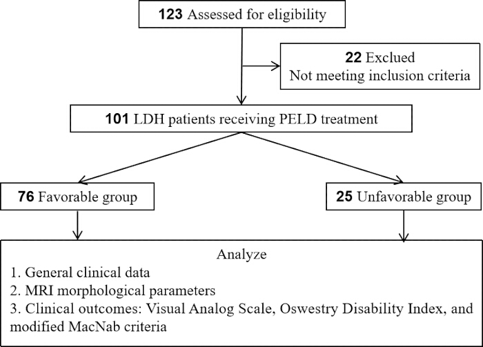
図1研究フローチャート 患者の選択およびグループ分けのフローチャート。評価対象となった123名の患者のうち、PELDを施行した単一レベルの腰椎間板ヘルニア(LDH)患者101名が組み入れられ、1年後の修正MacNab基準に基づき、良好群(n = 76)と不良群(n = 25)に分けられた。分析枠組みには、一般的な臨床データ、MRIパラメータ、および臨床成績(VAS、ODI、MacNab)が含まれた。 こちらの図の拡大版を表示するには、ここをクリックしてください。
組み入れ基準
年齢が18歳から75歳であること。臨床症状、身体診察、およびMRI/CT所見に基づき、L3/4、L4/5、またはL5/S1の単一レベルのLDHと診断されていること。神経根圧迫の明確な症状および徴候があり、少なくとも6週間の系統的な保存的治療(安静、薬物療法、理学療法などを含む)に反応しなかったこと。PELD手術を受けたこと。術後フォローアップ期間が1年以上であり、臨床データが完備していること。
除外基準
多レベルのLDHまたは腰椎手術の既往歴。腰椎すべり症、腰部脊柱管狭窄症、脊髄腫瘍、脊髄感染症、または脊髄骨折の併発。緊急手術を必要とする馬尾症候群の存在。正確な測定を妨げる術前MRI画像品質の不良。手術不適応となる重篤な併存疾患(例:心不全、肺不全、肝不全、または腎不全)。追跡不能または臨床データの不備。
手術手順
すべての手術は、同一の経験豊富な脊椎外科医チームによって実施されました。手術アプローチ(経椎間孔アプローチ:TF、または椎間板間アプローチ:IL)は、ヘルニアの形態と解剖学的制約の総合的な評価に基づいて決定されました。具体的には、椎間孔内、椎間孔外、または椎弓根レベルより下方に位置する中央型/傍中央型ヘルニアに対しては、TFアプローチを優先的に採用しました。逆に、高度に脱出した断片や分離片、特にL5椎弓根よりも頭側に脱出したもの、あるいは腸骨稜が高い患者に対しては、Choiら16によって以前に記述された方法に従い、ILアプローチを選択しました。この分化させた選択戦略により、手術経路を標的病変に最適に合わせることができました。
処置は局所麻酔下で、静脈内鎮静の有無にかかわらず、患者を腹臥位にして行った。標的となる椎間板腔はCアーム透視下で特定した。カンビンの三角または椎間孔間隙を通じて穿刺針を脊柱管内に進め、続いてガイドワイヤーを挿入し、段階的に拡張してワーキングチャンネルを構築した。ワーキング内視鏡(直径6.9 mm、視野角30°)を挿入し、直接視認下で脱出した髄核組織を除去して神経根の減圧を行った。高周波プローブを用いて環状縫合および止血を行った。その後、ワーキングチャンネルを抜去し、皮膚切開部を縫合した。
術前のDHIが25%以下であるか、またはMRIで重度の椎間孔狭窄を呈する症例では、Kambin's working zoneを拡大してチャンネル挿入を容易にするため、担当外科医の判断によりリーマーまたはハイスピードドリルを用いた追加の椎間孔形成術が実施されました。しかしながら、本研究はレトロスペクティブな設計であるため、この椎間孔形成術の実施は厳格に標準化されておらず、手術記録に体系的に記載もされていなかったため、本コホートにおける臨床成績への具体的な影響を定量的に分析することは不可能です。
観察指標
本研究では、T0を術前、T1を術後1週間、T2を術後3か月、T3を術後1年と定義した。
一般的臨床データ
年齢、性別、ボディマス指数(BMI)、罹患期間、喫煙歴、糖尿病の既往歴、高血圧の既往歴、手術レベル、および手術アプローチを含む患者データを収集した。
MRI形態学的パラメータの測定
すべての患者に対し、矢状断T1強調像、T2強調像、および軸位断T2強調像を含む術前腰椎MRI検査を実施した。2名の経験豊富な放射線科医が二重盲検法を用いて独立してパラメータを測定し、その平均値を解析に使用した。Pfirrmannグレード:椎間板の信号強度、構造、高さ、および髄核と線維輪の区別に基づき、矢状断T2強調像で評価し、1〜5のグレードに分類した。11Modic変化:椎体終板および隣接する骨髄の信号変化として評価し、「なし」、「タイプI(T1強調像で低信号、T2強調像で高信号)」、「タイプII(T1強調像で高信号、T2強調像で等信号または軽度高信号)」、または「タイプIII(T1強調像とT2強調像の両方で低信号)」に分類した。12・ヘルニアのタイプ:軸位断T2強調像に基づき、膨隆(bulging)、突出(protrusion)、脱出(extrusion)、または分離(sequestration)に分類する17・椎間板脱出部位:中心型、傍中心型、椎間孔型、または極外側のいずれかに分類される4脱出範囲:矢状断T2強調画像において、脱出した断片の上端または下端からディスクレベルまでの垂直距離として測定し、Leeらによる分類に従って分類した。18 遊走なし(グレード0)、低グレードの遊走(<椎間板高の50%未満(グレード1~2)、または高度脱出(椎間板高の50%以上、グレード3~4)。脊柱管占有率:最大脱出レベルの軸位像T2強調像(T2WI)にて、(脱出部の前後径 / 脊柱管の前後径)として算出。 × 100%19,20DHI:矢状断T2強調画像(T2WI)を用いて、[(前方椎間板高 + 後方椎間板高) / 2] / [(上位椎体的前後径 + 下位椎体的前後径) / 2] として測定する。 × 100%21椎間孔面積:ImageJソフトウェアを用い、椎間孔が最大となるレベルの軸位断T2強調画像(T2WI)上で手動で描出および測定した。22,23最大ヘルニア面積:最大ヘルニア部位における軸位断T2強調像(T2WI)上のヘルニア脱出片の断面積として測定した。24.
臨床転帰の評価
疼痛評価:疼痛強度は、腰痛(VAS-Back)および下肢痛(VAS-Leg)に対する視覚的アナログスケール(VAS)を用いて評価し、スコアは0(痛みなし)から10(想像しうる最悪の痛み)の範囲とした25,26。機能的障害:腰椎機能は、Oswestry Disability Index(ODI)を用いて評価した27,28。修正MacNab基準:1年後のフォローアップ時に評価を行い、転帰をexcellent(症状の完全消失、通常の活動への復帰)、good(時折痛みがあるが、仕事や日常生活に支障はない)、fair(症状は改善したが活動に制限あり)、またはpoor(改善なし、または症状が悪化)に分類した29,30。「excellent」および「good」の転帰を良好な予後と定義し、「fair」および「poor」の転帰を不良な予後と定義した。修正MacNab基準を主要評価項目として選択した理由は、疼痛緩和、機能回復、および日常生活への復帰を統合した、外科的手術の成功に関する包括的かつ患者中心の評価を提供できるためであり、これらの領域は単一の連続尺度よりも臨床的な意思決定において総体的に重要であるためである。さらに、MacNab分類はPELDの文献において広く利用され検証されており29,30、先行研究との直接的な比較を容易にする。
サンプルサイズの推定
サンプルサイズの推定にはG*Powerソフトウェア31を使用した。本研究は回顧的観察研究であるため、サンプルサイズは研究期間中に選択基準および除外基準を満たした患者の総数に基づいて決定し、その妥当性は事後パワー分析によって検証した。先行する関連研究8に基づき、PELD後の不良予後の発生率を約25%と想定し、椎間板脱出/遊離を主要な曝露因子として、両側α = 0.05、パワー(1-β)80%の場合、最低89名の参加者が必要であった。最終的に、統計的要件を満たす計101名の患者が本研究に含まれた。
統計解析
統計解析にはSPSSを使用した。連続変数の正規性はShapiro-Wilk検定により評価した。正規分布に従うデータは平均値 ± SDとして提示し、独立標本t検定および対応のあるt検定を用いて比較した。非正規分布のデータは中央値(Q1, Q3)として示し、Mann-Whitney U検定により解析した。カテゴリ変数についてはカイ二乗検定を用いて比較した。また、ピアソン相関分析および多変量ロジスティック回帰分析を適用した。すべての検定は両側検定とし、 P < 0.05を統計的に有意とみなした。
多変量ロジスティック回帰分析では、従属変数を良好な予後=1、不良な予後=0としてコード化した結果、陽性イベント数は76件であった。最終モデルに組み込む共変量の数は、EPV原則に従わなければならない。32MRIパラメータ間の多重共線性(マルチコリニアリティ)を評価するために分散拡大係数(VIF)を用い、VIF値を <5は許容範囲内であると考えられる。
結果
患者に関する一般情報
本研究には合計101名の患者が含まれた。表1に示すように、予後不良群の患者は有意に高齢であり(P < 0.05)、罹患期間が長かった(P < 0.05)。性別、BMI、喫煙歴、併存疾患、手術レベル、または手術アプローチに関しては、2群間に統計的な有意差は認められなかった(P > 0.05)。
| 変数 | 良好群 (n = 76) | 不利群 (n = 25) | 翻訳対象のテキストが提供されていません。翻訳する英文をご提示ください。χ²/Z | P |
| 年齢(歳) | 45.2 ± 11.8 | 51.6 ± 12.9 | 2.322 | 0.022 |
| オス | 42 (55.3) | 16 (64.0) | 0.587 | 0.444 |
| BMI (kg/m²)2) | 24.4 ± 3.0 | 25.4 ± 3.6 | 1.531 | 0.129 |
| 罹患期間(月) | 9 (8, 13) | 15 (12, 17) | 4.174 | <0.001 |
| 喫煙歴 | 18 (23.7) | 9 (36.0) | 1.457 | 0.227 |
| 糖尿病の歴史 | 8 (10.5) | 5 (20.0) | 0.779 | 0.337 |
| 高血圧の歴史 | 12 (15.8) | 6 (24.0) | 0.396 | 0.529 |
| 術中セグメント | ||||
| L3/4 | 11 (14.5) | 1 (4.0) | ||
| L4/5 | 40 (52.6) | 16 (64.0) | ||
| L5/S1 | 25 (32.9) | 8 (32.0) | 2.18 | 0.336 |
| 手術アプローチ | ||||
| 経椎間孔アプローチ | 59 (77.6) | 19 (76.0) | ||
| 層間アプローチ | 17 (22.4) | 6 (24.0) | 0.029 | 0.866 |
表 1:ベースライン特性[平均±SD, n (%), M(Q1, Q3)]。予後群別に層別化したベースライン特性。データは平均 ± SD、n (%)、または中央値 (Q1, Q3) で示す。Unfavorable群は有意に高齢であり(P = 0.02)、罹患期間がより長かった(P < 0.001)。その他の変数では、群間に有意な差は認められなかった。BMI:ボディマス指数。
術前MRI形態学的パラメータの比較
101名の患者の術前MRI形態学的パラメータを表2に示す。Modic変化の割合およびヘルニア型に関して、2群間に有意差(P < 0.05)が認められた。Favorable群のDHIおよび椎間孔面積は、Unfavorable群よりも有意に高かった(P < 0.05)。一方、Favorable群の脊柱管占有率および最大ヘルニア面積は、Unfavorable群よりも有意に低かった(P < 0.05)。Pfirrmannグレード、ヘルニア部位、および移行グレードについては、統計的な有意差(P > 0.05)は認められなかった。これらの結果は、上記のパラメータがPELDの術後予後の潜在的な予測指標となり得ることを示唆している。
| 良好群 (n = 76) | 不良群 (n = 25) | t/χ² | P | |
| Pfirrmann分類 | ||||
| I-II | 28(36.8) | 5(20.0) | ||
| III | 32(42.1) | 10(40.0) | ||
| IV-V | 16(21.1) | 10(40.0) | 4.27 | 0.18 |
| Modic変化 | ||||
| なし | 48(63.2) | 8(32.0) | ||
| タイプ I | 12(15.8) | 3(12.0) | ||
| タイプ II | 14(18.4) | 13(52.0) | ||
| タイプ III | 2(2.6) | 1(4.0) | 1.529 | 0.09 |
| 脱出型 | ||||
| 膨隆 | 8(10.5) | 1(4.0) | ||
| 突出 | 4(57.9) | 7(28.0) | ||
| 脱出 | 20(26.3) | 12(48.0) | ||
| 分離 | 4(5.3) | 5(20.0) | 1.605 | 0.09 |
| 脱出部位 | ||||
| 中心性 | 12(15.8) | 2(8.0) | ||
| 中心傍 | 48(63.2) | 18(72.0) | ||
| 椎間孔内 | 12(15.8) | 4(16.0) | ||
| 椎間孔外 | 4(5.3) | 1(4.0) | 1.1 | 0.75 |
| 遊離度 | ||||
| 遊離なし (グレード 0) | 25(32.9) | 6(24.0) | ||
| 低度遊離 (グレード 1–2) | 32(42.1) | 10(40.0) | ||
| 高度遊離 (グレード 3–4) | 19(25.0) | 9(36.0) | 1.326 | 0.515 |
| 脊柱管占有率 (%) | 35.6 ± 6.9 | 47.2 ± 8.4 | 6.892 | <0.01 |
| 脊柱管占有率 > 40% | 30(39.5) | 14(56.0) | ||
| DHI (%) | 28.5 ± 5.2 | 24.5 ± 4.8 | 3.392 | 0.01 |
| DHI > 25% | 58(76.3) | 1(44.0) | ||
| 椎間孔面積 (cm²) | 1.1 ± 0.3 | 0.8 ± 0.3 | 4.029 | <0.01 |
| 椎間孔面積 > 1 cm² | 49(64.5) | 13(52.0) | ||
| 最大脱出面積 (mm²) | 59.8 ± 1.9 | 86.0 ± 15.7 | 8.764 | <0.01 |
| 最大脱出面積 > 75 mm² | 21(27.6) | 13(52.0) |
表 2: 術前 MRI 形態学的パラメータの比較 [平均±標準偏差, n (%)]。群間での術前 MRI パラメータの比較。データは平均 ± 標準偏差または n (%) で示す。Modic 変化、ヘルニアのタイプ、移行グレード、脊柱管占有率、DHI、椎間孔面積、およびヘルニア面積において有意差が認められたが(すべて P < 0.05)、Pfirrmann グレードおよびヘルニアの部位には有意差は認められなかった。DHI:椎間板高指数。
臨床転帰指標の変化
表3に、修正MacNab基準を用いて1年間のフォローアップ評価を行った患者101例の臨床転帰を示す。その結果、51例(50.5%)が極めて良好(excellent)、25例(24.8%)が良好(good)であり、良好群は合計76例(75.2%)であった。また、16例(15.8%)で普通(fair)、9例(8.9%)で不良(unfavorable)な予後が観察され、不良群は25例(24.8%)であった。以上の結果は、全体として良好な臨床的有効性を示している。
| アウトカムグレード | 症例数 (n) | パーセンテージ (%) | 臨床アウトカムの定義 |
| 極めて優れた | 51 | 50.50% | 好調群 |
| 良好 | 25 | 24.80% | 良好群 |
| 公正な | 16 | 15.80% | 不利群 |
| 不十分な | 9 | 8.90% | 不適切群 |
表3:修正MacNab基準に基づく術後1年目の臨床的有効性評価 修正MacNab基準による術後1年目の転帰。良好(Excellent)または良好(Good)な転帰を良好群(Favorable group)(75.2%)とし、可(Fair)または不良(Poor)な転帰を不良群(Unfavorable group)(24.8%)とした。
表4に術後の臨床成績の動的な変化を示す。術前において、腰痛のVAS、下肢痛のVAS、およびODIスコアのいずれにおいても、2群間に統計的な有意差は認められなかった(P > 0.05)。術後のすべての時点において、両群のスコアは術前値と比較して有意な改善を示し(群内 P < 0.01)、PELDの全体的な有効性が示された。しかし、群間比較では、良好群が不良群と比較して、術後のすべての時点においてVAS(腰部および下肢)およびODIスコアが有意に良好であった(P < 0.001)。さらに、術後1年時点におけるODIの改善度は、不良群よりも良好群で有意に大きかった。これらの知見は、手術自体が有意な改善をもたらす一方で、術前MRIの形態学的特徴によって層別化された患者は、臨床的な回復軌跡において持続的な差異を示すことを証明しており、長期的な転帰に対する術前MRI特性の予測価値をさらに裏付けるものである。
| 時間 | 良好群 (n = 76) | 不良群 (n = 25) | P (群内) | P (群間) | |
| VAS-腰部 | T0 | 6(6, 7) | 7(6, 7) | - | 0.212 |
| T1 | 3(3, 4) | 4(4, 5) | <0.01 | <0.01 | |
| T2 | 2(1, 2) | 3(2, 4) | <0.01 | <0.01 | |
| T3 | 1(1, 1) | 2(2, 3) | <0.01 | <0.001 | |
| VAS-下肢 | T0 | 8(7, 9) | 8(7, 8) | - | 0.486 |
| T1 | 3(2, 3) | 5(4, 5) | <0.001 | <0.01 | |
| T2 | 2(1, 2) | 3(3, 4) | <0.01 | <0.01 | |
| T3 | 1(1, 1) | 3(2, 3) | <0.01 | <0.01 | |
| ODI(%) | T0 | 68.4 ± 9.6 | 70.2 ± 10.4 | - | 0.408 |
| T1 | 30.1 ± 7.4 | 42.4 ± 8.9 | <0.01 | <0.01 | |
| T2 | 18.7 ± 5.5 | 29.5 ± 6.4 | <0.01 | <0.001 | |
| T3 | 10.1 ± 4.0 | 21.8 ± 5.5 | <0.01 | <0.01 | |
| ODI変化量 | T3-T0 | 58.2 ± 10.9 | 48.5 ± 1.5 | - | <0.001 |
表 4:術前後の臨床転帰指標の変化 [平均 ± SD, M(Q1, Q3)]。VASおよびODIスコアの経時的変化。データは中央値 (Q1, Q3) または平均 ± SDで示す。両群ともに術後に有意に改善したが(群内 P < 0.01)、Favorable群はすべての時点において一貫して良好なスコアを示した(群間 P < 0.01)。群内比較にはWilcoxon符号付順位検定を用い、群間比較にはMann-Whitney U検定を用いた。
MRI形態学的パラメータと臨床転帰の相関分析 表 5 ピアソン相関分析の結果を示しており、脊柱管占有率および最大ヘルニア面積が、術後の腰痛に対するVAS、下肢痛に対するVAS、およびODIスコアと有意な正の相関があることが明らかになった(r: 0.375–0.538)。 P < 0.01)。これは、脊柱管狭窄の程度が重く、椎間板脱出量が多いほど、残存症状が顕著になり、機能回復が不良であることを示している。対照的に、DHIおよび椎間孔面積は、すべての臨床的アウトカム尺度と有意な負の相関を示した(r: -0.637~-0.240、 P < 0.05)。その中でも、椎間孔面積とODIスコアの相関は有意に近い値を示した(P=0.053)。これらの結果は、椎間板高の維持と十分な椎間孔スペースの確保が、術後の神経除圧と症状の改善を促進することを示唆している。これらの相関関係に関する知見は、MRIパラメータと予後との関連性の強さをさらに定量化するものである。DHIはVAS-Backと最も強い相関を示し(r = -0.637)、術後の機能回復に対する特筆すべき予測価値を有すること、および術前画像診断における定量的な根拠を提供することが明らかとなった。
| VAS-腰痛 | VAS-下肢痛 | ODI | ||||
| MRIパラメータ | r | P | r | P | r | P |
| 脊柱管占有率 | 0.501 | <0.001 | 0.52 | <0.01 | 0.404 | <0.01 |
| DHI | -0.637 | <0.001 | -0.608 | <0.01 | -0.323 | 0.01 |
| 椎間孔面積 | -0.24 | 0.016 | -0.298 | 0.02 | -0.193 | 0.053 |
| 最大ヘルニア面積 | 0.475 | <0.01 | 0.538 | <0.01 | 0.375 | <0.01 |
表5:術後1年におけるMRI形態学的パラメータと臨床転帰指標とのピアソン相関分析 MRIパラメータと1年後の転帰とのピアソン相関。脊柱管占有率およびヘルニア面積はVAS/ODIスコアと正の相関を示し(r = 0.457–0.653)、一方でDHIおよび神経孔面積は負の相関を示した(r = –0.627~–0.31)。すべて P < 0.01.
多変量ロジスティック回帰分析
術後1年時点での修正MacNab基準を従属変数(良好な予後 = 1、不良な予後 = 0)とし、MRI形態学的パラメータ(Modic変化、ヘルニアの形態、脱出範囲、脊柱管占有率、DHI、椎間孔面積、および最大ヘルニア面積)の単変量分析において P < 0.1であった変数を多変量ロジスティック回帰分析に組み込んだ。ヘルニアの形態と最大ヘルニア面積の間に共線性が認められ、また連続変数としての最大ヘルニア面積の方がより多くの情報を提供するため、ヘルニアの形態の代わりに最大ヘルニア面積をモデルに組み入れた。表6に示すように、多変量調整後、Modic type II変化(OR = 0.213, 95% CI: 0.062–0.728, P = 0.014)が不良な予後の独立したリスク因子として同定された一方、DHI > 25%(OR = 5.154, 95% CI: 1.62–16.376, P = 0.05)は良好な予後の独立した保護因子であった。表6より、すべての共変量のVIFが2未満であり、重大な共線性の問題がないことが示されている。
| オッズ比(OR)の95%信頼区間(CI) | ||||||||
| B | 標準誤差 | Wald | P | または | 低下 | 上部 | 分散拡大係数 | |
| Modic変化 type I | -0.542 | 0.853 | 0.404 | 0.525 | 0.581 | 0.109 | 3.095 | 1.082 |
| Modic変化タイプII | -1.547 | 0.627 | 6.082 | 0.014 | 0.213 | 0.062 | 0.728 | |
| Modic変化タイプIII | -0.802 | 1.452 | 0.305 | 0.581 | 0.449 | 0.026 | 7.72 | |
| 低グレード移動 | -0.539 | 0.707 | 0.583 | 0.445 | 0.583 | 0.146 | 2.329 | 1.073 |
| 高グレード遊走 | -0.953 | 0.738 | 1.669 | 0.196 | 0.386 | 0.091 | 1.637 | |
| 脊柱管占有率 > 40% | -0.813 | 0.563 | 2.084 | 0.149 | 0.444 | 0.147 | 1.337 | 1.016 |
| 直接毛髪移植(DHI) > 25% | 1.64 | 0.59 | 7.729 | 0.005 | 5.154 | 1.622 | 16.376 | 1.026 |
| 神経孔領域 > 1 cm² | 0.84 | 0.582 | 2.084 | 0.149 | 2.316 | 0.741 | 7.245 | 1.047 |
| 最大ヘルニア面積 > 75 mm² | -1.153 | 0.614 | 3.526 | 0.06 | 0.316 | 0.095 | 1.052 | 1.154 |
| 定数 | 1.596 | 0.782 | 4.173 | 0.041 | 4.936 | - | - | |
表 6: 術後臨床転帰の多変量ロジスティック回帰分析。良好な予後の予測因子に関する多変量ロジスティック回帰。Modic type IIは独立したリスク因子であり(OR = 0.213, P = 0.014)、一方でDHI > 25%は保護因子であった(OR = 5.154, P = 0.05)。VIF < 2であり、共線性は認められなかった(Hosmer-Lemeshow P = 0.933)。Hosmer-Lemeshow検定 χ2=3.017, P=0.93, Nagelkerke R2 = 0.353。
異なるタイプのModic変化を有する患者の予後解析
表7は、Modic変化のタイプ別に層別化した患者の予後結果を示している。VASスコアが4–6は中程度の疼痛を、ODIスコアが21%–40%は中程度の機能障害を示す。Modic type IIの変化を有する患者は、良好な予後の割合が最も低く(51.9%)、機能障害の割合が比較的高い(3.3%)ことが示された。
| モディック変化 | n | 良好な予後 | 中等度の背部痛 | 中等度の下肢痛 | 中等度障害 |
| 提供されたソーステキストがありません。翻訳が必要なテキストをご提示ください。 | 56 | 48(85.7) | 2(3.6) | 1(1.8) | 5(8.9) |
| I | 15 | 12(80.0) | 0 | 0 | 2(13.3) |
| II | 27 | 14(51.9) | 2(7.4) | 2(7.4) | 8(33.3) |
| III | 3 | 2(66.7) | 0 | 0 | 1(33.3) |
| χ²/F | - | 11.529 | 1.608 | 2.668 | 6.617 |
| P | - | 0.009 | 0.658 | 0.446 | 0.085 |
表 7:Modic変化のタイプ別臨床アウトカムの比較 [n (%)]。Modic変化のタイプ別の臨床アウトカム。データは n (%) で表示。Modic II 群で良好な予後の割合が最も低く (51.9%)、中等度の障害の割合が最も高かった (32.0%, P = 0.048)。
データ可用性:
本研究の結果を裏付けるすべてのデータは、本論文およびその補足ファイル 1に含まれています。
補足ファイル1:生データ こちらをクリックしてファイルをダウンロードしてください。
ディスカッション
This retrospective study analyzed 101 patients with single-level LDH who underwent PELD, aiming to investigate the correlation between preoperative MRI morphological features, clinical factors, and 1-year postoperative clinical outcomes. The findings demonstrated that PELD achieved favorable overall clinical efficacy for LDH, with an excellent-to-good rate (favorable group) of 75.2% at the 1-year follow-up. However, 24.8% of patients experienced unfavorable outcomes (unfavorable group). This result is highly consistent with recent multicenter large-sample studies reporting residual symptoms or recurrence rates ranging from 10% to 25% following PELD33,34. By comparing baseline data and morphological features between the two groups, this study confirmed that MRI morphological features, including Modic type II changes, are closely associated with unfavorable clinical outcomes.
To contextualize our findings within the existing body of literature, we summarized recent large-scale clinical series that investigated MRI predictors for outcomes after transforaminal PELD (Table 8)33,34,35. Compared to previous studies that predominantly focused on single parameters such as herniation type or Modic changes in isolation, our study provides a multidimensional morphological evaluation integrating qualitative (Modic subtypes, herniation grade) and quantitative metrics (DHI, foraminal area, and herniation cross-sectional area). This comprehensive approach allows for a more nuanced risk stratification, particularly highlighting the independent protective role of DHI > 25%, which has been underreported in earlier series. Therefore, the main contribution of this study is not only to identify individual MRI predictive factors, but also to establish a systematic and multidimensional preoperative evaluation framework that can accurately predict results and provide actionable guidance for tailoring surgical strategies based on the patient's specific imaging phenotype, including deciding to undergo intervertebral foramen shaping surgery or considering alternative methods.
| Author (Year) | N | MRI Parameters Evaluated | Key Prognostic Findings | Limitations |
| Shen et al. (2019) | 241 | Herniation type, Modic | Extrusion & Modic I as risk factors | No quantitative foraminal/DHI analysis |
| Li et al. (2021) | 582 | Canal occupation ratio | Occupation > 50% predicted poor outcome | Did not analyze Modic subtypes separately |
| Zhu et al. (2022) | 120 | Modic type II | Modic II linked to higher residual back pain | Focused only on Modic changes |
| Present study | 101 | Modic type, DHI, Foraminal area, Herniation area | Modic II (OR=0.213) risk; DHI>25% (OR=5.154) protective | Single-center, no post-op MRI, limited 1-year F/U |
Table 8: Summary of recent clinical series reporting MRI predictors for outcomes after transforaminal PELD. Comparison with recent PELD outcome studies. Unlike prior single-parameter studies, the present study integrates qualitative and quantitative MRI metrics, highlighting DHI > 25% as an independent protective factor. Shen et al. (2019)33; Li et al. (2021)34; Zhu et al. (2022)35.
Before further exploring the predictive value of individual MRI parameters, it is important to validate the clinical relevance of the outcome grouping itself. In our cohort, the MacNab-based classification demonstrated strong consistency with VAS and ODI changes. The favorable group (excellent/good MacNab) showed a mean ODI improvement from 68.4% preoperatively to 10.1% at 1 year (a reduction of 58.3 percentage points), whereas the unfavorable group improved from 70.2% to 21.8% (a reduction of 48.4 percentage points). These improvements substantially exceeded the established minimal clinically important difference (MCID) for ODI in lumbar disc surgery, generally accepted as a 10–15 percentage point reduction36. The marked between-group difference in ODI improvement (58.3% vs. 48.4%) further confirms that the modified MacNab criteria effectively distinguish patients with clinically meaningful functional recovery from those with suboptimal outcomes, reinforcing the validity of this grouping for subsequent prognostic analysis.
Univariate analysis revealed a significant association between disc extrusion/sequestration, maximum herniation area, and unfavorable postoperative outcomes, along with a strong positive correlation between maximum herniation area and postoperative pain and disability scores. These findings indicate that morphological features of disc herniation are closely related to functional recovery following surgery for LDH. From a pathophysiological standpoint, while a sequestered fragment may occasionally undergo spontaneous resorption37, the persistence of suboptimal outcomes following PELD is often rooted in structural pathologies extending beyond the herniated nucleus itself. Annular tears, cartilaginous endplate injuries, posterior ring apophyseal fractures (PRAF), and calcified disc components, all of which may be overlooked or inadequately addressed during standard endoscopic procedures, can impair disc healing, create non-resorbable fragments, and lead to incomplete decompression38,39. Furthermore, it is critical to recognize that the severity of clinical symptoms does not strictly correlate with herniation size40; rather, symptom severity and postoperative recovery are modulated by the local inflammatory response triggered by exposure of the nucleus pulposus to the epidural space. While some studies have identified extrusion/sequestration and fragment weight as predictors of residual symptoms and slower recovery41,42, others have found no direct correlation between herniation size and symptom severity39, reflecting the complex interplay of mechanical compression, anatomical disruption, and chemical radiculopathy. Nevertheless, as an objective baseline parameter, maximum herniation area retains predictive value for postoperative prognosis.
In this study, Modic type II changes were identified as an independent risk factor for unfavorable outcomes (OR = 0.213, P = 0.014), consistent with previous research35,43. While our findings establish a clear statistical association, the underlying mechanisms remain incompletely understood. Based on prior literature, several hypotheses have been proposed. First, Rajasekaran et al.38 demonstrated that endplate damage is the most common anatomical failure mode in lumbar disc herniation, impairing disc nutrition and predisposing to irreversible degeneration. Second, Kjaer et al.43 reported that Modic changes are closely associated with chronic low back pain, suggesting that endplate-derived pain may persist even after adequate nerve root decompression. Third, a meta-analysis by Luo et al.44 (n = 5,446) reported that Modic changes increased the risk of recurrence after PELD by 2.48-fold (OR = 2.48, 95% CI: 1.54–3.99). Additionally, Zhu et al.35 specifically found that patients with Modic type II changes had significantly higher rates of persistent low back pain and recurrence following PELD compared to those without. In summary, our study confirms the prognostic relevance of Modic type II changes as an imaging biomarker, but the mechanistic pathways linking these changes to unfavorable prognosis are multifactorial and not fully elucidated. Future prospective studies incorporating histologic or biochemical analyses are needed to clarify the causal mechanisms.
DHI and foraminal area were significantly reduced in the unfavorable group and showed negative correlations with postoperative outcomes, with DHI demonstrating the strongest correlation with low back pain VAS (r = -0.637). Disc height loss triggers a cascade of anatomical changes, such as facet subluxation, ligamentum flavum buckling, and secondary foraminal stenosis, thereby reducing Kambin's triangle and increasing surgical difficulty45,46. Moreover, the resulting stenosis is osseous or ligamentous in nature; PELD primarily targets soft disc tissue, and without adequate foraminoplasty, removing the herniated fragment alone cannot fully address the stenotic volume47,48. These findings support the routine use of power instruments for foraminoplasty or consideration of alternative approaches when DHI is severely reduced or foraminal stenosis is present.
This study inevitably has certain limitations. First, as a single-center retrospective observational study, inherent selection bias may exist, and the findings may not be representative of broader population characteristics. Second, the follow-up duration was limited to one year. While this captures short- to medium-term clinical outcomes, it is insufficient for evaluating long-term recurrence rates and adjacent segment degeneration, necessitating future studies with 5- to 10-year follow-up periods. Third, all MRI morphological parameters were manually measured on two-dimensional images by two radiologists, with averaged values used for analysis. Despite the double-blind design, subjective measurement errors and variations in image slice selection may have influenced the results. Fourth, we did not routinely perform postoperative MRI to verify the adequacy of decompression. Thus, we cannot definitively exclude the possibility that incomplete resection of occult calcified fragments, missed PRAF, or untreated secondary stenosis contributed to the unfavorable outcomes. Fifth, although two radiologists independently measured all MRI parameters, we did not formally calculate interobserver reliability coefficients (ICC or kappa). This represents a methodological limitation, as reproducibility of imaging measurements cannot be quantified. Sixth, this study did not incorporate potentially confounding factors such as individual history of heavy occupational labor and postoperative rehabilitation adherence into the multivariate model, as these variables are difficult to quantify, which may have had some impact on the findings. Seventh, this study has several additional methodological constraints that should be considered. The single-center retrospective design inherently carries selection bias, and the relatively modest sample size (101 patients) limits the generalizability of our findings. Moreover, the absence of an external validation cohort precludes assessment of the reproducibility and transportability of our proposed multidimensional MRI evaluation model. Finally, there is the issue of selection bias related to surgical indications. Because the same surgical team believes that all patients are suitable candidates for PELD, this may introduce confusion that cannot be fully adjusted in our analysis.
In summary, this study confirms that preoperative MRI features are significantly associated with 1-year clinical outcomes, yet these findings should not be interpreted deterministically. Instead, they serve as a roadmap for surgical decision-making. For patients presenting with Modic type II changes, DHI ≤ 25%, or large calcified/sequestered fragments, the surgeon must recognize the inherent technical limitations of standard transforaminal PELD. In this high-risk phenotype, achieving the endpoint of decompression, which is the ultimate determinant of surgical success, may require assisted foraminal reconstruction, switching to interlayer or UBE approaches, or even open microdissection. Therefore, preoperative MRI empowers the surgeon not merely to predict outcomes, but to critically assess whether the chosen approach can adequately address both the disc fragment and the concomitant secondary stenotic pathology.
Preoperative MRI morphological features, particularly Modic type II changes (independent risk factor) and DHI > 25% (protective factor), are closely linked to 1-year outcomes after PELD. However, the clinical value of these imaging biomarkers lies in their ability to guide proactive and adaptable surgical planning rather than serving as static prognosticators. The pivotal lesson for spine surgeons is to respect the limitations of their chosen approach and to prioritize the achievement of objective decompression endpoints. If standard PELD is unlikely to achieve these endpoints based on preoperative MRI traits (e.g., severe foraminal stenosis, calcified discs), alternative strategies such as UBE or fusion should be considered. Future prospective studies with mandatory post-operative imaging are warranted to validate whether adapting surgical aggressiveness based on these MRI features can effectively convert a predicted “unfavorable” prognosis into a favorable one.
開示事項
著者らは、金銭的な利益相反がないことを言明します。
材料
| 名前 | 会社 | カタログ番号 | コメント |
|---|---|---|---|
| C-arm Fluoroscope | Philips | 718133 | Intraoperative localization of target disc space |
| G*Power | Heinrich-Heine-Universität Düsseldorf | Version 3.1.9.7 | Performed post-hoc power analysis for sample size. |
| ImageJ Software | National Institutes of Health (NIH) | Version 1.54r | Measure foraminal area & herniation area |
| MRI Scanner (3.0T Siemens Skyra) | Siemens Healthineers, Erlangen, Germany | syngo MR E11 | Preoperative lumbar MRI: sagittal T1WI, T2WI, and axial T2WI sequences using a dedicated spine coil. |
| Radiofrequency System | Elliquence | IEC6-SU170 | Intraoperative annuloplasty and hemostasis |
| Statistical Package for the Social Sciences (SPSS) | IBM | Version 25.0 | Used for all statistical analyses. |
参考文献
- Hoy D, et al. The global burden of low back pain: estimates from the Global Burden of Disease 2010 study. Ann Rheum Dis. 2014;73(6):968-974.
- Konstantinou K, Dunn KM. Sciatica: review of epidemiological studies and prevalence estimates. Spine (Phila Pa 1976). 2008;33(22):2464-2472.
- Lan W, Cui H, Zhou D, Xiao X, Xiong H. Advances in minimally invasive surgical techniques for lumbar disc herniation: a comprehensive review. Front Surg. 2025;12.
- Yeung AT, Tsou PM. Posterolateral endoscopic excision for lumbar disc herniation: surgical technique, outcome, and complications in 307 consecutive cases. Spine (Phila Pa 1976). 2002;27(7):722-731.
- Ruetten S, Komp M, Merk H, Godolias G. Full-endoscopic interlaminar and transforaminal lumbar discectomy versus conventional microsurgical technique: a prospective, randomized, controlled study. Spine (Phila Pa 1976). 2008;33(9):931-939.
- Pan M, et al. Percutaneous endoscopic lumbar discectomy: indications and complications. Pain Physician. 2020;23(1):49-56.
- Choi KC, Kim JS, Park CK. Percutaneous endoscopic lumbar discectomy as an alternative to open lumbar microdiscectomy for large lumbar disc herniation. Pain Physician. 2016;19(2)-E300.
- Yao Y, et al. Risk factors for recurrent herniation after percutaneous endoscopic lumbar discectomy. World Neurosurg. 2017;100:1-6.
- Yin S, et al. Prevalence of recurrent herniation following percutaneous endoscopic lumbar discectomy: a meta-analysis. Pain Physician. 2018;21(4):337-350.
- Krishnan A, et al. End-points of decompression of in lumbar transforaminal endoscopic spine surgery: a narrative review of objective and subjective criteria to prevent failures. J Minim Invasive Spine Surg Tech. 2022;7(1):68-83.
- Pfirrmann CW, Metzdorf A, Zanetti M, Hodler J, Boos N. Magnetic resonance classification of lumbar intervertebral disc degeneration. Spine (Phila Pa 1976). 2001;26(17):1873-1878.
- Modic MT, Steinberg PM, Ross JS, Masaryk TJ, Carter JR. Degenerative disk disease: assessment of changes in vertebral body marrow with MR imaging. Radiology. 1988;166(1 Pt 1):193-199.
- Choi G, et al. Percutaneous endoscopic lumbar herniectomy for high-grade down-migrated L4-L5 disc through an L5-S1 interlaminar approach: a technical note. Minim Invasive Neurosurg. 2010;53(3):147-152.
- Guo Y, D X, Liu M, Liu D, Wang J. Robotic-assisted systems in minimally invasive neurosurgical techniques: a review. Neurosci Res Clin Pract. 2025;1(1):27-40.
- Wen B, Zhang G, Zhan C, Chen C, Yi H. The 2024 revision of the Declaration of Helsinki: a modern ethical framework for medical research. Postgrad Med J. 2025;101(1194):371-382.
- Choi KC, Kim JS, Lee DC, Park CK. Percutaneous endoscopic lumbar discectomy: minimally invasive technique for multiple episodes of lumbar disc herniation. BMC Musculoskelet Disord. 2017;18.
- Fardon DF, et al. Lumbar disc nomenclature: version 2.0: recommendations of the combined task forces of the North American Spine Society, the American Society of Spine Radiology and the American Society of Neuroradiology. Spine J. 2014;14(11):2525-2545.
- Lee S, et al. Percutaneous endoscopic lumbar discectomy for migrated disc herniation: classification of disc migration and surgical approaches. Eur Spine J. 2007;16(3):431-437.
- Kalidindi KKV, Sath S, Vishwakarma G, Chhabra HS. Magnetic resonance imaging findings in intervertebral disc herniation: comparison of canal compromise and canal size in patients with and without cauda equina syndrome. Surg Neurol Int. 2020;11:171.
- Korse NS, Kruit MC, Peul WC, Vleggeert-Lankamp CLA. Lumbar spinal canal MRI diameter is smaller in herniated disc cauda equina syndrome patients. PLoS One. 2017;12(10).
- Shiwen L, et al. MRI changes and clinical results of low energy semiconductor percutaneous laser disc decompression (LS-PLDD) for lumbar disc herniation in adolescents. Lasers Med Sci. 2025;40(1):481.
- Lee CK, Rauschning W, Glenn W. Lateral lumbar spinal canal stenosis: classification, pathologic anatomy and surgical decompression. Spine (Phila Pa 1976). 1988;13(3):313-320.
- Inufusa A, et al. Anatomic changes of the spinal canal and intervertebral foramen associated with flexion-extension movement. Spine (Phila Pa 1976). 1996;21(21):2412-2420.
- Yuan S, Tang Q, Wang X, Kong K. Significance of spinal canal and dural sac dimensions in predicting treatment of lumbar disc herniation. Acta Orthop Belg. 2014;80(4):575-581.
- Huskisson EC. Measurement of pain. Lancet. 1974;2(7889):1127-1131.
- Aun C, Lam YM, Collett B. Evaluation of the use of visual analogue scale in Chinese patients. Pain. 1986;25(2):215-221.
- Fairbank JC, Pynsent PB. The Oswestry Disability Index. Spine (Phila Pa 1976). 2000;25(22):2940-2952.
- Liu H, Tao H, Luo Z. Validation of the simplified Chinese version of the Oswestry Disability Index. Spine (Phila Pa 1976). 2009;34(11):1211-1216.
- Macnab I. Negative disc exploration: an analysis of the causes of nerve-root involvement in sixty-eight patients. J Bone Joint Surg Am. 1971;53(5):891-903.
- Gugliotta M, et al. Surgical versus conservative treatment for lumbar disc herniation: a prospective cohort study. BMJ Open. 2016;6(12).
- Kim N, Fischer AH, Dyring-Andersen B, Rosner B, Okoye GA. Research techniques made simple: choosing appropriate statistical methods for clinical research. J Invest Dermatol. 2017;137(10)-e178.
- Peduzzi P, Concato J, Kemper E, Holford TR, Feinstein AR. A simulation study of the number of events per variable in logistic regression analysis. J Clin Epidemiol. 1996;49(12):1373-1379.
- Shen Z, et al. Predictors for poor outcomes after percutaneous endoscopic lumbar discectomy: a retrospective study of 241 patients. World Neurosurg. 2019;126-e431.
- Li Y, et al. Adjuvant surgical decision-making system for lumbar intervertebral disc herniation after percutaneous endoscopic lumber discectomy: a retrospective nonlinear multiple logistic regression prediction model based on a large sample. Spine J. 2021;21(12):2035-2048.
- Zhu H, et al. Percutaneous endoscopic lumbar discectomy for lumbar disc herniation with Type II Modic changes. World Neurosurg. 2022;164-e149.
- Copay AG, et al. Minimum clinically important difference: current trends in the orthopaedic literature, Part II lower extremity—a systematic review. JBJS Rev. 2018;6.
- Krishnan P. Spontaneous complete absorption of a large prolapsed lumbar intervertebral disc with extrusion and cranial migration. J Neurosci Rural Pract. 2017;8(2):307-309.
- Rajasekaran S, Bajaj N, Tubaki V, Kanna RM, Shetty AP. ISSLS Prize winner: the anatomy of failure in lumbar disc herniation: an in vivo, multimodal, prospective study of 181 subjects. Spine (Phila Pa 1976). 2013;38(17):1491-1500.
- Dunsmuir RA, Nisar S, Cruickshank JA, Loughenbury PR. No correlation identified between the proportional size of a prolapsed intravertebral disc with disability or leg pain. Bone Joint J. 2022;104-B(6):715-720.
- Sriri H, et al. Postoperative chemical radiculitis mimicking recurrent disc herniation: a case report. Cureus. 2026;18(4).
- Yu Y, et al. Prognostic factors for residual symptoms following percutaneous endoscopic lumbar discectomy. Neurosurg Rev. 2024;47(1).
- Mariajoseph FP, et al. Relationship between herniated intervertebral disc fragment weight and pain in lumbar microdiscectomy patients. J Clin Neurosci. 2022;102:75-79.
- Kjaer P, Korsholm L, Bendix T, Sorensen JS, Leboeuf-Yde C. Modic changes and their associations with clinical findings. Eur Spine J. 2006;15(9):1312-1319.
- Luo M, et al. Risk factors for lumbar disc herniation recurrence after percutaneous endoscopic lumbar discectomy: a meta-analysis of 58 cohort studies. Neurosurg Rev. 2023;46(1):159.
- Yusof MI, Salim AA, Johari J, Rajagopal AR. Determination of the entry point for lower lumbar intradiscal procedure using transforaminal technique: cross-sectional study using magnetic resonance imaging. Spine Surg Relat Res. 2022;6(6):689-695.
- Sinha MB, Rachel John M, Abraham Poonuraparampil J, Prasad Sinha H. Dimensions of bony Kambin’s triangle pertaining to endoscopic procedure. Med J Armed Forces India. 2024;80-S134.
- Choi KC, et al. Advanced Techniques of Endoscopic Lumbar Spine Surgery. Springer Singapore; 2020:103-113.
- Nurmukhametov R, et al. Exploring pathways for pain relief in treatment and management of lumbar foraminal stenosis: a review of the literature. Brain Sci. 2024;14(8):740.