This manuscript describes a workflow to assess platelet-associated Tissue Factor (TF)-dependent thrombin generation in isolated intact human platelets using the calibrated automated thrombogram (CAT). By avoiding exogenous TF and integrating contact pathway inhibition, phospholipid normalization, and TF / Tissue Factor Pathway Inhibitor (TFPI)modulation, this approach enables a specific and physiologically relevant functional assessment of endogenous platelet TF.
方法論記事
Protocol for the Assessment of Endogenous Platelet-Associated Tissue Factor-Dependent Thrombin Generation in Human Platelets Using the Calibrated Automated Thrombogram
* These authors contributed equally
この記事について
サマリー
要約
Platelet-associated Tissue Factor (TF) is increasingly recognized as a functionally active determinant of platelet procoagulant activity, enabling platelets to initiate and sustain thrombin generation beyond their established role as a phospholipid surface for coagulation factor assembly. However, its specific functional assessment remains challenging, as conventional Calibrated Automated Thrombogram (CAT) assays rely on exogenous TF to trigger thrombin generation, thereby masking the endogenous contribution of platelet-associated TF.
Here, we describe a Calibrated Automated Thrombogram (CAT)-based workflow that enables the selective assessment of TF-dependent thrombin generation in isolated intact human platelets under conditions that preserve the contribution of endogenous platelet-associated TF. The protocol integrates key methodological steps, including inhibition of contact pathway activation through the use of microvesicle-free plasma pool containing a contact pathway inhibitor, normalization of the procoagulant membrane surface by exogenous phospholipids, and functional validation through neutralizing antibodies against TF and Tissue Factor Pathway Inhibitor (TFPI). The workflow also addresses key pre-analytical variables related to blood collection and platelet-rich plasma (PRP) preparation. Under these controlled conditions, thrombin generation kinetics -particularly LagTime- reflect the functional activity of platelet-associated TF, as demonstrated by their modulation upon TF and TFPI inhibition and their association with TFpos-platelets levels measured by flow cytometry. By avoiding the addition of exogenous TF, this approach overcomes key methodological limitations that have contributed to the ongoing controversy surrounding platelet-associated TF, providing a functional readout that complements antigen-based measurements. This protocol therefore represents a robust and reproducible framework for investigating platelet-associated TF in both mechanistic and clinical studies.
概要
For decades, platelets have been regarded as passive contributors to coagulation, primarily providing a negatively charged phospholipid surface that supports the assembly of coagulation factor complexes1. This view has progressively evolved, and platelets are now recognized as active participants in coagulation not only amplifying thrombin generation but also contributing to its initiation2,3. Among the mechanisms underlying this paradigm shift, the expression of Tissue Factor (TF) by a subset of circulating platelets has emerged as a particularly relevant concept2,4,5,6,7. Although the detection of TF in platelets has been controversial for many years8,9, likely due to methodological issues10, experimental evidence indicates that platelet-associated TF is functionally active and able to bind factor VII/VIIa thereby initiating thrombin generation2,7,10,11. However, its biological and clinical relevance remains incompletely characterized, in part due to limitations in current methodological approaches for its detection and functional assessment.
TF expression is dynamically regulated upon platelet activation and is functionally active2,11. These observations suggest that platelets are autonomous procoagulant cells that can initiate and sustain coagulation, rather than merely being a catalytic membrane surface. In addition to TF, platelets also carry Tissue Factor Pathway Inhibitor (TFPI), the main physiological inhibitor of the TF/FVIIa complex12. Therefore, the overall procoagulant potential of platelets reflects the dynamic balance between TF-driven thrombin generation and TFPI-mediated inhibition. This balance can be assessed using global functional assays that capture both procoagulant and anticoagulant pathways13.
The thrombin generation assay is a global coagulation test that provides a comprehensive assessment of hemostatic balance by continuously measuring thrombin formation and inhibition over time13. The most widely used method is the Calibrated Automated Thrombogram (CAT), in which thrombin generation is typically triggered by exogenous TF and phospholipids14,15. While this approach ensures assay robustness and standardization16,17, it inherently masks the contribution of endogenous TF, thereby limiting its applicability for studying TF-expressing cells such as platelets.
These considerations highlight the need for methodological approaches that enable the selective evaluation of platelet-associated TF under controlled conditions. In particular, accurate assessment requires careful preparation of platelet-rich plasma (PRP)10, minimization of the contribution of the intrinsic pathway18,19, standardization of the availability of procoagulant phospholipid surfaces17, and functional validation of TF-dependent activity.
In this context, we present a workflow based on the CAT assay that enables the specific assessment of TF-dependent thrombin generation in isolated intact human platelets without the addition of exogenous TF. By integrating control of pre-analytical variables, inhibition of contact activation, and functional modulation through TF and TFPI neutralization, this protocol allows the evaluation of platelet-associated TF as a biologically active contributor to thrombin generation.
Importantly, this approach provides a functional readout that complements antigen-based measurements, such as the detection of TFpos-platelets by flow cytometry20, thereby offering a more comprehensive framework to investigate the role of platelet-associated TF in physiological and pathological conditions.
プロトコル
The protocol was approved by the Institutional Ethics Committee (approval no. R1814/23-CCM 1934) and conducted in accordance with the Declaration of Helsinki and institutional regulations. Written informed consent was obtained from all blood donors prior to participation. The workflow for the preparation of human platelet samples for the thrombin generation assay is divided into three main sections (Figure 1). Section 1 outlines the pre-analytical procedures that must be considered prior to conducting thrombin generation experiments. This section includes (1) reagents and (2) TICA-containing plasma pool preparation. Section 2 describes the (3) blood collection, (4) platelet-rich plasma (PRP) preparation and platelet recovery calculation and (5) sample preparation. Section 3 then defines the (6) CAT assay setup, and (7) data analysis.
Details of the reagents and equipment used are listed in the Table of Materials.
1. Pre-analytical procedures and reagents preparation
- Ensure that all reagents are prepared in advance and available for the entire study before starting thrombin generation experiments to minimize inter-assay variability.
- Use the same batch of all CAT assay reagents and antibodies for the entire study. This includes the MP-reagent (4 µM synthetic phospholipids), the thrombin calibrator, the FluCa, the Prostaglandin E1 (PGE1) and the anti-TF and anti-TFPI monoclonal antibodies.
- Prepare the thermostable inhibitor of contact activation (TICA)-containing plasma pool as described in paragraph 2 and 3, at least one day before starting the thrombin generation experiments, ensuring that a sufficient volume is produced to cover the entire study.
- Prepare a stock solution of human recombinant Tissue Factor (hrTF) to be used as a positive control, ensuring that sufficient quantity is prepared for the entire study.
NOTE: Aliquots of hrTF can be stored at -20 °C until use.
2. Preparation of the TICA-containing plasma pool
NOTE: Prepare a plasma pool recruiting at least 20 healthy donors, with a balanced sex ratio and an age range appropriate for the intended study population. Exclude individuals who have taken non-steroidal anti-inflammatory drugs (NSAIDs) or antiplatelet drugs within the previous 7 days. Also consider excluding smokers and individuals taking oral contraceptives due to their increased prothrombotic potential21,22.
- Reconstitute TICA in deionized water to a working concentration of 2.5 mg/mL.
NOTE: Once reconstituted, TICA can be aliquoted and stored at -80°C. TICA can be replaced with Corn Trypsin Inhibitor (CTI)18. - Add 40 µL of TICA to each 2.7 mL vacutainer (containing 0.129 M sodium citrate) to obtain a final concentration of 37 µg/mL.
NOTE: Add TICA using a microliter syringe without opening the tube to avoid loss of vacuum. Prepare TICA-containing vacutainers just before blood collection.
3. Blood withdrawal for TICA-containing plasma pool preparation
- Perform peripheral venous blood collection using a 19G butterfly needle. Apply the tourniquet only until venous access is obtained, then release it to minimize platelet activation.
- Collect 2 mL of blood into an EDTA vacutainer for blood cell count. Gently invert the tube 5 times to ensure mixing.
- Collect blood into 0.129 M sodium citrate tubes containing TICA. Gently invert the tubes 5 times to ensure mixing.
NOTE: Process blood samples within 60 min after collection. - Centrifuge the TICA-containing tubes at 2,500 x g for 15 min at room temperature, without brake.
- Transfer the supernatant plasma to a polypropylene tube using a plastic Pasteur pipette, avoid disturbing the buffy coat.
- Centrifuge the plasma again at 2,500 x g for 15 min at room temperature, without brake.
- Transfer the resulting platelet-free plasma (PFP) to a new polypropylene tube using a plastic Pasteur pipette, taking care to avoid the bottom of the tube, where an invisible pellet may remain.
- Combine plasma from all donors into a single pooled sample.
- Measure residual platelet contamination using a blood cell counter.
NOTE: Platelet count in PFP must be zero. If residual platelets are detected, repeat the centrifugation steps. - Prepare 1 mL aliquots of pooled PFP in 1.5 mL tubes and centrifuge them at 20,000 x g for 20 min at room temperature, with brake, to deplete microvesicles (MVs).
NOTE: Because MVs can trigger and support thrombin generation, their removal is required to avoid interference with the assay. - Transfer the MVs-free pooled plasma to a new tube using a P1000 pipette.
- Aliquot and store at -80 °C until use.
NOTE: Avoid repeated freezing-thawing cycles to prevent alterations in coagulation factors.
4. Preparation of Platelet-Rich Plasma (PRP)
- Prepare 0.129 M sodium citrate tube containing TICA as described in step 2.2.
- Perform peripheral venous blood collection as described in step 3.1.
- Collect blood into a 0.129 M sodium citrate tube containing TICA for PRP preparation. Gently invert the tube 3 times to ensure mixing.
NOTE: Approximately 1.5 mL of PRP can be obtained from one 2.7 mL tube. Collect at least 2 tubes if a larger PRP volume is required. - For collection through a needle-cannula, do not apply a tourniquet during sampling. Flush the cannula with 0.9% saline before blood withdrawal and discard the first 5 mL of blood.
- Gently invert the sodium citrate tube 5 times and measure platelet count twice using a blood cell counter.
- Centrifuge the sodium citrate tube at 100 x g for 10 min at room temperature, without brake.
NOTE: Process blood samples within 15 min after collection to minimize in vitro platelet activation. If delays are expected, add Prostaglandin E1 (PGE1) to whole blood in order to limit ex vivo platelet activation23,24. - Transfer the PRP to a 10 mL polypropylene tube and record the recovered volume.
NOTE: Do not use polystyrene tubes, as they may promote platelet activation. - Measure the platelet count in the PRP sample twice and calculate the platelet recovery rate according to the following formula20:

NOTE: If the recovery rate is less than 65%, the platelet population is not representative of the real composition of whole blood. In this case, the sample is not suitable for testing.
5. Preparation of samples and controls for CAT assay
- Sample preparation
- Prepare a plate layout including triplicate samples, positive and negative controls, and thrombin calibrators (Table 1).
- Reconstitute PGE1 in deionized water to a stock concentration of 1 mM.
- Dilute PGE1 in 1X PBS to a working concentration of 28 µM.
NOTE: Aliquots can be stored at -20 °C until use. - Add 1 µL of working PGE1 solution per 1 mL of PRP to obtain a final concentration of 28 nM.
- Prepare 3 tubes corresponding to the following experimental conditions (Table 2):
a)Untreated platelets;
b)Platelets incubated with anti-TF monoclonal antibody;
c)Platelets incubated with anti-TFPI monoclonal antibody;
d)Platelets incubated with an irrelevant IgG₁. - Calculate the PRP volume required to obtain 40 x 106 platelets/well, according to the measured platelet count, and dispense the corresponding volume into each tube.
NOTE: Prepare sufficient volume for triplicate measurements (80 µL × 3 wells), plus one additional volume as excess, for a final volume of 320 µL. - Centrifuge PRP at 1,000 x g for 10 min at room temperature, with brake, to obtain platelet pellets.
- Remove the supernatant and gently resuspend each pellet in 320 µL of TICA-containing plasma pool.
NOTE: Avoid vigorous pipetting, which may activate platelets. - Add anti-TF monoclonal antibody to Tube b to obtain a final concentration of 8 µg/mL.
- Add anti-TFPI monoclonal antibody to Tube c to obtain a final concentration of 8 µg/mL.
- Add an irrelevant IgG1 to Tube d as isotype-matched control to obtain a final concentration of 8 µg/mL.
- Preparation of the assay controls
NOTE: Include both positive and negative controls in each experiment to monitor the reproducibility and performance of the assay. In this protocol, 1 pM hrTF is used as the positive control while TICA-containing plasma pool is used as the negative control.- Preparation of hrTF as the positive control
- Reconstitute 1 vial of hrTF in deionized water to a final concentration of 8.5 nM.
- Gently swirl or invert the vial until the reagent has completely dissolved.
NOTE: Do not vortex. Leave the solution to stand at room temperature for at least 30 min to allow it to reconstitute completely. - Dilute the solution in Hepes buffer saline (HBS) with 0.5% BSA to a final concentration of 60 pM.
- Dilute the 60 pM solution in HBS 0.5% BSA to a final concentration of 3 pM. Aliquot and store at -20 °C.
- Immediately before use, dilute the 3 pM solution in TICA-containing plasma pool to obtain a final concentration of 1 pM in a final volume of 320 µL.
NOTE: In our experimental conditions, 1 pM hrTF generates thrombin kinetics comparable to those obtained with 40 x 106 platelets.
- Preparation of TICA-containing plasma pool as the negative control
- Transfer 320 µL of TICA-containing plasma pool to a tube and keep it at room temperature until use.
NOTE: This sample is used as a baseline reference for thrombin generation in each experiment.
- Transfer 320 µL of TICA-containing plasma pool to a tube and keep it at room temperature until use.
- Preparation of hrTF as the positive control
6. Thrombin generation assay by Calibrated Automated Thrombogram
NOTE: The following procedure refers to the Fluoroskan Ascent instrument using the CAT method. Minor modifications may be required for other instruments. Use round-bottom 96-well plates only. Do not use flat-bottom plates, as reagent mixing is inadequate during instrument shaking. Do not use white or black plates, as they may interfere with fluorescence reading. Use TICA-containing plasma pool as calibrator for platelet samples and plasma pool diluted 1:2 in deionized water as calibrator for the positive control.
- Instrument set-up and reagent preparation
- Turn on the instrument at least 30 min before analysis to allow a stable temperature of 37 °C.
- Open the Thrombinoscope software and select the measurement mode.
- Set up the plate layout in the software according to Table 1, assigning sample wells and calibrator wells as appropriate.
NOTE: Assign one calibrator well to each sample well. - Add groups and rename them according to the experimental design.
- Set the acquisition parameters to 90 min total reading time with shaking every 20 s.
- Set thrombin calibrator activity according to the value reported on the vial label.
NOTE: Repeat this step when using a new calibrator batch. - Wash the fluidics at least 3 times with deionized or Milli-Q water to remove residual solution and air bubbles.
- Reconstitute MP-reagent in 1 mL of deionized water and allow it to dissolve for at least 30 min, according to the manufacturer's instructions.
NOTE: Once reconstituted, MP-reagent is stable for 3 h at room temperature. - Reconstitute the thrombin calibrator in 1 mL of deionized water and allow it to dissolve for at least 30 min, according to the manufacturer's instructions.
NOTE: Once reconstituted, the thrombin calibrator is stable for 3 h at room temperature.
- Plate preparation
NOTE: Avoid bubble formation during the plate preparation. If bubbles are present, remove them with a pipette tip before starting the measurement.- Add 20 µL of MP-reagent to each sample well and 20 µL of thrombin calibrator to the calibrator wells according to the plate layout (Table 1).
- Add 80 µL of the samples prepared in step 5.1 to each well according to the plate layout (Table 1).
- Add 80 µL of hrTF prepared in step 5.2.1 and of TICA-containing plasma pool prepared in step 5.2.2 according to the plate design (Table 1).
- Add 80 µL of TICA-containing plasma pool in the calibrator wells.
- Load the multi-well plate into the Fluoroskan holder and allow it to incubate at 37 °C for 10 min before initiating the CAT experiment.
- Prepare FluCa by diluting 40 µL of fluorogenic substrate in 1 vial of buffer according to the manufacturer's instructions, and warm it to 37 °C until use.
- Thrombin generation measurement
NOTE: The following procedure refers to the Thrombinoscope v5.0 software; therefore, the reported steps should be considered representative. The assay should only be started once the plate has been incubated.- Click "Start" to start the measurement and follow software’s instructions.
- Prime the dispenser with warm distilled or Milli-Q water.
- Empty the dispenser by removing the filling tube from the water.
- Fill the system with the FluCa solution prepared in step 6.2.5, taking care to avoid introducing bubbles.
NOTE: To prevent bubble formation, keep the filling tube at the bottom of the vial and the dispenser nozzle just below the liquid surface. If bubbles are introduced, stop and repeat the priming procedure. - Insert the dispenser into the hole marked “M”.
NOTE: Only use the hole marked “M”, as the system is calibrated for dispensing from that position. - Allow the instrument to automatically dispense FluCa into the wells defined in the plate setup.
NOTE: Thrombin generation measurement begins immediately after the end of FluCa distribution. - At the end of the measurement, save the file.
- Wash the fluidics at least 3 times with warm deionized or Milli-Q water before shutdown to prevent clogging. Remove the dispenser nozzle and store it in deionized water.
- Close the software and switch off the instrument.
7. Data Analysis
NOTE: The following procedure refers to Thrombinoscope v5.0 and should be considered representative. Thrombin generation curves are described by the following parameters:
- LagTime (min): time between trigger addition and start of generation of thrombin.
- Peak (nM): maximal thrombin concentration generated during the reaction.
- Time to peak (min): time required to reach the thrombin peak.
- ETP (nM × min): endogenous thrombin potential, defined as the area under the curve.
- Velocity index (nM/min): maximum rate of thrombin formation between LagTime and peak height.
- Open the Thrombinoscope software.
- Select the analysis mode and load the data file. A window displaying the thrombin generation curves for all samples will appear.
- Review the triplicate curves for each condition and check that they overlap. If one replicate clearly differs from the others, de-flag it in the software and exclude it, then proceed with the remaining samples.
NOTE: The exclusion criteria for discordant replicates must be predefined and applied consistently throughout the study. If no effect is observed following antibody treatment, please verify the consistency of the results -provided that positive and negative controls are as expected- by repeating the assay in order to determine whether the measured thrombin generation is TF- or TFPI-dependent. Confirm antibody integrity and storage conditions before repeating the experiment. - Once analysis is complete, export the raw data by selecting “File” and then “Export to Excel”. The exported file will include mean values, standard deviations, and time points for curve reconstruction. The exported data can also be transferred to a database for centralized data storage and subsequent analysis.
- Save all changes and close the software.
結果
Optimization of the assay conditions
Setup to assess the extrinsic pathway contribution only. To specifically assess TF-dependent thrombin generation, it is essential to prevent activation of the intrinsic pathway18,19. As shown in Figure 2A, thrombin generation LagTime was 1.4-fold shorter when platelets were resuspended in plasma without TICA compared to plasma containing TICA (p = 0.0002), reflecting the contribution of FXII-dependent contact activation. Thus, all experiments were performed using TICA-containing plasma pool25.
Phospholipid contribution. Thrombin generation is also influenced by the availability of phospholipid surfaces. As shown in Figure 2B-E, the addition of exogenous phospholipids significantly accelerated thrombin generation formation, resulting in increased peak, shortened time-to-peak (ttPeak), and increased velocity (p < 0,0001 for all the parameters), while LagTime -the parameter most sensitive to TF activity- was only minimally affected.
Of note, the LagTime was found to be significantly correlated with the percentage of TFpos-platelets measured by flow cytometry20 -in parallel experiments- following ADP stimulation (p = 0.0039, r2 = 0.054; Figure 2E).
To ensure that platelet-associated TF was the main rate-limiting factor in the assay, all subsequent experiments were performed in the presence of exogenous phospholipids (4 µM), thereby normalizing the contribution of the membrane surface.
Proof of platelet TF's and TFPI’s contribution. To analyse the relative contribution of TF and its physiological inhibitor TFPI, thrombin generation was assessed in the presence of either an anti-TF or an anti-TFPI antibodies. Representative thrombin generation curves are shown in Figure 3A, in which samples were reported alongside positive (hrTF, 1 pM) and negative controls (TICA-containing plasma pool alone).
LagTime was significantly prolonged (Δ = 5.2 ± 4.5 min, p < 0.0001) when samples were preincubated with the anti-TF antibody compared to untreated samples (Figure 3B). Conversely, sample preincubated with the anti-TFPI antibody significantly reduced LagTime (Δ = 5 ± 4.1 min, p < 0.0001; Figure 3C), revealing the total TF-driven platelet procoagulant potential.
Platelet concentration. In order to run the assay with a platelet counts comparable to those present in whole blood (200-400,000 platelet/µL), a platelet concentration of 40 × 106 platelets/well was used.
For samples with reduced platelet counts, such as those from thrombocytopenic patients, the assay was performed using 10 × 106 platelets/well. Under these conditions, LagTime was 1.6-fold shorter than that of the negative control (data not shown), yet still allowed for the detection of the inhibitory effect of the anti-TF antibody. With platelet concentration as low as 10 × 106 platelets/well thrombin generation kinetics decreased, with a significant prolongation of LagTime compared to that observed with 40 × 106 platelets/well (Δ = 4.4 ± 2.3 min, p < 0.0391; Figure 3D). Similarly, ETP, peak thrombin and velocity were all significantly lower at lower platelet concentrations (ETP: −10%, p = 0.016; peak: −25%, p = 0.005; velocity: −31%, p = 0.0003)
Platelet thrombin generation correlates with platelets recovery
Since TF is expressed primarily in a subset of platelets characterized by the largest size within the whole population, the correct preparation of PRP is essential to prevent the loss of this subpopulation10. Figure 4A shows a positive correlation between platelet yield and the effect of the anti-TF antibody used as a proof of TF presence in the sample (p = 0.0009, r2 = 0.1901).
When samples were divided according to platelet recovery using a 65% cut-off, a significant correlation between TF-dependent thrombin generation (expressed as the effect of a neutralizing anti-TF antibody on LagTime) and TFpos-platelets was observed only in samples with adequate recovery (p = 0.0331, r2 = 0.04294; Figure 4B). This correlation was lost when samples with a recovery below 65% were analyzed (Figure 4C), which further highlights the critical impact of pre-analytical variables on the assay performance. Figure 4 also provides a representative example of a suboptimal assay outcome resulting from insufficient platelet recovery.
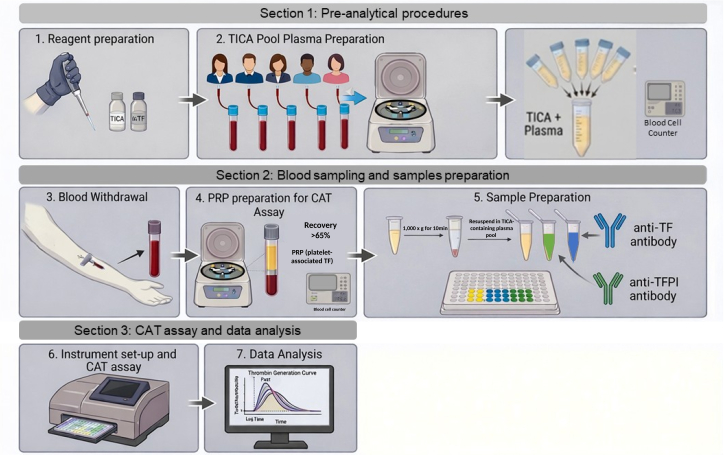
Figure 1. Workflow for the platelet-associated thrombin generation assay. The illustration shows the workflow for platelet-associated thrombin generation, divided into three sections. Section 1 (pre-analytical procedure) includes the preparation of reagents and TICA-containing plasma pool from multiple donors. Section 2 (blood sampling and sample preparation) covers venous blood withdrawal and the preparation of platelet-rich plasma (PRP) for the CAT assay. Section 3 (the CAT assay and data analysis) includes setting up the instrument and acquiring thrombin generation curves. Figure 1 was generated using the AI-based tool Gemini (Google). Please click here to view a larger version of this figure.
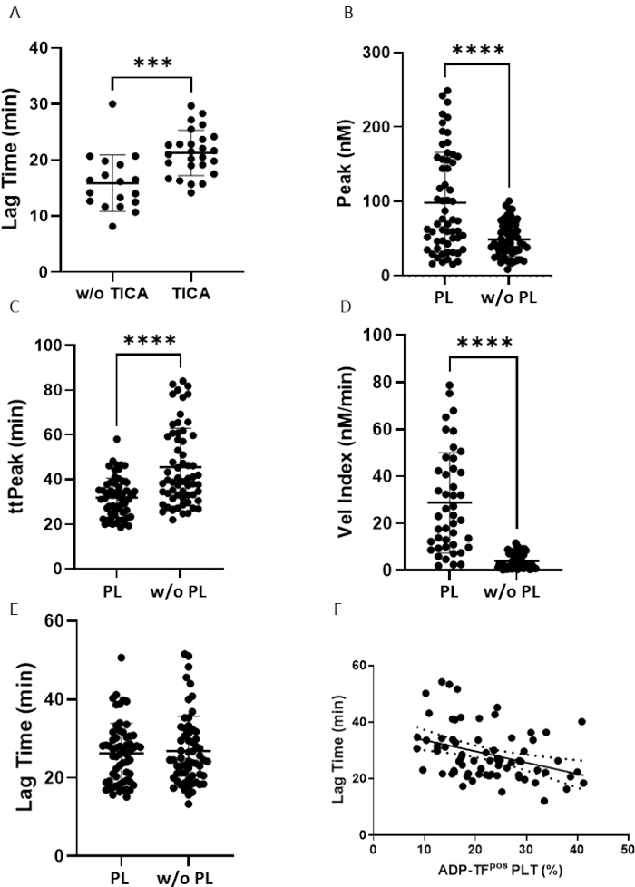
Figure 2. Effects of contact activation and phospholipid availability on thrombin generation kinetics. (A) Comparison of LagTime in platelet samples analyzed in the absence or presence of TICA, highlighting the contribution of FXII-dependent contact activation to thrombin generation (n = 18). (B–D) Amplification phase parameters measured in the presence or absence of exogenous phospholipids (PL), including peak thrombin (B), time-to-peak (ttPeak; C), and velocity index (D) showing that addition of exogenous phospholipids accelerates thrombin generation kinetics (n = 61). (E) Comparison of LagTime in platelet preparations analyzed with or without (w/o) exogenous phospholipids, showing minimal dependence of LagTime on phospholipid availability (n = 61). (F) Correlation between LagTime and the percentage of TFpos-platelets measured by flow cytometry following stimulation with adenosine diphosphate (ADP; 10 µM) for 15 min at RT (n=73), indicating an association between thrombin generation kinetics and platelet-associated TF expression. In all panels, n indicates the number of independent platelet preparations obtained from healthy subjects. Each data point represents the mean of three technical replicates performed within the same CAT assay run. Results are reported as individual data. Normal distribution of data was assessed using the Shapiro-Wilk test. Statistical significance was assessed using the Mann-Whitney test (A) and Paired t-test (B-E), *** p < 0.001, **** p < 0.0001. Please click here to view a larger version of this figure.
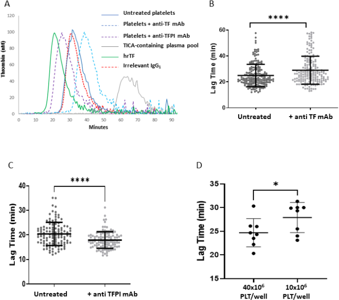
Figure 3. Thrombin generation kinetic is TF and TFPI dependent and related to platelet concentration. (A) Representative thrombin generation curves obtained from untreated platelets (blue line), platelets incubated with a neutralizing anti-TF monoclonal antibody (blue dotted line), platelets incubated with a neutralizing anti-TFPI monoclonal antibody (violet dotted line), TICA-containing pooled plasma (negative control, grey line), human recombinant TF (hrTF; positive control, green line) and irrelevant IgG1 (isotype control, red dotted line). (B-C) LagTime measured after sample preincubation with an anti-TF (B) or an anti-TFPI antibody (C), demonstrating that platelet-associated thrombin formation is regulated by both TF and its physiological inhibitor TFPI (n = 118). (D) Comparison of LagTime at two different platelet concentrations (PLT/well), showing the dependence of thrombin generation kinetics on platelet count (n = 8). In all panels, n indicates the number of independent platelet preparations obtained from healthy subjects. Each data point represents the mean of three technical replicates performed within the same CAT assay run. Results are reported as individual data. Normal distribution of data was assessed using the Shapiro-Wilk test. Statistical significance was assessed using Paired t-test, * p < 0.05, **** p < 0.0001. Please click here to view a larger version of this figure.
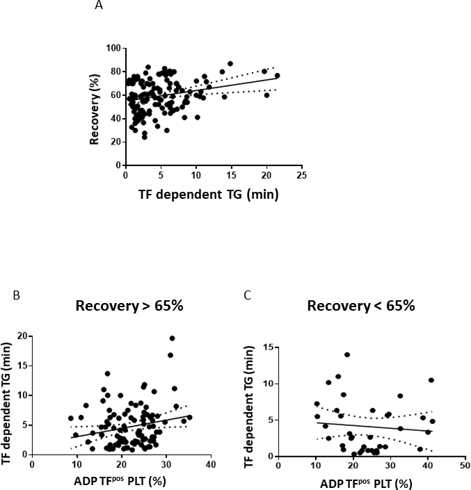
Figure 4. TF-dependent thrombin generation is influenced by platelet recovery and correlates with TFpos-platelets. (A) Correlation between platelet recovery and TF-dependent thrombin generation, expressed as the effect of a neutralizing anti-TF antibody on LagTime (n = 134). (B-C) Correlation between the percentage of TFpos-platelets, measured by flow cytometry after stimulation with 10 µM adenosine diphosphate (ADP) and TF-dependent thrombin generation, expressed as the difference in LagTime in the presence versus absence of the anti-TF antibody, in samples with platelet recovery >65% (B, n = 106) and <65% (C, n = 37). In all panels, n indicates the number of independent platelet preparations obtained from healthy subjects. Please click here to view a larger version of this figure.
| 1 | 2 | 3 | |
| A | Untreated platelets | Untreated platelets | Untreated platelets |
| B | Platelets with anti-TF monoclonal antibody | Platelets with anti-TF monoclonal antibody | Platelets with anti-TF monoclonal antibody |
| C | Platelets with anti-TFPI monoclonal antibody | Platelets with anti-TFPI monoclonal antibody | Platelets with anti-TFPI monoclonal antibody |
| D | hrTF 1pM | hrTF 1pM | hrTF 1pM |
| E | TICA-containg plasma pool | TICA-containg plasma pool | TICA-containg plasma pool |
| F | Platelets with irrelevant IgG1 | Platelets with irrelevant IgG1 | Platelets with irrelevant IgG1 |
| G | Sample Calibrator | Sample Calibrator | Sample Calibrator |
| H | hrTF Calibrator | hrTF Calibrator | hrTF Calibrator |
Table 1: Plate layout for CAT assay.
| ID | sample |
| Tube a | Untreated platelets |
| Tube b | Platelets with anti-TF monoclonal antibody |
| Tube c | Platelets with anti-TFPI monoclonal antibody |
| Tube d | Platelets with irrelevant IgG1 |
Table 2: Sample preparation for CAT assay.
ディスカッション
This study outlines a comprehensive workflow for assessing Tissue Factor (TF)-dependent thrombin generation in intact human platelets using the Calibrated Automated Thrombogram (CAT) assay. It provides a detailed description of each step, beginning with the pre-analytical ones, which are critical for the success of the assay.
Although the abundance and biological relevance of platelet-associated TF remain a matter of discussion, accumulating evidence supports its functional contribution to thrombin generation and thrombosis. Platelets are now recognized as active contributors to coagulation, providing a procoagulant phospholipid surface1 and also expressing functional TF2,3. These features enable platelets to initiate thrombin generation.This concept has important pathophysiological implications, particularly in conditions characterized by increased levels of TFpos-platelets and enhanced thrombotic risk26,27,28,29,30. TFpos-platelets have been indeed reported as independent predictors of cardiovascular mortality in patients with coronary artery disease31. TFPI is the physiological inhibitor of the TF-driven coagulation cascade12. Therefore, the overall platelet procoagulant potential also depends on the contribution of TFPI. Consequently, functional assays that measure thrombin generation while accounting for the balance between TF and TFPI13 are highly informative in complementing antigenic measurements, such as those obtained from the analysis of TFpos-platelets by flow cytometry20. In this context, our data show that thrombin generation parameters -particularly the LagTime- reflect the functional contribution of platelet-associated TF, as demonstrated by their correlation with the percentage of TFpos-platelets. These results suggest that, when performed under the right conditions, the CAT assay provides an indication of the activity of platelet-associated TF rather than simply measuring thrombin formation.
Conventional thrombin generation assays were primarily developed for plasma analysis and rely on the addition of exogenous TF to evaluate the contribution of coagulation factors14. While this approach ensures the robustness and standardization of the test16, it may mask the contribution of endogenous TF. This is a significant drawback when the aim of the study is to examine the TF-dependent procoagulant activity of the sample. Indeed, assays designed to evaluate the thrombin-generating capacity of TFpos-cells, such as tumor cells32, monocytes33, or polymorphonuclear leukocytes34 are performed without the addition of exogenous TF. Consistent with this approach, our data show that platelets are able to sustain thrombin generation in the absence of exogenously added TF.
To best highlight the contribution of the TF-dependent pathway, the outlined protocol involves the combined use of a plasma pool containing a contact pathway inhibitor and blocking antibodies. The addition of a contact pathway inhibitor (TICA or CTI) during blood collection for plasma pool preparation limits thrombin generation to the TF/FVIIa axis by suppressing FXII-dependent activation18,19. This is particularly important in low-TF conditions, where contact activation could otherwise contribute significantly to thrombin generation and hinder the specific assessment of platelet-associated TF activity18,19. The use of neutralizing antibodies further supports the specificity of the assay10. Inhibiting TF results in a marked prolongation of LagTime, which confirms that thrombin generation is driven by platelet-associated TF. Conversely, inhibiting TFPI significantly shortens LagTime, confirming its role in regulating TF-dependent platelet-driven coagulation. Overall, these findings emphasise that platelet-dependent coagulation reflects the dynamic balance between TF and TFPI, rather than TF activity alone.
Another relevant feature of this protocol is that thrombin generation was assessed using fresh, intact whole platelets. This preserves the organization of the membrane, the surface receptors, and the intrinsic spatial relationship between TF, TFPI, and the procoagulant phospholipid surface35. This is important because these elements jointly determine the procoagulant behavior of platelets under physiological conditions. In contrast, while platelet lysates may be useful in certain experimental settings32, they do not fully reproduce the structural and functional context of intact circulating platelets. For this reason, the present assay was primarily designed using intact cells, to ensure that the resulting thrombin generation profile more accurately reflects the contribution of platelet to coagulation in vivo.
To further resemble physiological conditions, the assay was optimized using a platelet concentration reflecting that found in circulation (200-400 ×103/µL). Importantly, the protocol can also be adapted for use in cases of thrombocytopenia, where platelet counts can be as low as 100 × 103/µL or even lower, thus extending its applicability to this clinical setting.
Another key consideration is the impact of pre-analytical variables17, including blood collection procedures and the time interval between sampling and processing to prevent platelet activation, paying particular attention to preparation of PRP10. Since TF is primarily expressed in a subset of platelets characterized by the largest size within the whole population, it is essential to preserve a platelet population that accurately reflects the composition of whole blood and does not lose this subset10.
However, even under optimized centrifugation conditions -also accounting for variables such as hematocrit, which can influence platelet yield in PRP-, platelet recovery may remain suboptimal, which can preclude reliable thrombin generation measurements. Our data consistently show that platelet recovery correlates directly with TF-dependent thrombin generation, indicating that inadequate sample preparation may result in an underestimation of platelet-associated TF activity. These findings emphasise the importance of carefully controlling PRP preparation to ensure consistent and reliable results. In addition, biological variability in platelet-associated TF expression, platelet activation status, and platelet recovery may contribute to inter-individual differences in thrombin generation and should be considered when comparing results across subjects.
From a practical standpoint, particularly in the context of clinical studies, it is essential to ensure consistency across experiments16. To minimize inter-assay variability, it is advisable to use the same batch of reagents for all samples so that they can be analysed under identical experimental conditions. This is particularly important when evaluating large cohorts of people, as variability in reagents can significantly affect thrombin generation measurements16.
Another key feature of this protocol is the use of a plasma pool obtained from healthy donors. The use of pooled plasma rather than autologous plasma was a deliberate methodological choice aimed at minimizing inter-individual variability in coagulation factor levels and endogenous plasma-derived modulators of thrombin generation. This approach allows the assay to focus on the platelet component under investigation while maintaining a standardized coagulation environment across samples. The pooled plasma therefore serves as a consistent source of coagulation factors for all samples. Additionally, the protocol incorporates a series of internal controls to ensure the reliability of the assay. A positive control consisting of human recombinant TF is included to monitor test performance, while the plasma pool itself serves as a reference condition to standardize the plasma component across all experiments.
Furthermore, the inclusion of biological controls is strongly recommended when the assay is applied in clinical settings or in studies involving specific patient populations. Samples from healthy subjects can be analyzed alongside patient samples to provide a physiological reference, for example. Ideally, matched healthy controls would be used to further strengthen data interpretation.
A limitation of the protocol is that the pooled plasma preparation, although subjected to double centrifugation and large extracellular vesicle depletion, cannot be considered completely free of residual platelets or extracellular vesicles. However, plasma-only controls consistently generated negligible thrombin generation under the experimental conditions used, indicating that any residual procoagulant contribution from the plasma matrix is functionally minimal compared with that of the experimentally added platelet preparation. Therefore, while complete elimination of all platelet- or vesicle-derived material cannot be guaranteed, its influence on the assay readout is expected to be limited, as also supported by the negligible thrombin generation observed in plasma-only control samples.
For broader clinical implementation, further studies will be required to assess inter-laboratory reproducibility and to establish standardized operating procedures across centers. Nevertheless, several features of the present workflow, including the use of pooled plasma, predefined platelet recovery criteria, and functional TF/TFPI controls, may facilitate future harmonization efforts and support multicenter application of the assay.
In conclusion, this workflow enables the assessment of platelet-associated TF-dependent thrombin generation under conditions that preserve the physiological interplay between procoagulant and anticoagulant pathways. By avoiding the addition of exogenous TF and integrating control of pre-analytical variables, contact pathway inhibition, and functional validation through TF and TFPI modulation, this approach allows a more specific evaluation of platelet-driven coagulation.
Importantly, the thrombin generation parameters obtained under these conditions -particularly LagTime- provide a functional readout that reflects the activity of platelet-associated TF, rather than merely measuring thrombin formation. This feature makes the assay particularly suitable to complement antigen-based approaches, such as flow cytometric detection of TFpos-platelets.
By addressing key methodological limitations that have historically hindered the study of platelet-associated TF10, this protocol contributes to a more consistent and reproducible assessment of its functional role. As such, it provides a robust framework for both mechanistic investigations and clinical studies exploring the contribution of platelet-associated TF to thrombotic risk. This approach may help to bridge the gap between experimental observations and clinical translation of platelet-associated TF.
開示事項
Figure 1 was generated using the AI-based tool Gemini (Google) and subsequently reviewed and modified by the Authors to ensure scientific accuracy and consistency with the experimental protocol. The use of AI was limited to figure 1 preparation and did not involve data generation, analysis, or interpretation.
The Authors declare no conflict of interest.
謝辞
This study was supported by research funding Italian Ministry of Health (CUP B43C22001110008 - PNRR-MAD-2022-12376277 - PNRR-M6C2I2.1 NextGenerationEU) and by funds from University of Milan -Piano di Sostegno alla Ricerca 2025-Linea 2 (to M.C.).
材料
| 名前 | 会社 | カタログ番号 | コメント |
|---|---|---|---|
| Biotube 10 mL | Biosigma | BSP048 | To collect PRP |
| Butterfly Needle G19 | PIKDARE | 2044019030010 | For blood withdrawal |
| Dade Innovin | Siemens Healthcare | B4212-50 | Human recombinant Tissue Factor used as positive control |
| FluCa buffer | STAGO | 86197 | Fluo substrate and buffer for thrombin generation assay |
| Fluoroskan Ascent plate reader | Thermo Fisher | 1506450 | For thrombin generation assay |
| Immulon 2HB Plate (x50) | STAGO | 86175 | Plate for thrombin generation assay |
| Microliter syringe | Hamilton | 80600/00 | To add TICA to vacutainer |
| MP-reagent | STAGO | 86222 | Phospholipids for thrombin generation assay |
| Murine MAb anti-human TFPI Kunitz domain 1, clone 2H8 | ImmBioMed GmbH&Co KG | ADG4903 | anti Tissue Factor Pathway Inhibitor antibody |
| Murine Monoclonal Antibody anti TF, clone HTF-1 | Invitrogen | 16-142-29-82 | anti Tissue Factor antibody |
| P10 pipette | Gilson | F144055M | For adding antibodies to the samples |
| P20 pipette | Gilson | F144056M | For plate preparation |
| P200 pipette | Gilson | F144058M | For sample preparation |
| P1000 pipette | Gilson | F144059M | To transfer PRP and sample preparation |
| Pipette tips 10 µL | Gilson | F161630 | For adding antibodies to the samples |
| Pipette tips 200 µL | Gilson | F161930 | For sample and plate preparation |
| Pipette tips 1000 µL | Gilson | F161670 | For TICA-containing plasma pool, PRP and sample preparation |
| Plastic Pasteur pipet | Merck | BR747765 | To transfer PFP |
| Prostaglandin E1 | Sigma | P5515 | To avoid platelet activation |
| Sodium Chloride 0.9 % | BD | 306575 | To wash the needle-cannula |
| Thermostable Inhibitor of Contact Activation (TICA) | University of Maastricht | Non-catalog item | To inhibit Factor XII activation |
| Thrombin Calibrator | STAGO | 86192 | Calibrator for thrombin generation assay |
| Thrombinoscope BV | STAGO | Non-catalog item | For thrombin generation assay analysis |
| Tube 1.5 mL | Eppendorf | 120086 | For PFP centrifugation and sample preparation |
| Tube 50 mL | Corning | 352098 | To collect PFP |
| Vacutainer EDTA | BD | 368857 | To collect blood for blood cell count |
| Vacutainer multiple sample luer adapter | BD | 367300 | For blood withdrawal |
| Vacutainer one use holder | BD | 364815 | For blood withdrawal |
| Vacutainer sodium citrate 0.129M | BD | 363079 | To collect blood for TICA-pool plasma and PRP preparation |
| XN-1000 Automated Hematology Analyzers | Dasit | Non-catalog item | To count blood cells |
参考文献
- Reddy EC, et al. Procoagulant phosphatidylserine-exposing platelets in vitro and in vivo. Front Cardiovasc Med. 2020;7:15.
- Camera M, et al. Platelet activation induces cell-surface immunoreactive Tissue Factor expression, which is modulated differently by antiplatelet drugs. Arterioscler Thromb Vasc Biol. 2003;23(9):1690-6.
- Camera M. The coagulative cascade. Direct oral anticoagulants: from pharmacology to clinical practice: Springer, Cham; 2021. p. 1-18.
- Siddiqui FA, et al. The presence and release of Tissue Factor from human platelets. Platelets. 2002;13(4):247-53.
- Muller I, et al. Intravascular Tissue Factor initiates coagulation via circulating microvesicles and platelets. FASEB J. 2003;17(3):476-8.
- Panes O, et al. Human platelets synthesize and express functional Tissue Factor. Blood. 2007;109(12):5242-50.
- Brambilla M, et al. Human megakaryocytes confer Tissue Factor to a subset of shed platelets to stimulate thrombin generation. Thromb Haemost. 2015;114(3):579-92.
- Bouchard BA, et al. Platelet Tissue Factor is not expressed transiently after platelet activation. Blood. 2012;119(18):4338-9; author reply 4339-41.
- Osterud B, et al. Human platelets do not express Tissue Factor. Thromb Res. 2013;132(1):112-5.
- Brambilla M, et al. Do methodological differences account for the current controversy on Tissue Factor expression in platelets? Platelets. 2018;29:406–14.
- Camera M, et al. Tissue Factor expression on platelets is a dynamic event. Blood. 2010;116(23):5076-7.
- Maroney SA, et al. Platelet Tissue Factor Pathway Inhibitor modulates intravascular coagulation. Thromb Res. 2012;129 Suppl 2(Suppl 2):S21-2.
- Depasse F, et al. Thrombin generation assays are versatile tools in blood coagulation analysis: A review of technical features, and applications from research to laboratory routine. J Thromb Haemost. 2021;19(12):2907-17.
- Hemker HC, et al. Calibrated automated thrombin generation measurement in clotting plasma. Pathophysiol Haemost Thromb. 2003;33(1):4-15.
- Hemker HC, Kremers R. Data management in thrombin generation. Thromb Res. 2013;131(1):3-11.
- Dargaud Y, et al. Evaluation of a standardized protocol for thrombin generation measurement using the calibrated automated thrombogram: an international multicentre study. Thromb Res. 2012;130(6):929-34.
- Subcommittee on Control of Anticoagulation of the SSC-ISTH. Towards a recommendation for the standardization of the measurement of platelet-dependent thrombin generation. J Thromb Haemost. 2011;9(9):1859-61.
- Luddington R, et al. Clinical measurement of thrombin generation by calibrated automated thrombography requires contact factor inhibition. J Thromb Haemost. 2004;2(11):1954-9.
- Dargaud Y, et al. Elimination of contact factor activation improves measurement of platelet-dependent thrombin generation by calibrated automated thrombography at low-concentration Tissue Factor. J Thromb Haemost. 2006;4(5):1160-1.
- Brambilla M, et al. Flow cytometry analysis of Tissue Factor expression in human platelets. J Vis Exp. 2024(213).
- Tchaikovski SN, et al. Effect of oral contraceptives on thrombin generation measured via calibrated automated thrombography. Thromb Haemost. 2007;98(6):1350-6.
- Csordas A, et al.. The biology behind the atherothrombotic effects of cigarette smoke. Nat Rev Cardiol. 2013;10(4):219-30.
- Hawker RJ, Turner VS, Mitchell SG. Use of prostaglandin E1 during preparation of platelet concentrates. Transfus Med. 1996;6(3):249-54.
- Valeri CR, et al. Prostaglandins in the preparation of blood components. Science. 1972;175(4021):539-42.
- Berg TVD. Novel Thermostable Inhibitor of Contact Activation Tica effectively blocks contact activation in low Tissue Factor thrombin generation. Blood. 2019;Volume 134:1146.
- Brambilla M, et al. Tissue Factor in patients with acute coronary syndromes: expression in platelets, leukocytes, and platelet-leukocyte aggregates. Arterioscler Thromb Vasc Biol. 2008;28(5):947-53.
- Falanga A, et al. Cancer tissue procoagulant mechanisms and the hypercoagulable state of patients with cancer. Semin Thromb Hemost. 2015;41(7):756-64.
- Canzano P, et al. Platelet and endothelial activation as potential mechanisms behind the thrombotic complications of COVID-19 patients. JACC Basic Transl Sci. 2021;6(3):202-18.
- Trabattoni D, et al. Migraine in patients undergoing PFO closure: characterization of a platelet-associated pathophysiological mechanism: The LEARNER Study. JACC Basic Transl Sci. 2022;7(6):525-40.
- Tsantes AG, et al. Sepsis-induced coagulopathy: an update on pathophysiology, biomarkers, and current guidelines. Life (Basel). 2023;13(2).
- Camera M, et al. Tissue Factor, a membrane-associated marker of platelet activation, predicts 5-year cardiovascular mortality in coronary artery disease patients. Thromb Haemost. 2025; TH-25-08-0472.
- Marchetti M, et al. Characterization of the thrombin generation potential of leukemic and solid tumor cells by calibrated automated thrombography. Haematologica. 2012;97(8):1173-80.
- Ernofsson M, et al. Monocyte Tissue Factor expression, cell activation, and thrombin formation during cardiopulmonary bypass: a clinical study. J Thorac Cardiovasc Surg. 1997;113(3):576-84.
- Maugeri N, et al. Tissue Factor expressed by neutrophils: another piece in the vascular inflammation puzzle. Semin Thromb Hemost. 2015;41(7):728-36.
- Monroe DM, et al. Platelets and thrombin generation. Arterioscler Thromb Vasc Biol. 2002;22(9):1381-9.
再版と許可
この記事は公開されました
動画は近日公開