本レビューでは、免疫調節および移植拒絶におけるDNAメチル化の役割についてまとめます。DNAメチル化は、免疫細胞の発生と分化を制御し、免疫細胞の機能を調節し、さらにグラフト生存率を向上させるためのバイオマーカーや治療標的として機能する可能性があります。
レビュー記事
DNAメチル化の免疫学的調節と同種移植片拒絶におけるその意義
64 回視聴
⸱
2026年9月11日
この記事について
サマリー
要約
移植拒絶反応は、依然として移植片の長期生存における大きな障壁となっており、免疫応答と分子調節機構の間の複雑な相互作用によって引き起こされます。主要なエピジェネティック修飾であるDNAメチル化は、免疫細胞の発生、分化、成熟、および機能の調節において重要な役割を果たしています。DNAメチル化の異常は、免疫関連遺伝子の発現を変化させ、免疫応答の性質や強度に影響を及ぼす可能性があります。移植においては、これらの変化が移植拒絶に関与するT細胞、B細胞、および自然免疫細胞に影響を与えると考えられています。また、DNAメチル化は、免疫活性化、免疫寛容、および炎症反応に関連するシグナル伝達経路の調節にも寄与しています。したがって、メチル化パターンの変化は、拒絶反応の進行と移植片機能の維持の両方に影響を与える可能性があります。本レビューでは、免疫調節におけるDNAメチル化に関する現在の知見をまとめ、特に胸腺の発達、ヘルパーT細胞および細胞傷害性T細胞、制御性T細胞、B細胞、自然免疫細胞、およびシグナル伝達経路に重点を置いて、移植拒絶への影響を検討します。さらに、移植におけるバイオマーカーおよび治療ターゲットとしてのDNAメチル化の潜在的な有用性についても考察します。これらのエピジェネティックなメカニズムを理解することで、固形臓器移植後の拒絶リスクの評価、免疫応答の調節、および移植片の長期的な転帰の改善に向けた、より精密なアプローチの開発が期待されます。
概要
免疫学は、免疫系の構造と機能に関する科学的分野であり、免疫応答の基礎となるメカニズムや、自己成分と非自己成分の認識後の恒常性維持などが含まれます。また、この分野は免疫機能の調節不全から生じる病理学的プロセスも網羅しています。免疫系は、防御、監視、恒常性維持という3つの基本的な機能を担っています。これらの機能が連携することで、外部病原体や有害物質の侵入を防ぎ、体内の非自己成分を排除し、安定した内部免疫環境を維持しています。免疫応答の特性に基づき、免疫は自然免疫または獲得免疫に分類されます。自然免疫は外来病原体に対して迅速かつ非特異的な応答を提供し、一方で獲得免疫は特定の抗原に向けられた高度に特異的な応答を提供します1,2。免疫応答の間、外部および内部の要因が免疫機能に関連する遺伝子の発現を調節します。このような調節には、古典的な遺伝学的メカニズムだけでなくエピジェネティックな修飾も関与しており、複雑でダイナミックな転写調節ネットワークを形成しています3。
伝統的な遺伝学はDNA配列の変化に起因する遺伝子機能の変化に焦点を当てていますが、エピジェネティクスは、基礎となるDNA配列を修飾することなく起こる、可逆的かつ遺伝的な遺伝子発現の変化を調査します。感染、化学物質への曝露、医薬品などの環境刺激や、サイトカインやホルモンなどの内部要因は、遺伝子のエピジェネティックな状況を変化させることができます。これらの変化は遺伝子発現を調節し、最終的に細胞表現型と生物学的機能を形成します。遺伝子発現を制御する主要なエピジェネティック・メカニズムには、DNAメチル化、ヒストン修飾、クロマチンリモデリング、および非コードRNA介在性調節が含まれます。胚発生の初期段階において、DNAメチル化の標識はほぼ完全に消去され、その後の分化の過程で再構築されます4,5。これらのエピジェネティック修飾の正確な除去と再構築は、正常な細胞の発達、分化、および成熟にとって不可欠です。対照的に、異常なエピジェネティック変化は遺伝子機能を乱し、疾患の発症および進行に寄与する可能性があります6。DNAメチル化パターンは、T細胞およびB細胞の発達と分化の間にも動的に変化します7,8。これらの知見は、骨髄造血幹細胞が多様な免疫細胞系へと分化する方向性を決定する上で、エピジェネティック調節が重要な役割を果たしていることを強調しています。ヒストン修飾は、単球/マクロファージおよびヘルパーT細胞の分化と機能的可塑性を調節し9,10,11,12,13,14、Toll様受容体経路を含む主要なシグナル伝達経路に影響を与えます15,16。クロマチンリモデル因子であるMi-2betaは胸腺細胞に高度に発現しており、CD4遺伝子の発現とT細胞の発達を直接的に調節しています17。非コードRNAもまた、免疫応答の重要な調節因子として機能します。例えば、インターフェロン-γ (IFN-γ) は細胞内細菌感染に対する宿主防御において中心的な役割を果たしますが、miR-21はIFN-γ mRNAを直接標的にすることでIFN-γの産生を抑制できます18。総合すると、これらの知見は免疫応答の調節におけるエピジェネティック・メカニズムの中心的な役割を強調するものです。
免疫細胞は免疫機能の中核をなしており、移植片の生存を決定付ける主要な要因である移植拒絶反応の重要な推進力となります19,20,21。移植拒絶反応の根底にあるメカニズムについては広範な免疫学的研究が行われてきましたが、依然として多くの側面が十分に解明されていません22。また、免疫細胞の発達、分化、および成熟において、エピジェネティックなメカニズムが極めて重要な役割を果たすという証拠も増えています23,24。したがって、エピジェネティックな観点から移植拒絶反応の分子基盤を検討することは、臓器移植後の移植拒絶反応や慢性同種移植腎症のより早期の介入および予防を後押しし、長期的な移植片の生存に寄与する可能性があります。さまざまなエピジェネティックな修飾の中でも、DNAメチル化は哺乳類ゲノムにおける転写を制御する最も安定したメカニズムです25。そこで本レビューでは、免疫細胞の発達、分化、および機能におけるDNAメチル化の役割と、臓器移植後の急性拒絶反応への関与について検討します。
レビューと展望
1. DNAメチル化およびシーケンシング技術
DNAメチル化では、S-アデノシルメチオニンがメチル基供与体として利用され、DNAメチルトランスフェラーゼがCpGジヌクレオチド(シトシン-リン酸-グアニン)内のシトシンの5位炭素へメチル基を共有結合的に付加することで、シトシンを5-メチルシトシン(5mC)に変換します。この修飾により、DNA配列そのものとは別に、遺伝子発現にさらなる調節層が加わります。一般に、メチル化レベルが低い領域ではタンパク質の結合が可能となり、能動的な遺伝子転写を促進するクロマチン構造が維持されます26。対照的に、メチル化されたDNA領域では、メチルCpG結合ドメイン(MBD)タンパク質のリクルート、ヒストン修飾およびクロマチンリモデリング複合体の促進、あるいは転写因子やその他のDNA結合タンパク質の結合を直接的に阻害することによって、遺伝子の転写および発現が抑制されます27,28,29。しかし、DNAメチル化は遺伝子の活性化に関連する場合もあり、特に遺伝子本体(gene body)領域においては、遺伝子発現と正の相関を示します。このような関係性は頻度は低いものの、普遍的に発現する遺伝子において遺伝子本体のメチル化は一般的です30,31。
DNAメチル化の生物学的機能は、ゲノム上のコンテキストの中で解釈されなければならない。CpGジヌクレオチドは、CpGアイランドと呼ばれる領域にクラスターを形成することが多い。CpGアイランドは、長さが200塩基対(bp)以上で、G+C含有率が50%以上であり、かつ観測値/期待値(Obs/Exp)比が0.6以上であるDNAセグメントとして定義される32。哺乳類ゲノムにおいて、CpGジヌクレオチドは比較的稀であり、ゲノム全体の約1%に過ぎない。ヒトの遺伝子プロモーターの約60%はCpGアイランドに関連しており、これらの領域は通常、正常細胞では非メチル化状態である。初期発生時または組織分化時にメチル化されるのは、ごく一部(約6%)である33,34。CpGショア(shores)とは、CpGアイランドの上流および下流2,000 bp以内の領域を指し、そのメチル化状態は転写サイレンシングと密接に相関している。メチル化リプログラミングにおいて、差次的にメチル化された領域の約70%はCpGショアに存在する23,35。さらに、組織特異的なメチル化の大部分は、CpGアイランドではなくショアで起こる34,35。CpGシェルフ(shelves)は、CpGショアの上流および下流2,000 bp以内に位置し、一方で「オープンシー(open sea)」という用語は、CpGアイランド、ショア、シェルフ以外のすべてのゲノム領域を指す。
DNAメチル化は、主に維持メチル化とde novoメチル化という2つのプロセスを通じて起こります。維持メチル化は主に半保存的DNA複製時に起こり、維持メチルトランスフェラーゼが既存のメチル化パターンを娘鎖にコピーします。DNMT1は主要な維持メチルトランスフェラーゼとして機能し、ヘミメチル化DNAを優先的に標的とし、細胞内で最も豊富に存在するメチルトランスフェラーゼであり、細胞周期のS期に活発に転写されます36。対照的に、de novoメチル化は、これまでメチル化されていなかった部位を標的にすることで、DNA複製とは独立して新しいメチル化パターンを構築します。DNMT3AとDNMT3Bが主要なde novoメチルトランスフェラーゼとして機能します。これらは胚性幹細胞で高く発現しており、胚発生過程におけるメチル化パターンの構築において重要な役割を果たしますが、細胞の分化に伴いその発現は低下します27。DNMT3ファミリーにはDNMT3Lも含まれており、これは触媒活性を欠いていますが、母親由来のゲノムインプリンティングの確立に不可欠です37。DNMT3LはDNMT3AおよびDNMT3Bの活性を高め、それによってDNAメチル化の効率を向上させます38。ゲノム全体において、多くのメチル化シトシンが、特に遺伝子本体領域においてDNA脱メチル化を受けます。脱メチル化は能動的および受動的なメカニズムを通じて起こります。能動的脱メチル化では、TET酵素によって5mCがその酸化誘導体に酸化されます39。一方、受動的脱メチル化は、維持メチルトランスフェラーゼ活性がない状態でDNA複製が行われる際に、メチル化シトシンが徐々に希釈されることで起こります40。
DNAメチル化は遺伝子の転写および発現の調節において中心的な役割を果たすため、メチル化検出技術の進歩も相まって、過去10年間にわたり主要な研究焦点となっています。サンプルの前処理の違いに基づき、DNAメチル化検出アプローチは大きく3つのカテゴリーに分類できます41。第一のアプローチはメチル化感受性制限エンドヌクレアーゼを利用するもので、メチル化されたDNA領域を特異的に切断しないため、メチル化状態に応じて異なるサイズの断片が生成されます。第二のアプローチは、5-メチルシトシンを特異的に認識するメチル化結合タンパク質または抗体を用いて、メチル化DNA断片を濃縮する方法です。第三のアプローチは、重亜硫酸塩処理を用いて、メチル化されたシトシンはそのままに、非メチル化シトシンをウラシルに変換することで、メチル化部位と非メチル化部位を精密に識別する方法です。現在、重亜硫酸塩変換と次世代シーケンシングを組み合わせた手法が、DNAメチル化解析において最も広く利用されている戦略となっています。Table 1に、主要なメチル化検出アプローチの原理、利点、限界、および移植研究における潜在的な応用についてまとめています。DNAメチル化検出技術の継続的な進歩は、エピジェネティック研究の技術的ツールキットを拡大させ、新たなエピジェネティック治療戦略の開発を後押ししています41,42,43,44,45。
2. 初期の胸腺細胞の発達および分化におけるメチル化特性
移植拒絶反応には、細胞性免疫および抗体介在性免疫反応の両方を含む複雑なメカニズムが関与しています。移植免疫における現在の知見は、T細胞介在性免疫が移植片拒絶において中心的な役割を果たすことを示しています46,47。胸腺は、T細胞の発生、分化、および成熟のための主要な器官として機能します48。T細胞の前駆段階である胸腺細胞の発生過程において、DNAメチル化パターンは継続的かつダイナミックに再構築されます。これらの変化には、T細胞の転写を制御するゲノム領域における脱メチル化とメチル化の両方が含まれますが、これらのプロセスは完全に均衡して起こるわけではありません。発生の初期段階では、CD3、RUNX3、RORC、RAG1、およびLCKを含む、主要なT細胞受容体(TCR)構成要素および必須の調節因子をコードする遺伝子が主に脱メチル化されます7。より広義には、免疫細胞の分化過程では、メチル化よりも脱メチル化の方が頻繁に起こります49,50。対照的に、de novo メチル化は比較的限定的であり、主に胸腺細胞発生の初期コミットメント段階で起こります7。de novo メチル化は、幹細胞の多能性と関連する遺伝子をサイレンシングし、それによって系統決定と分化を可能にするためにも重要です7,51。胸腺細胞のDNAメチル化のもう一つの重要な特徴は、ほとんどのメチル化の変化が分化中に確立される一度形成されると、安定し、大部分が不可逆的であることです。しかし、RAG遺伝子などの一部の遺伝子はダイナミックな調節を示し、免疫細胞発生の初期に脱メチル化され、その後、後期の段階で再メチル化されます7。
3. 免疫細胞の分化と機能におけるDNAメチル化
DNAメチル化は、多様な免疫細胞集団の分化、機能、および可塑性を調節する上で重要な役割を果たしており、それによって移植免疫応答の結果に影響を与えます。
- 獲得免疫細胞
- ヘルパーT細胞 (CD4+ T細胞)
CD4+ T細胞は、急性臓器移植拒絶反応における主要なエフェクター細胞として機能する。52,53ナイーブT細胞(Tn)は、複数のCD4+ T細胞へと分化する。+ 遺伝的、エピジェネティック、およびその他の調節メカニズムの協調的な作用によって、T細胞サブセットが形成されます。特定のシグナルによる刺激を受けると、ナイーブT細胞のエピジェネティックな景観が変化し、それによって遺伝子の転写および発現が変化します。これらの変化は、最終的に移植後の免疫拒絶反応のタイプと強度の両方に影響を及ぼす可能性があります。T細胞の活性化後、 IL2 遺伝子は急速に脱メチル化され、脱メチル化状態に維持される。このエピジェネティックな変化は、T細胞機能をサポートする遺伝子の転写および発現を増強させるために不可欠である。54.
DNAメチル化は、ナイーブT細胞がTh1およびTh2サブセットへと分化する方向性を制御する上でも重要な役割を果たしている。55,56未刺激のCD4++ T細胞は、 IFNG この遺伝子は高度にメチル化されているが、Th1分化の過程で特異的に脱メチル化され、一方でTh2細胞では高度にメチル化したままとなる。57対照的に、 IL-4 この遺伝子はナイーブT細胞およびTh1細胞では強くメチル化されているが、Th2細胞では部分的な脱メチル化が起こる。58外部要因は、DNAメチルトランスフェラーゼ(DNMT)の活性を調節することで、これらのメチル化パターンをさらに制御することができます。例えば、ビタミンD欠乏はDNMT活性を上昇させ、以下のハイパーメチル化を促進します。 IFNG 遺伝子である一方で、ビタミンDの補給はこれらの変化を改善させることができる59生物学的老化もまた、DNAメチル化パターンに影響を及ぼします。Yuらによる研究を含む諸研究では、DNMT3a mRNAの発現および、以下のメチル化が... IL4 遺伝子プロモーターは加齢とともに増加するが、一方でそのメチル化は IFNG 遺伝子プロモーターの減少60これらの知見は、加齢に伴うDNAメチル化の変化が、Th1細胞とTh2細胞の応答バランスを変動させる可能性を示唆している。
ナイーブT細胞の分化には、さらに他の2つの重要なCD4+ T細胞サブセットが関与しています。+ T細胞サブセット:Th17および制御性T(Treg)細胞61,62DNAメチル化はこのプロセスを厳密に調節している。Th17特異的な転写因子RORの発現はγT細胞およびサイトカインIL-17の産生は、それぞれの遺伝子プロモーター領域のメチル化状態に依存している63,64,65移植組織内のTh17細胞は、移植拒絶反応を悪化させ、血管傷害を促進させる可能性がある。66DNAメチル化は、Th17細胞の機能的可塑性にも影響を及ぼします。慢性炎症条件下では、 IFNG Th17細胞における当該遺伝子は脱メチル化状態にあり、これによりこれらの細胞はIFN-を産生することが可能となる。γその結果、Th17細胞はTh1およびTh17の両方の表現型の特性を獲得し得る。67さらに、 in vitro 特定の刺激条件下で、Th17細胞を強力な免疫抑制能を持つ機能的なTreg細胞へと分化させることができることが研究で示されている。68.
対照的に、Treg細胞は免疫寛容の維持および移植片内におけるホメオスタシスの保持において重要な役割を果たしている。69これらの細胞は胸腺で発生するか、あるいはTGF-βの影響下で末梢で分化することがあります。βその起源にかかわらず、Treg細胞は一貫して転写因子Foxp3を発現している。70エピジェネティックな制御、特にDNAメチル化は、以下を含むTreg機能に関連する遺伝子の発現制御において重要な役割を果たしている。 Foxp3 および Ctla4、それによってそれらの転写活性に直接的に影響を及ぼす71,72,73,74胸腺におけるTreg細胞の分化過程において、 Foxp3 遺伝子は段階的な脱メチル化を受ける。この脱メチル化状態は、細胞が末梢組織へ遊走した後も持続し、適切な強度の持続的なTCRシグナルを必要とする。62,75,76,77DNMT1が欠損している場合、胸腺および末梢組織の両方において、TCR刺激によってFoxp3陰性T細胞にFoxp3の発現を効果的に誘導することができる。しかし、TCR刺激によって十分なエピジェネティックな再構成が誘導されない場合、細胞は安定したFoxp3の発現を維持できず、Treg表現型の保持に不可欠な状態を維持することができない。62,75,78Foxp3の安定的な発現は、主にシス調節エレメント、特に保存された非翻訳領域(CNS)におけるエピジェネティックな修飾によって制御されている。71,79,80これらのうち、Treg特異的脱メチル化領域(TSDR)のメチル化状態は、安定したTreg集団と不安定なTreg集団を区別するための信頼性の高いマーカーとして機能します。71,81,82,83,84,85,86天然に存在するナイーブ調節性T細胞(nTreg)では、TSDRは非メチル化状態にあり、Stat5、NFAT、Runx1、CREBなどの転写因子の動員を可能にすることで、安定したFoxp3の発現を維持している。対照的に、誘導性調節性T細胞(iTreg)では、通常TSDRがメチル化されており、その結果、Foxp3の発現は不安定になる。72,74,87,88,89さらに、TET1やTET2などの脱メチル化酵素の喪失は、Foxp3遺伝子座の過剰メチル化を招き、これによりTreg機能が損なわれ、自己免疫疾患の発症が促進される。90臨床研究により、不顕性拒絶反応を伴う腎移植片生検検体において、Foxp3の脱メチル化レベルが高いほど、移植片の予後が改善することがさらに示されている。82,84.
同種移植片の拒絶反応を防止し、免疫寛容を促進するための手段として、Treg細胞の研究がますます進められている。91移植拒絶を引き起こすエフェクターT細胞を効果的に抑制し、免疫寛容を維持するためには、十分な数のTreg細胞が移植片内に存続していなければならない。92しかしながら、Treg細胞はある程度の表現型の不安定性を示します。特定の条件下では、これらは次のようなエピジェネティックな変化を起こすことがあります。すなわち、~の脱メチル化を含む。 RORC 遺伝子であり、炎症性サイトカインIL-17の発現を誘導する93,94この変化はTreg細胞の可塑性を浮き彫りにしており、治療への応用における課題となっています。さらに、エピジェネティックな調節によってTreg機能を強化できることが実験的研究で示唆されています。例えば、Guoらによれば 他 動物モデルにおいて、DNAメチル化阻害剤とIL-35を併用することで、Foxp3の発現が増加し、Treg細胞の分化が促進され、移植片の生存率が向上することが示された。95これらの進展にもかかわらず、Treg細胞に関連するメカニズムの理解は不十分なままである。今後の研究では、Tregの分化を促進し、Treg表現型を安定させる条件を明確にすることが求められており、これらの知見を免疫介在性疾患の治療に応用することが目標となる。 - 細胞傷害性T細胞(CD8+)+ T細胞)
活性化後、ナイーブCD8+ T細胞は、細胞傷害性Tリンパ球(CTL)として知られる細胞傷害性エフェクター細胞へと分化します。これらの細胞は一般に2つのサブセットに分類されます。一つはIFN-γを産生するTc1細胞であり、γ、およびIL-4とIL-5を産生するTc2細胞96臨床研究により、CD69の割合が+/CD8+ 心臓移植患者の末梢血中のT細胞は、重度の拒絶反応と相関している。加えて、CD8+ 心筋組織内のT細胞はCD69を発現している+ 活性化表現型を呈し、パフォリン介在性の細胞毒性活性を示す97,98さらに、実験的根拠によってCD8の役割が裏付けられている。+ 移植拒絶反応におけるT細胞。Jones 他 胸腺切除およびanti-CD4/CD8処理により作製したT細胞欠損マウスにおいて、TCR抗原特異的なCD8の移入により、~であることを示した。+ T細胞単独で、完全に不一致な同種心移植片の拒絶反応を誘導するのに十分である99これらの知見は、CD8の極めて重要な寄与を強調するものである。+ 免疫介在性移植拒絶反応におけるT細胞。また、DNAメチル化はCD8をも調節している。+ T細胞の発達と機能。DNMT1が欠損すると、胸腺細胞の生存能が低下する100さらに、成熟CD8 T細胞におけるDNMT3aの欠損により+ T細胞は、末端エフェクター分化を抑制しつつ、メモリー細胞への分化を促進する。この効果の一部はT細胞因子1(Tcf1)の発現上昇によって媒介されており、これはDNMT3aがTcf7遺伝子のプロモーター領域を標的とし、初期エフェクターCD8 T細胞において新規メチル化を誘導するためである。+ T細胞101さらに、急性ウイルス感染のマウスモデルにおいて、T細胞におけるTET2の条件付き欠損は、メモリーCD8 T細胞の形成を促進します。+ T細胞102以上の結果を総合すると、DNAメチル化がCD8 T細胞の発達、分化、および機能的応答を決定的に制御していることが示されました。+ T細胞 - B細胞
移植拒絶においてT細胞が中心的な役割を果たすが、B細胞もまた細胞性および液性拒絶反応の両方に寄与している。103,104体液性拒絶反応は、主にBリンパ球によって媒介されます。活性化されたB細胞は形質細胞へと分化し、大量の特異的抗体を産生・分泌します。これにより、移植片に炎症性損傷が引き起こされる可能性があります。105DNAメチル化は、さらにB細胞の免疫応答を調節している。Lee 他 B細胞の初期発生過程において、特に遺伝子本体領域および非CpGアイランド領域で広範なDNA脱メチル化が起こることが示された。対照的に、B細胞分化の後期段階では、DNAメチル化による制御の影響は弱くなる。106さらに、Shaknovichは ら 胚中心B細胞とナイーブB細胞の間で、DNAメチル化プロファイルの有意な差を同定した107胚中心B細胞は脱メチル化を経て、機能遺伝子の発現が変化し、NF-を含む主要なシグナル伝達経路に影響を及ぼす。κBおよびMAPK経路107,108機能研究により、DNAメチル化がB細胞の活性を制限することがさらに示されている。de novoメチルトランスフェラーゼであるDnmt3aおよびDnmt3bの欠損(Dnmt3欠損)は、B細胞の発達および成熟を促進する。同時に、B細胞の活性化を増強し、胚中心B細胞および形質細胞の拡大を支持する。これらの知見は、de novo DNAメチル化が通常、B細胞の活性化を制限し、形質細胞への分化を抑制するように作用していることを示している。109.
- ヘルパーT細胞 (CD4+ T細胞)
- 自然免疫細胞
固形臓器移植後の免疫応答には、自然免疫と獲得免疫の協調的な相互作用が関与している。自然免疫系には、樹状細胞、マクロファージ、ナチュラルキラー細胞、好中球などの造血系および非造血系細胞が含まれ、これらすべてが移植後の免疫拒絶に寄与し得る。110.
樹状細胞は、自然免疫と獲得免疫を橋渡しする重要な調節因子として機能し、免疫活性化または免疫寛容のいずれかを促進する能力を備えている。111,112単球から分化する際、樹状細胞ではDNAメチル化の広範な変化が起こり、多くのゲノム領域においてメチル化レベルが著しく低下することを特徴とする。これらの変化は、主に系統決定および免疫活性化に不可欠なエンハンサー領域および転写因子結合部位で起こる。113,114,115この分化過程の特徴は、細胞表面におけるCD14の下方制御とCD209の上方制御である。この変化は、~のプロモーター領域内にあるCpG部位の脱メチル化と相関している。 CD209 遺伝子116さらに、DNAメチル化抑制剤による処理は、CD40やCD86などの共刺激分子の発現を有意に増加させ、それによって抗原提示能を高め、樹状細胞の機能的な成熟を促進します。これらの知見は、DNAメチル化が樹状細胞の機能および免疫活性を直接的に制御していることを示しています。117.
単球およびマクロファージは、異常細胞や侵入した病原体の食作用と除去を媒介するため、免疫系における不可欠な構成要素である。多くの証拠により、マクロファージは早期の急性移植拒絶反応に見られる炎症性障害だけでなく、後期の慢性拒絶反応における線維化の進行にも寄与することが示されている。118,119M1マクロファージ(古典的活性化)とM2マクロファージ(代替活性化)のバランスは、免疫応答を極めて重要なレベルで調節している。M1マクロファージが優位な状態へのシフトは、炎症と組織損傷を促進する。対照的に、DNAメチル化阻害剤または遺伝子ノックアウトによってDNMT1の発現を低下させると、ペルオキシソーム増殖剤活性化受容体のメチル化が低下する。 γ1 (PPARγ1) プロモーター。この変化はM2マクロファージの活性化を促進し、免疫介在性の損傷を抑制するのに寄与する。120DNAメチル化パターンの相違は、マクロファージ機能におけるエピジェネティック制御の役割をさらに浮き彫りにしている。冠動脈疾患を有する個体と有さない個体の末梢血単球/マクロファージにおいて、特定の遺伝子のプロモーターメチル化に有意な変動があることが研究により明らかになっている。121さらに、単球からマクロファージへの分化には、転写因子結合部位におけるメチル化の相当な変化が伴っており、これは主要な調節領域におけるDNAメチル化が、マクロファージの分化において正確かつ重要な役割を果たしていることを示唆している。122.
ナチュラルキラー(NK)細胞は、移植拒絶においてエフェクター機能と調節機能の両方を担っている。NK細胞は移植片の拒絶を促進させる一方で、免疫寛容の形成にも寄与することができる。骨髄造血幹細胞からNK細胞への分化過程において、~のメチル化レベルが低下し、 キラー細胞免疫グロブリン様受容体 この遺伝子はKIR発現の増加を導き、それが免疫調節において重要な役割を果たします。加えて、DNAメチル化阻害剤による治療は、〜の脱メチル化を誘導します。 キラー細胞免疫球状凝集反応(KIR) 遺伝子であり、NK細胞におけるその発現を促進する123,124これらの知見は、DNAメチル化がNK細胞機能の主要な調節因子であることを示している。
好中球もまた、移植拒絶反応に積極的に関与している。活性化した好中球はさまざまなメディエーターを放出し、抗原提示細胞をリクルートして活性化させる。これにより、抗原特異的なT細胞反応が開始され、免疫拒絶が増幅される。125,126,127好中球に発現するアンカータンパク質であるPRV-1は、生理的および病理的条件の両方において、そのプロモーター領域におけるDNAメチル化によって制御されている。128.
4. 主要なシグナル伝達経路におけるDNAメチル化
DNAメチル化は、機能性遺伝子の転写を直接的に制御するだけでなく、主要なシグナル伝達経路を調節することで細胞の挙動にも影響を及ぼします。例えば、分泌型Frizzled関連タンパク質(SFRP)遺伝子のプロモーター領域の過剰メチル化は、遺伝子のサイレンシングを引き起こします。このサイレンシングはWnt/β-カテニンシグナル伝達経路の正常な調節を乱し、その結果、異常な活性化を招き、大腸がんの発症を促進します129。同様に、免疫細胞におけるmTORシグナル伝達経路の上流の負の調節因子の過剰メチル化は、それらの発現を低下させ、過剰なmTOR活性化を招き、糖尿病性腎臓における炎症性傷害に寄与します。デシタビンやRNA干渉などの介入により、DNMT1を阻害してこれらの調節遺伝子のメチル化を減少させることができ、これにより糖尿病性腎症の進行を抑制することが可能です130。さらに、DNAメチルトランスフェラーゼ阻害剤は、NF-κBやJAK/STATなどのシグナル伝達経路を調節することで細胞増殖を抑制し、それによって疾患の発症および進行に影響を与えます131,132(図1)。
5. 臨床的意義:免疫抑制剤とDNAメチル化の相互作用
標準的な免疫抑制剤は免疫細胞におけるDNAメチル化パターンに影響を与えることが示されているが、その効果は薬剤やゲノム上のコンテキストによって異なる。T細胞におけるIFNγプロモーターのメチル化に関する研究では、ミコフェノール酸はT細胞活性化によって誘導される脱メチル化を打ち消すが、タクロリムスはこの特定の遺伝子座において同様の効果を示さないことが実証されている133。対照的に、シロリムスはmTORシグナル伝達経路への影響を通じてグローバルなメチル化プロファイルを調節することが示されており134、また、DNAメチル化自体がmTOR経路を調節し、それによって腎移植後の同種移植片の生存および急性拒絶反応に影響を与えることが証明されている135。さらに、デキサメタゾンなどのグルココルチコイドは、末梢免疫系において広範なDNAメチル化のリモデリングを誘導する。好中球デキサメタゾンメチル化指数は、グルココルチコイド曝露の感受性の高い薬力学的マーカーとして機能し、免疫抑制的な免疫プロファイルと関連している136。ATG導入療法も腎移植レシピエントのDNAメチロームを変化させることが報告されており、メチル化ベースのモデルはトランスクリプトームプロファイルよりも効果的に感染リスクを予測できることが示されている137。個々のエピジェネティックな変動がこれらの治療に対する患者の反応に影響を与える可能性があり、メチル化バイオマーカーが個別化された免疫抑制レジメンの指針となる可能性が示唆されている136,137。これらの薬剤とエピゲノムの相互作用は、長期的な移植片の予後を最適化する上で重要な意味を持つ可能性があるため、さらなる調査が必要である。
6. 課題と展望:エピジェネティック修飾と免疫学的安全性のバランス
移植における有用性が期待されているものの、アザシチジンやデシタビンなどのDNMT阻害剤(DNMTis)は、感染症および腫瘍形成のリスクを伴います。これらの薬剤による治療は重篤な敗血症性合併症に関連しており138,139、また、低メチル化剤への全身的な曝露は、二次的な急性骨髄性白血病への転化リスクの増加に関連しており、罹患した患者の全生存期間の中央値は5か月未満であることが示されています140。薬剤関連の毒性以外に、細胞型特異性の欠如が重要な課題となっています。全身的な脱メチル化は、エフェクターT細胞を活性化させる一方でTreg画分を不安定にさせ、拒絶反応を悪化させる可能性があり141、また、CMVなどの日和見感染症に対するCD8+メモリーT細胞による保護機能を損なう可能性があります101,102。
これらの制限を克服するために、細胞型および遺伝子座特異的な調節を実現する新しい戦略が検討されています。ナノ粒子プラットフォームを用いることで、DNMTを移植浸潤細胞に優先的に送達することが可能です142,143。CRISPR/dCas9エピゲノム編集は、全般的な影響を及ぼすことなくFoxp3-TSDRの標的脱メチル化を可能にし、Tregを安定化させます144,145。また、バイオマーカーによるモニタリングは、より安全な臨床応用を支援する可能性があります146。
結論
移植片の生存率を向上させることは、臓器移植研究における主要な目標であり続けています。移植拒絶反応は複雑な免疫学的プロセスであり、ドナー臓器とレシピエントの両方からのエピジェネティックな修飾がダイナミックに相互作用し、全体的なエピジェネティックな景観を形成します。レシピエントの免疫系は、移植片拒絶の主要な要因となります。エピジェネティックなメカニズムは、免疫細胞の発達、分化、および機能を決定的に制御し、それによって移植後の拒絶反応や移植片の生存に影響を与えます。加えて、免疫抑制剤、感染症、および移植後の環境などの要因が、エピジェネティックな制御と免疫応答の関係をさらに複雑にします。これらの要因はエピジェネティックなパターンを変化させ、免疫細胞の挙動を修飾するため、移植結果にさらなる複雑さをもたらします。したがって、臓器移植における新たなバイオマーカーや治療標的を特定し、診断アプローチおよび治療戦略を改善するためには、エピジェネティックなメカニズムと移植拒絶反応との関係についてさらなる調査が必要です。
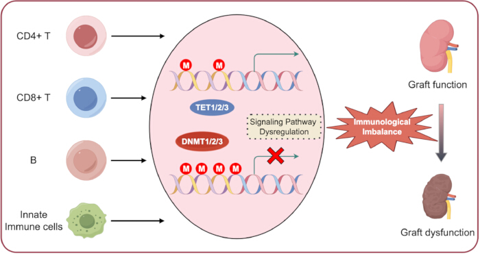
図1移植免疫におけるDNAメチル化。免疫細胞は、その寿命を通じて固有の異なるDNAメチル化パターンを維持している。 この生理学的プロセスの破綻は、異常なDNAメチル化と下流のシグナル伝達経路の調節不全を誘発し、最終的に免疫恒常性の低下および同種移植片の機能不全を引き起こす可能性がある。こちらの図の拡大版を表示するには、ここをクリックしてください。
| 方法 | 原理 | 利点 | 限界点 | 移植における応用 |
| MeDIP-seq⁴¹ | 抗5mC抗体濃縮 + NGS | ゲノムワイド解析におけるコスト効率に優れ、大規模なサンプルコホートに適している | 低解像度(約10–30 bp);5mCと5hmCを区別できない | 移植レシピエント由来末梢血単核細胞(PBMC)における全ゲノムメチル化変化のスクリーニング |
| 全ゲノムビスルフィトシーケンシング (WGBS)⁴² | 重亜硫酸塩コンバージョン + NGS | 単一塩基解像度、ゲノムワイドなカバー率 | 高コストであり、大量のデータを必要とする | 移植生検組織の包括的なメチロームマッピング:拒絶反応に関連する新規DMRの同定 |
| RRBS⁴³ | 制限酵素濃縮 + ビスルファイト変換 + NGS | 費用対効果が高く、CpGリッチ領域を広範囲にカバーしている | non-CpG領域およびCpG低頻度領域におけるカバレッジの不足 | シーケンシングコストを低減した移植組織における標的メチローム解析 |
| メチル化特異的PCR⁴ | 重亜硫酸塩変換 + メチル化特異的プライマーを用いたPCR | 高感度であり、簡便かつ迅速で低コストである | 特定の遺伝子座のみを標的とする;定性的または限定的な定量的解析 | 探索コホートから得られた候補DMRの検証;臨床バイオマーカーの開発 |
| パイロシーケンシング⁴⁵ | 重亜硫酸塩変換 + 合成によるシーケンシング | 定量的;標的座位における1塩基解像度 | 中程度のスループットであり、事前のターゲット同定が必要。 | Treg細胞におけるFOXP3 TSDRなど、特定のCpG部位におけるメチル化レベルの精密定量 |
表1:主要なDNAメチル化検出法のまとめ。 この表は、一般的に用いられているDNAメチル化検出法、その原理、利点、限界、および移植研究における潜在的な応用についてまとめたものである。
開示事項
著者らは、競合する利益または潜在的な利益相反がないことを宣言します。
謝辞
著者は、図の作成におけるFigdrawの支援に感謝いたします。
参考文献
- Güell-Alonso R, Cabrera-Pérez R, Vives J. Immunocompatibility in transplantation: adapting to a changing therapeutic landscape. Front Immunol. 2025;16:1648534.
- Li XQ, Ding RD, Cai JZ. Organ transplantation: current status, challenges, and future prospects. MedComm. 2026;7(1):e70567.
- Medzhitov R, Horng T. Transcriptional control of the inflammatory response. Nat Rev Immunol. 2009;9:692–703.
- Bagci H, Fisher AG. DNA demethylation in pluripotency and reprogramming: the role of tet proteins and cell division. Cell Stem Cell. 2013;13:265–269.
- Gu TP, et al. The role of Tet3 DNA dioxygenase in epigenetic reprogramming by oocytes. Nature. 2011;477:606–610.
- Cedar H, Bergman Y. Programming of DNA methylation patterns. Annu Rev Biochem. 2012;81:97–117.
- Rodriguez RM, et al. Regulation of the transcriptional program by DNA methylation during human αβ T-cell development. Nucleic Acids Res. 2015;43:760–774.
- Rauch TA, et al. A human B cell methylome at 100-base pair resolution. Proc Natl Acad Sci U S A. 2009;106:671–678.
- Hardbower DM, et al. Ornithine decarboxylase regulates M1 macrophage activation and mucosal inflammation via histone modifications. Proc Natl Acad Sci U S A. 2017;114:E751–E760.
- De Santa F, et al. The histone H3 lysine-27 demethylase Jmjd3 links inflammation to inhibition of polycomb-mediated gene silencing. Cell. 2007;130:1083–1094.
- Antignano F, Zaph C. Regulation of CD4 T-cell differentiation and inflammation by repressive histone methylation. Immunol Cell Biol. 2015;93:245–252.
- Wei G, et al. Global mapping of H3K4me3 and H3K27me3 reveals specificity and plasticity in lineage fate determination of differentiating CD4+ T cells. Immunity. 2009;30:155–167.
- Liu Z, et al. The histone H3 lysine-27 demethylase Jmjd3 plays a critical role in specific regulation of Th17 cell differentiation. J Mol Cell Biol. 2015;7:505–516.
- He H, et al. Histone methylation mediates plasticity of human FOXP3(+) regulatory T cells by modulating signature gene expressions. Immunology. 2014;141:362–376.
- Menden H, et al. Histone deacetylase 6 regulates endothelial MyD88-dependent canonical TLR signaling, lung inflammation, and alveolar remodeling in the developing lung. Am J Physiol Lung Cell Mol Physiol. 2019;317:L332–L346.
- Irizarry-Caro RA, et al. TLR signaling adapter BCAP regulates inflammatory to reparatory macrophage transition by promoting histone lactylation. Proc Natl Acad Sci U S A. 2020;117:30628–30638.
- Williams CJ, et al. The chromatin remodeler Mi-2beta is required for CD4 expression and T cell development. Immunity. 2004;20:719–733.
- Yang F, et al. MiR-21 is remotely governed by the commensal bacteria and impairs anti-TB immunity by down-regulating IFN-γ. Front Microbiol. 2020;11:512581.
- Nankivell BJ, Kuypers DR. Diagnosis and prevention of chronic kidney allograft loss. Lancet. 2011;378:1428–1437.
- Zununi Vahed S, et al. Genetics and epigenetics of chronic allograft dysfunction in kidney transplants. Iran J Kidney Dis. 2016;10:1–9.
- Goldberg RJ, Weng FL, Kandula P. Acute and chronic allograft dysfunction in kidney transplant recipients. Med Clin North Am. 2016;100:487–503.
- Wood KJ, Bushell A, Hester J. Regulatory immune cells in transplantation. Nat Rev Immunol. 2012;12:417–430.
- Ji H, et al. Comprehensive methylome map of lineage commitment from haematopoietic progenitors. Nature. 2010;467:338–342.
- Suarez-Alvarez B, Rodriguez RM, Fraga MF, Lopez-Larrea C. DNA methylation: a promising landscape for immune system-related diseases. Trends Genet. 2012;28:506–514.
- Eckhardt F, et al. DNA methylation profiling of human chromosomes 6, 20 and 22. Nat Genet. 2006;38:1378–1385.
- Thomson JP, et al. CpG islands influence chromatin structure via the CpG-binding protein Cfp1. Nature. 2010;464:1082–1086.
- Esteller M. Epigenetic gene silencing in cancer: the DNA hypermethylome. Hum Mol Genet. 2007;16(Spec No 1):R50–R59.
- Lopez-Serra L, Esteller M. Proteins that bind methylated DNA and human cancer: reading the wrong words. Br J Cancer. 2008;98:1881–1885.
- Kuroda A, et al. Insulin gene expression is regulated by DNA methylation. PLoS One. 2009;4:e6953.
- Hellman A, Chess A. Gene body-specific methylation on the active X chromosome. Science. 2007;315:1141–1143.
- Jones PA. Functions of DNA methylation: islands, start sites, gene bodies and beyond. Nat Rev Genet. 2012;13:484–492.
- Esteller M. Epigenetics in evolution and disease. Lancet. 2008;372:S90–S96.
- Straussman R, et al. Developmental programming of CpG island methylation profiles in the human genome. Nat Struct Mol Biol. 2009;16:564–571.
- Irizarry RA, et al. The human colon cancer methylome shows similar hypo- and hypermethylation at conserved tissue-specific CpG island shores. Nat Genet. 2009;41:178–186.
- Doi A, et al. Differential methylation of tissue- and cancer-specific CpG island shores distinguishes human induced pluripotent stem cells, embryonic stem cells and fibroblasts. Nat Genet. 2009;41:1350–1353.
- Portela A, Esteller M. Epigenetic modifications and human disease. Nat Biotechnol. 2010;28:1057–1068.
- Bourc'his D, et al. Dnmt3L and the establishment of maternal genomic imprints. Science. 2001;294:2536–2539.
- Gowher H, et al. Mechanism of stimulation of catalytic activity of Dnmt3A and Dnmt3B DNA-(cytosine-C5)-methyltransferases by Dnmt3L. J Biol Chem. 2005;280:13341–13348.
- Wu X, Zhang Y. TET-mediated active DNA demethylation: mechanism, function and beyond. Nat Rev Genet. 2017;18:517–534.
- Bochtler M, Kolano A, Xu GL. DNA demethylation pathways: additional players and regulators. Bioessays. 2017;39:1–13.
- Laird PW. Principles and challenges of genomewide DNA methylation analysis. Nat Rev Genet. 2010;11:191–203.
- Lister R, et al. Human DNA methylomes at base resolution show widespread epigenomic differences. Nature. 2009;462:315–322.
- Meissner A, et al. Reduced representation bisulfite sequencing for comparative high-resolution DNA methylation analysis. Nucleic Acids Res. 2005;33:5868–5877.
- Herman JG, et al. Methylation-specific PCR: a novel PCR assay for methylation status of CpG islands. Proc Natl Acad Sci U S A. 1996;93:9821–9826.
- Colella S, et al. Sensitive and quantitative universal pyrosequencing methylation analysis of CpG sites. Biotechniques. 2003;35:114–120.
- Nankivell BJ, Alexander SI. Rejection of the kidney allograft. N Engl J Med. 2010;363:1451–1462.
- Gelman AE, et al. CD4+ T lymphocytes are not necessary for the acute rejection of vascularized mouse lung transplants. J Immunol. 2008;180:4754–4762.
- Seo W, Taniuchi I. Transcriptional regulation of early T-cell development in the thymus. Eur J Immunol. 2016;46:531–538.
- Calvanese V, et al. A promoter DNA demethylation landscape of human hematopoietic differentiation. Nucleic Acids Res. 2012;40:116–131.
- Lee ST, et al. A global DNA methylation and gene expression analysis of early human B-cell development reveals a demethylation signature and transcription factor network. Nucleic Acids Res. 2012;40:11339–11351.
- Challen GA, et al. Dnmt3a is essential for hematopoietic stem cell differentiation. Nat Genet. 2011;44:23–31.
- Chitilian HV, et al. The strength of cell-mediated xenograft rejection in the mouse is due to the CD4+ indirect response. Xenotransplantation. 1998;5:93–98.
- Pierson RN, et al. Xenogeneic skin graft rejection is especially dependent on CD4+ T cells. J Exp Med. 1989;170:991–996.
- Bruniquel D, Schwartz RH. Selective, stable demethylation of the interleukin-2 gene enhances transcription by an active process. Nat Immunol. 2003;4:235–240.
- Sanders VM. Epigenetic regulation of Th1 and Th2 cell development. Brain Behav Immun. 2006;20:317–324.
- Wilson CB, Rowell E, Sekimata M. Epigenetic control of T-helper-cell differentiation. Nat Rev Immunol. 2009;9:91–105.
- Schoenborn JR, et al. Comprehensive epigenetic profiling identifies multiple distal regulatory elements directing transcription of the gene encoding interferon-gamma. Nat Immunol. 2007;8:732–742.
- Santangelo S, et al. Chromatin structure and DNA methylation of the IL-4 gene in human T(H)2 cells. Chromosome Res. 2009;17:485–496.
- Jiao X, et al. Vitamin D deficiency during pregnancy affects the function of Th1/Th2 cells and methylation of IFN-γ gene in offspring rats. Immunol Lett. 2019;212:98–105.
- Xia Y, et al. Age-related changes in DNA methylation associated with shifting Th1/Th2 balance. Inflammation. 2016;39:1892–1903.
- Muranski P, Restifo NP. Essentials of Th17 cell commitment and plasticity. Blood. 2013;121:2402–2414.
- Ohkura N, et al. T cell receptor stimulation-induced epigenetic changes and Foxp3 expression are independent and complementary events required for Treg cell development. Immunity. 2012;37:785–799.
- Janson PCJ, et al. Profiling of CD4+ T cells with epigenetic immune lineage analysis. J Immunol. 2011;186:92–102.
- Thomas RM, Sai H, Wells AD. Conserved intergenic elements and DNA methylation cooperate to regulate transcription at the il17 locus. J Biol Chem. 2012;287:25049–25059.
- Huang Y, et al. Decipherer manifestations and Treg/Th17 imbalance in multi-staging rheumatoid arthritis and correlation with TSDR/RORC methylation. Mol Immunol. 2020;127:1–11.
- Yuan X, et al. A novel role of CD4 Th17 cells in mediating cardiac allograft rejection and vasculopathy. J Exp Med. 2008;205:3133–3144.
- Cohen CJ, et al. Human Th1 and Th17 cells exhibit epigenetic stability at signature cytokine and transcription factor loci. J Immunol. 2011;187:5615–5626.
- Ye J, et al. Human tumor-infiltrating Th17 cells have the capacity to differentiate into IFN-γ+ and FOXP3+ T cells with potent suppressive function. Eur J Immunol. 2011;41:936–951.
- Issa F, Chandrasekharan D, Wood KJ. Regulatory T cells as modulators of chronic allograft dysfunction. Curr Opin Immunol. 2011;23:648–654.
- Josefowicz SZ, Rudensky A. Control of regulatory T cell lineage commitment and maintenance. Immunity. 2009;30:616–625.
- Baron U, et al. DNA demethylation in the human FOXP3 locus discriminates regulatory T cells from activated FOXP3(+) conventional T cells. Eur J Immunol. 2007;37:2378–2389.
- Kim HP, Leonard WJ. CREB/ATF-dependent T cell receptor-induced FoxP3 gene expression: a role for DNA methylation. J Exp Med. 2007;204:1543–1551.
- Morikawa H, et al. Differential roles of epigenetic changes and Foxp3 expression in regulatory T cell-specific transcriptional regulation. Proc Natl Acad Sci U S A. 2014;111:5289–5294.
- Polansky JK, et al. Methylation matters: binding of Ets-1 to the demethylated Foxp3 gene contributes to the stabilizationization of Foxp3 expression in regulatory T cells. J Mol Med (Berl). 2010;88:1029–1040.
- Toker A, et al. Active demethylation of the Foxp3 locus leads to the generation of stable regulatory T cells within the thymus. J Immunol. 2013;190:3180–3188.
- Jordan MS, et al. Thymic selection of CD4+CD25+ regulatory T cells induced by an agonist self-peptide. Nat Immunol. 2001;2:301–306.
- Kawahata K, et al. Generation of CD4(+)CD25(+) regulatory T cells from autoreactive T cells simultaneously with their negative selection in the thymus and from nonautoreactive T cells by endogenous TCR expression. J Immunol. 2002;168:4399–4405.
- Josefowicz SZ, Wilson CB, Rudensky AY. Cutting edge: TCR stimulation is sufficient for induction of Foxp3 expression in the absence of DNA methyltransferase 1. J Immunol. 2009;182:6648–6652.
- Floess S, et al. Epigenetic control of the foxp3 locus in regulatory T cells. PLoS Biol. 2007;5:e38.
- Polansky JK, et al. DNA methylation controls Foxp3 gene expression. Eur J Immunol. 2008;38:1654–1663.
- Barzaghi F, et al. Demethylation analysis of the FOXP3 locus shows quantitative defects of regulatory T cells in IPEX-like syndrome. J Autoimmun. 2012;38:49–58.
- Bestard O, et al. Intragraft regulatory T cells in protocol biopsies retain foxp3 demethylation and are protective biomarkers for kidney graft outcome. Am J Transplant. 2011;11:2162–2172.
- Liu J, et al. T regulatory cells in cord blood—FOXP3 demethylation as reliable quantitative marker. PLoS One. 2010;5:e13267.
- Wieczorek G, et al. Quantitative DNA methylation analysis of FOXP3 as a new method for counting regulatory T cells in peripheral blood and solid tissue. Cancer Res. 2009;69:599–608.
- Zhuo C, et al. Higher FOXP3-TSDR demethylation rates in adjacent normal tissues in patients with colon cancer were associated with worse survival. Mol Cancer. 2014;13:153.
- Lal G, et al. Epigenetic regulation of Foxp3 expression in regulatory T cells by DNA methylation. J Immunol. 2009;182:259–273.
- Zhou L, Chong MM, Littman DR. Plasticity of CD4+ T cell lineage differentiation. Immunity. 2009;30:646–655.
- Burchill MA, et al. IL-2 receptor beta-dependent STAT5 activation is required for the development of Foxp3+ regulatory T cells. J Immunol. 2007;178:280–290.
- Kanamori M, et al. Induced regulatory T cells: their development, stability, and applications. Trends Immunol. 2016;37:803–811.
- Yang R, et al. Hydrogen sulfide promotes Tet1- and Tet2-mediated Foxp3 demethylation to drive regulatory T cell differentiation and maintain immune homeostasis. Immunity. 2015;43:251–263.
- Issa F, Robb RJ, Wood KJ. The where and when of T cell regulation in transplantation. Trends Immunol. 2013;34:107–113.
- Issa F, Wood KJ. CD4+ regulatory T cells in solid organ transplantation. Curr Opin Organ Transplant. 2010;15:757–764.
- Suarez-Alvarez B, Baragano Raneros A, Ortega F, Lopez-Larrea C. Epigenetic modulation of the immune function: a potential target for tolerance. Epigenetics. 2013;8:694–702.
- Schmidl C, et al. Epigenetic reprogramming of the RORC locus during in vitro expansion is a distinctive feature of human memory but not naive Treg. Eur J Immunol. 2011;41:1491–1498.
- Guo H, et al. Inhibiting cardiac allograft rejection with interleukin-35 therapy combined with decitabine treatment in mice. Transpl Immunol. 2013;29:99–104.
- Betts RJ, Kemeny DM. CD8+ T cells in asthma: friend or foe? Pharmacol Ther. 2009;121:123–131.
- Schwengerdt KO, Fricker FJ, Bahjat KS, Kuntz ST. Increased expression of the lymphocyte early activation marker CD69 in peripheral blood correlates with histologic evidence of cardiac allograft rejection. Transplantation. 2000;69:2102–2107.
- Rutella S, et al. Induction of CD69 antigen on normal CD4+ and CD8+ lymphocyte subsets and its relationship with the phenotype of responding T-cells. Cytometry. 1999;38:95–101.
- Jones ND, et al. T-cell activation, proliferation, and memory after cardiac transplantation in vivo. Ann Surg. 1999;229:570–578.
- Lee PP, et al. A critical role for Dnmt1 and DNA methylation in T cell development, function, and survival. Immunity. 2001;15:763–774.
- Ladle BH, et al. De novo DNA methylation by DNA methyltransferase 3a controls early effector CD8+ T-cell fate decisions following activation. Proc Natl Acad Sci U S A. 2016;113:10631–10636.
- Carty SA, et al. The loss of TET2 promotes CD8(+) T cell memory differentiation. J Immunol. 2018;200:82–91.
- Schiffer L, et al. Chemokine CXCL13 as a new systemic biomarker for B-cell involvement in acute T cell-mediated kidney allograft rejection. Int J Mol Sci. 2019;20(10):2552. doi:10.3390/ijms20102552.
- Xu H, He X, Xu R. B cell activating factor, renal allograft antibody-mediated rejection, and long-term outcome. J Immunol Res. 2018;2018:5251801.
- Schmitz R, et al. B cells in transplant tolerance and rejection: friends or foes? Transpl Int. 2020;33:30–40.
- Lee ST, et al. A global DNA methylation and gene expression analysis of early human B-cell development reveals a demethylation signature and transcription factor network. Nucleic Acids Res. 2012;40:11339–11351.
- Shaknovich R, et al. DNA methyltransferase 1 and DNA methylation patterning contribute to germinal center B-cell differentiation. Blood. 2011;118:3559–3569.
- Caron G, et al. Cell-cycle-dependent reconfiguration of the DNA methylome during terminal differentiation of human B cells into plasma cells. Cell Rep. 2015;13:1059–1071.
- Barwick BG, et al. B cell activation and plasma cell differentiation are inhibited by de novo DNA methylation. Nat Commun. 2018;9:1900.
- Kaur BP, Secord E. Innate immunity. Pediatr Clin North Am. 2019;66:905–911.
- Steinman RM, Banchereau J. Taking dendritic cells into medicine. Nature. 2007;449:419–426.
- Merad M, Collin M, Bromberg J. Dendritic cell homeostasis and trafficking in transplantation. Trends Immunol. 2007;28:353–359.
- Zhang X, et al. DNA methylation dynamics during ex vivo differentiation and maturation of human dendritic cells. Epigenetics Chromatin. 2014;7:21.
- Pacis A, et al. Bacterial infection remodels the DNA methylation landscape of human dendritic cells. Genome Res. 2015;25:1801–1811.
- Klug M, et al. Active DNA demethylation in human postmitotic cells correlates with activating histone modifications, but not transcription levels. Genome Biol. 2010;11:R63.
- Bullwinkel J, Lüdemann A, Deberry J, Singh PB. Epigenotype switching at the CD14 and CD209 genes during differentiation of human monocytes to dendritic cells. Epigenetics. 2011;6:45–51.
- Frikeche J, et al. Impact of the hypomethylating agent 5-azacytidine on dendritic cells function. Exp Hematol. 2011;39:1056–1063.
- van den Bosch TP, et al. CD16+ monocytes and skewed macrophage polarization toward M2 type hallmark heart transplant acute cellular rejection. Front Immunol. 2017;8:346.
- Wang YY, et al. Macrophage-to-myofibroblast transition contributes to interstitial fibrosis in chronic renal allograft injury. J Am Soc Nephrol. 2017;28:2053–2067.
- Wang X, et al. Epigenetic regulation of macrophage polarization and inflammation by DNA methylation in obesity. JCI Insight. 2016;1:e87748.
- Bakshi C, Vijayvergiya R, Dhawan V. Aberrant DNA methylation of M1-macrophage genes in coronary artery disease. Sci Rep. 2019;9:1429.
- Dekkers KF, et al. Human monocyte-to-macrophage differentiation involves highly localized gain and loss of DNA methylation at transcription factor binding sites. Epigenetics Chromatin. 2019;12:34.
- Li G, Yu M, Weyand CM, Goronzy JJ. Epigenetic regulation of killer immunoglobulin-like receptor expression in T cells. Blood. 2009;114:3422–3430.
- Schenk A, Bloch W, Zimmer P. Natural killer cells—an epigenetic perspective of development and regulation. Int J Mol Sci. 2016;17:326.
- Scapini P, et al. The neutrophil as a cellular source of chemokines. Immunol Rev. 2000;177:195–203.
- Chertov O, Yang D, Howard OM, Oppenheim JJ. Leukocyte granule proteins mobilize innate host defenses and adaptive immune responses. Immunol Rev. 2000;177:68–78.
- Hirayama S, et al. Prevention of neutrophil migration ameliorates rat lung allograft rejection. Mol Med. 2006;12:208–213.
- Jelinek J, et al. Epigenetic control of PRV-1 expression on neutrophils. Exp Hematol. 2007;35:1677–1683.
- Suzuki H, et al. Epigenetic inactivation of SFRP genes allows constitutive WNT signaling in colorectal cancer. Nat Genet. 2004;36:417–422.
- Chen G, et al. Aberrant DNA methylation of mTOR pathway genes promotes inflammatory activation of immune cells in diabetic kidney disease. Kidney Int. 2019;96:409–420.
- Shang D, Han T, Xu X, Liu Y. Decitabine induces G2/M cell cycle arrest by suppressing p38/NF-κB signaling in human renal clear cell carcinoma. Int J Clin Exp Pathol. 2015;8:11140–11148.
- Zhu Z, et al. STAT3 signaling pathway is involved in decitabine induced biological phenotype regulation of acute myeloid leukemia cells. Am J Transl Res. 2015;7:1896–1907.
- Peters F, et al. Interferon-gamma DNA methylation is affected by mycophenolic acid but not by tacrolimus after T-cell activation. Front Immunol. 2017;8:822.
- Kraig E, et al. A randomized control trial to establish the feasibility and safety of rapamycin treatment in an older human cohort: immunological, physical performance, and cognitive effects. Exp Gerontol. 2018;105:53–69.
- Zhu C, et al. DNA methylation modulates allograft survival and acute rejection after renal transplantation by regulating the mTOR pathway. Am J Transplant. 2021;21(2):567–581.
- Wiencke JK, et al. DNA methylation as a pharmacodynamic marker of glucocorticoid response and glioma survival. Nat Commun. 2022;13(1):5537.
- Hsu FM, et al. DNA methylation predicts infection risk in kidney transplant recipients. Life Sci Alliance. 2025;8(7):e202403124. doi:10.26508/lsa.202403124.
- Bai JF, et al. Efficacy and safety of venetoclax and azacitidine for acute myeloid leukemia in China: a real-world single-center study. BMC Cancer. 2025;25(1):990.
- Arora S, et al. Tumor lysis syndrome and infectious complications during treatment with venetoclax combined with azacitidine or decitabine in patients with acute myeloid leukemia. Leuk Res. 2022;117:106844.
- Batra A, et al. Characteristics and survival of secondary acute myeloid leukemia from myelodysplastic syndromes in older adults: a population analysis. Clin Lymphoma Myeloma Leuk. 2025;25(6):e417–e423.
- Bai L, Hao X, Keith J, Feng Y. DNA methylation in regulatory T cell differentiation and function: challenges and opportunities. Biomolecules. 2022;12(9):1282.
- Trinh A, Huang Y, Shao H, et al. Targeting the ADPKD methylome using nanoparticle-mediated combination therapy. APL Bioeng. 2023;7(2):026111.
- Yuan N, Ma Z, Xue Z, et al. DNMT1-targeted macrophages delivering drug-loaded nanoparticles attenuate heart allograft fibrosis via PTPRD-STAT3/6 axis. Cell Death Discov. 2026. doi:10.1038/s41420-026-03295-5.
- Cameron J, Martino P, Nguyen L, Li X. Cutting edge: CRISPR-based transcriptional regulators reveal transcription-dependent establishment of epigenetic memory of Foxp3 in regulatory T cells. J Immunol. 2020;205(11):2953–2958.
- Liu XS, et al. Rescue of fragile X syndrome neurons by DNA methylation editing of the FMR1 gene. Cell. 2018;172:979–992.e6.
- Velut Y, Poulet G, Bersez T, et al. Epigenetic signatures on plasma cell-free DNA to detect kidney allograft rejection in a non-invasive way: development of a 10-plex digital PCR assay. Biomark Res. 2025;13:118.