本研究では、5xFADマウスにおけるQi-fu-yin(QFY)治療の効果を評価しました。その結果、QFYの投与により、学習および記憶能力の向上、脳内Aβ蓄積と細胞老化の減少、および大脳皮質におけるシナプス構造損傷の軽減が認められました。
研究記事
5xFADマウスの脳における認知機能障害の改善および細胞老化の抑制に対するQi-fu-yinの効果
* These authors contributed equally
この記事について
サマリー
要約
伝統的な中医学の処方であるQi-fu-yin(QFY)のメカニズムはまだ完全には解明されていませんが、アルツハイマー病(AD)に対する治療的な可能性が示されています。本研究は、5xFADマウスにおけるQFY治療と認知機能障害との関連性を調査することを目的としました。握力、 歩行解析、コロニーネスティング、シャトルボックス、モーリス水迷路試験、およびELISAを用いて、5xFADマウスの脳内におけるアミロイドβタンパク質1-42(Aβ1-42)、Aβ1-40、成長関連タンパク質43(GAP-43)、シナプトフィジン(SYN)、およびポストシナプス密度タンパク質95(PSD-95)のレベルを評価しました。皮質における老化関連分泌表現型(SASP)レベルの定量には、Luminexサイトカイン分析を用いました。また、老化関連β-ガラクトシダーゼ(SA-β-Gal)量、Aβ蓄積、および樹状突起スパインは、それぞれSA-β-Gal染色、免疫組織化学染色、およびゴルジCox染色により測定しました。免疫蛍光試験の結果、QFY治療は5xFADマウスの皮質におけるAβプラーク負荷を劇的に減少させ、p21の発現を低下させました。5xFADマウスにおいて、QFY療法は学習および記憶能力を著しく向上させ、 老化を軽減し、マウス皮質におけるGAP-43、PSD-95、およびSYNタンパク質の量を増加させました。QFY治療後、脳内のAβプラークとSA-β-Gal活性、ならびに海馬におけるAβ1-42およびAβ1-42/Aβ1-40レベルが有意に減少しました。また、5xFADマウスの皮質において、IL-1α、IL-1β、IL-6、IL-17A、およびIFN-γのレベルがかなり低下していました。主成分分析(PCA)およびピアソンの相関分析により、SASPサイトカインとシナプスタンパク質レベルの間に強い負の相関があることが示されました。これらの知見は、QFY治療が5xFADマウスにおいて、シナプス構造損傷の軽減とともに、加齢に伴う認知機能低下および細胞老化表現型の改善に関連していることを示唆しています。以上の結果は、アルツハイマー病の潜在的な治療候補としてのQFYのさらなる研究を支持するものです。
概要
Alzheimer's disease (AD)1 is the most prevalent form of dementia, characterized by amyloid-β protein (Aβ), hyperphosphorylated tau proteins (P-Tau), synaptic loss, and chronic inflammation. Currently available medications for AD have a number of side effects and are unable to stop the disease's progression2,3,4,5.
Senescence, which manifests as organism-wide disruptions across cellular and interstitial microenvironments rather than localized organ-specific alterations, is a key risk factor in AD pathogenesis. This systemic dysregulation distinguishes AD progression from single tissue-pathology models by fundamentally changing homeostatic mechanisms across biological systems6,7. Although cellular senescence initially serves as a homeostatic regulatory mechanism, the accumulation of senescent cells eventually causes progressive tissue degradation and persistent inflammatory cascades, fundamentally disrupting physiological homeostasis8. The ensuing inflammation may harm synapses and impair cognitive function9,10.
Cellular senescence is a complex cellular stress response characterized by telomere shortening, permanent cell-cycle arrest, and altered secretory activity11. Additional hallmarks include enlarged, flattened cell morphology, positive senescence-associated β-galactosidase (SA-β-Gal) staining, lipofuscin accumulation, and secretion of SASP factors12. Memory formation is affected by the notable decline in the levels of proteins associated with synaptic plasticity of axons and dendrites, which occurs with cellular senescence13,14. Because synaptic plasticity is essential for learning and memory, disruptions in synaptic proteins contribute to cognitive impairment. These proteins include GAP-43, SYN, and PSD-95. Undoubtedly, synaptic function is affected by reductions in dendritic numbers and functional abnormalities in the brains of the elderly15.
QFY is a traditional Chinese medicine formula derived from Jing Yue Quan Shu. It is composed of Panax ginseng C.A.Mey (Araliaceae), Polygala tenuifolia Willd (Polygalaceae), Rehmannia glutinosa (Gaertn.) Libosch. ex Fisch. & C. A. Mey (Orobanchaceae), Angelica sinensis (Oliv.) Diels (Apiaceae), Glycyrrhiza uralensis Fisch. ex DC. (Fabaceae), Atractylodes macrocephala Koidz (Asteraceae), Ziziphus jujuba Mill. var. spinosa (Bunge) Hu ex H.F. Chou (Rhamnaceae). Research has demonstrated that QFY significantly ameliorates cognitive dysfunction in rats with diabetes-induced brain injury16. Our previous study found that QFY could improve cognitive learning and memory functions impairment in 5xFAD and APP/PS1 mice. In addition, QFY demonstrated high safety in non-clinical studies, and no obvious acute, subacute, or long-term toxic reactions were observed17,18,19. Analysis of the clinical therapeutic effect of QFY showed that AD patients experienced a significant increase in their capacity for activities of daily living and MMSE scores after QFY treatment20. Following three consecutive days of QFY administration in male Sprague-Dawley rats, ten prototype QFY-derived components, including butylidenephthalide, butylphthalide, 20(S)-ginsenoside Rh1, 20(R)-ginsenoside Rh1, and zingibroside R1, were detected in cerebrospinal fluid21,22. However, the therapeutic mechanism of QFY in AD is still unclear. In this study, we investigated the association between QFY treatment, cellular senescence, and AD-related pathological changes in 5xFAD mice.
Although previous studies have demonstrated the neuroprotective effects of QFY in AD models, the specific involvement of cellular senescence in mediating these effects remains unexplored. Moreover, whether QFY confers cognitive benefits through senescence‑associated synaptic preservation, rather than solely through amyloid‑β reduction, has not been addressed. The present study was therefore designed to investigate whether QFY treatment attenuates cellular senescence and whether this attenuation is associated with improved synaptic plasticity in 5xFAD mice.
プロトコル
すべての動物実験は、山東中医学大学の実験動物福祉倫理委員会によって承認されました(SDUTCM20221101001)。
薬剤
QFY顆粒ドライクリームパウダーは、鲁南製薬候铺研究開発センターによって調製および試験されました。DonepezilおよびMemantineは、Shanghai Yuanye Technologyから購入しました。本研究では、初期体重が19–27 gの雄の5xFADマウスを用いました。QFYの投与量は、臨床的なヒト投与量である43 g/dに基づき、QFY顆粒ドライクリームパウダーへの換算後、16.297 g/dとして算出されました。本実験の中用量は、臨床的なヒト投与量に基づいて計算されました(16.297 g/70 kg x 9.1 = 2.11861 g/kg/d)。高用量は中用量の2倍(4.24 g/kg/d)、低用量は中用量の半分(1.06 g/kg/d)に設定しました。すべての薬剤溶液は、体重10 gあたり0.1 mLの容量で投与しました。
陽性対照として、ドネペジルと塩酸メマンチンの併用投与を用いた。投与量は、現在の臨床応用量(それぞれ 10 mg/d および 20 mg/d)および当研究グループの過去のデータを参照して決定し、ドネペジル 1.0 mg/kg/d とメマンチン 2.8 mg/kg/d の併用、合計投与量 3.8 mg/kg/d のレジメンとした。ドネペジル・メマンチン混合液の最終濃度は、3.8 mg/kg を体重 10 g あたり 0.1 mL の投与量で除して算出し、0.38 mg/mL として調製した。
低、中、高用量のQFY溶液を以下のように調製した:QFY乾燥粉末5.3 g、10.6 g、および21.2 gをそれぞれ別々の100 mLビーカーに入れ、30 mLの精製水を加えて十分に攪拌し、メスシリンダーに移して精製水で最終容量を50 mLに調整した。これにより、最終質量濃度をそれぞれ0.106 g/mL、0.212 g/mL、および0.424 g/mLとした。陽性対照については、5 mgのdonepezilと14 mgのmemantineを精製水に溶解し、最終容量を50 mLに調整した。各群の動物に対し、対応する薬剤溶液を1日1回、経口投与した。
動物
5xFADトランスジェニックマウス [B6. Cg-Tg (APPSwFlLon, PSEN1* M146L* L286V)6799Vas/Mmjax] を研究室内で繁殖させた。動物はすべて山東中医薬大学のSPF環境下で、温度 22–24 oC、湿度 50–60%、12時間の明暗サイクルにて飼育した。飼料と水は常に自由に摂取できるように提供した。
生後2.5ヶ月の雄性5xFADマウスを、未処理5xFAD群、ドネペジル+メマンチン群、QFY低用量群、QFY中用量群、QFY高用量群の5群にランダムに割り付けた。年齢を合わせた雄性野生型(WT)の同腹マウスをWT対照群とした。治療は1日1回、220日間投与した(図1)。
行動テスト
握力測定テスト
マウスの前肢骨格筋力は、握力計を用いて測定した23。実験中、マウスがテーブル表面と平行に保たれるよう、測定装置を水平に配置した。装置の電源を入れた後、マウスをグリッププレートの上にそっと置いた。マウスがプレートをしっかりと掴んだところで、マウスがグリップを離すまで、尾を後方に一定の力で引いた。最大握力を記録した。この測定を3回繰り返し、これらの値の平均をマウスの握力として算出した。
歩行テスト
マウスの歩行規則性をCatWalk XTを用いて測定した。実験はトレーニング期間とテスト期間に分けて実施した24。実験1日前、実験用マウスを実験室に移送し、新しい環境に慣れさせた。トレーニング期間中、マウスをスタート地点に置き、通路に沿ってエンドポイントまで走らせた。各マウスにつき1日3回のトレーニングを行った。3日間のトレーニング後(各マウスがスムーズに通路を通過できるようになるまで)、マウスをケージに戻した。テスト期間はトレーニング期間と同様の手順で行い、コンピュータソフトウェアを用いて各移動軌跡を自動的に解析した。軌跡が適格であれば緑色の点が、不適格であればオレンジ色の点が表示され、1匹あたり少なくとも3つの緑色の点が確認されたものを適格としてCatWalkXT 10.0ソフトウェアで解析した。
シャトルボックステスト
マウスの条件付き能動的回避能力を評価するために、シャトルボックス試験を用いた25。装置は、遮音チャンバー内に設置されたシャトルボックス(18 cm x 18 cm)で構成された。 実験は、4日間の学習期間と、それに続く1日間のテスト期間で構成された。 各学習日において、トレーニング開始前にマウスをシャトルボックス内で120 sの間適応させた。各試行は、6 sの条件刺激(70 dB、1000 Hzのトーンと500 luxの光を併用)から始まり、続いて格子状の床を通じて3 s、0.3 mAのフットショックを負荷し、12 sの試行間インターバルで終了した。条件刺激の間にマウスが反対側の区画に移動した場合、ショックは省略され、その反応を能動的回避として記録した。条件刺激の間には移動しなかったが、フットショックの間に移動した場合は、 受動的回避反応として記録した。試行を通じて移動しなかった場合は、 脱出失敗として記録した。トレーニングは、1日30試行を4日間連続して繰り返した。テスト期間(5日目)では、他のすべてのパラメータは学習期間と同じとしたが、フットショックは省略した。能動的回避反応の回数を、学習および記憶の主要な指標として記録した。
モーリス水迷路
実験動物の空間学習および記憶能を評価するために、モリス水迷路試験を用いた26。実験は、直径120 cm、深さ50 cmの水槽で行った。水槽には濁った白水を満たし、水温は21 °Cに維持した。直径10 cmのプラットフォームを、いずれかのクアドラントの水面下1 cmに設置した。マウスのための空間的指標として、プールの周囲に視覚的手がかりを配置した。学習プロセスは4日間連続して行い、各日のセッションは3つのクアドラントのいずれかから開始した。開始位置は、4つの基本方位の間で擬似ランダムに変化させた。各試行において、動物がプラットフォームに到達した時点で手順を終了とした。最大試行時間は60 sと設定し、この時間内に動物がプラットフォームを見つけられなかった場合は、慎重にプラットフォームまで誘導した。プラットフォーム到達後、動物に10 sの短い休息を与えてからホームケージに戻した。学習期間の終了から24時間後にテスト期間を開始した。5日目に、マウスをプラットフォームの対角にあるクアドラントから投入した。テスト期間中の脱出潜時、標的クアドラント内での移動時間、および元の水中プラットフォーム領域を通過した回数を記録した。
コロニーネスティング試験
コロニーネスティングテストを用いて、実験動物の日次活動を評価した。27実験は24時間にわたって行われ、1群あたり3匹の動物を用いた。薬剤投与と同日に新しいケージを設置し、巣作り用に6 gのソフトペーパーを敷いた。その後、6時間、12時間、24時間後に写真撮影を行った。実験終了時、ペーパーがケージ内に散乱しており、噛み跡がない場合は1点とした。ペーパーがケージの端に集まっているが、緩やかで巣の形を成しておらず、明らかな破断や折り畳みが見られない場合は2点とした。ペーパーが集まり、折り畳まれて形を成しているが、比較的平坦で明らかな破断が見られない巣は3点とした。ペーパーが広範囲に細かく切り刻まれ、深い巣が形成されている場合は4点とした。 実験の終了時に、残りの 断裂していない組織 (重量が~の紙片として定義される) >(シュレッドされず、また巣に組み込まれなかった)0.1 gの試料を注意深く回収し、風乾させた後、電子天秤(精度:0.01 g)を用いて重量を測定した。
サンプリングおよび試料採取
WT、5xFAD、およびQFY(2.12 g/kg/d)群からのみサンプルを採取した。CO2 による安楽死後、頭部を切り離し、右脳半球を速やかに開いて海馬と皮質を分離し、リン酸緩衝生理食塩水(PBS)で洗浄した後、1.5 mLの凍結チューブに入れて液体窒素中で処理した。その後、生化学的試験のため-80 oCで保存した。左脳半球は4%パラホルムアルデヒドに24時間浸漬して固定し、絶対エタノールに浸漬して脱水し、パラフィンワックスに埋め込み、5 µmの厚さで切片を作成し、免疫組織化学的検出のために脱パラフィン処理を行った。
酵素結合免疫吸着法(ELISA)
脳組織中の標的タンパク質レベルを定量するためにELISAを実施した。海馬ホモジネート中のAβ1–40およびAβ1–42含有量は市販のキットを用いて測定し、皮質におけるGAP-43、PSD-95、およびSYNのレベルはJiangsu Enzyme Immunoassay Industry社製のキットを用いて測定した。すべての手順は、製造元のプロトコルに厳密に従って実施した。標準系列は、キットの指示に従い段階希釈により調製した。マイクロプレートのレイアウトには、ブランクウェル、標準ウェル、5xFADサンプルウェル、QFY投与(2.12 g/kg/d)サンプルウェル、および野生型ウェルを含めた。分注前に、すべてのサンプルを付属の希釈液で2倍に希釈した。試薬はピペットチップをウェルの底に対して垂直に保持して添加し、その後、十分に混合されるように緩やかに撹拌した。
プレートを密封した後、37 °Cで30分間の一次インキュベーションを行った。この間に、濃縮ストック液を超純水で1:29の比率(濃縮液1 mLに対し水29 mL)で希釈し、洗浄バッファーを調製した。インキュベーション後、シーリングフィルムを取り除き、液を廃棄してプレートを吸水紙で拭き取った。その後、各ウェルに200 µLの洗浄バッファーを満たし、30秒間静置してから液を抜き出した。このサイクルを5回繰り返した。次に、ブランクを除くすべてのウェルに酵素標識試薬(50 µL)を添加し、プレートを再び密封して37 °Cで30分間インキュベートし、その後、同様に5サイクルの洗浄ステップを行った。
発色させるため、各ウェルに50 µLの呈色剤溶液Aを加え、続いて50 µLの呈色剤溶液Bを加え、各添加後に緩やかに混合した。プレートを37 °Cの暗所で10分間インキュベートした。各ウェルに50 µLの停止液を加えて酵素反応を停止させると、青色が速やかに黄色に変化した。吸光度(光学密度)をマイクロプレートリーダーを用いて450 nmで測定し、ブランクウェルをリファレンスとした。すべての測定値は、反応停止後15分以内に取得した。
Luminex
皮質におけるSASP因子のレベルをBio‑Plex Pro Mouse Cytokine 23‑plex Assayキットを用いて測定し、すべての手順は製造元の指示に従って実施した。試薬を4 °Cの保存状態から取り出し、使用前に30分間室温で平衡化した。キットのプロトコルに従い、標準液ストックを連続希釈して、1つのブランクウェルを含む7点の標準曲線を作成した。磁気ビーズをあらかじめ混合し、推奨される作業濃度まで希釈した。検出抗体を作業溶液として調製し、洗浄バッファー濃縮液を脱イオン水で1xに希釈した。PE‑ストレプトアビジン複合体についても、付属の希釈バッファーを用いて1xに希釈した。
アッセイでは、プレートレイアウトに従い、ビーズ懸濁液、標準物質、クオリティコントロール、および希釈した組織試料(5.6 µg/µL)を各50 µLずつ指定のウェルに順次添加した。その後、プレートを密閉し、800 rpmのオービタルシェーキングを加えながら4 °Cで一晩インキュベートした。インキュベーション後、各ウェルに200 µLの洗浄バッファーを用いてビーズを3回洗浄した。続いて、調製した検出抗体を各ウェルに50 µLずつ添加し、800 rpmのシェーキングを加えながら室温で1時間インキュベートした。さらに3回の洗浄サイクルを行った後、PE‑streptavidinワーキングソリューションを50 µL添加し、同様のシェーキング条件下で室温にてさらに30分間インキュベートした。最後に洗浄ステップ(200 µLの洗浄バッファーで3回)を行い、各ウェルに150 µLの洗浄バッファーを添加し、室温で800 rpmにて2分間シェーキングした後、Bio‑Plex装置で測定した。
SA-β-Gal染色および免疫組織化学染色
SA-β-Gal染色は、Senescence β-Galactosidase Staining Kitに従って実施した。SA-β-Gal染色後、切片をクエン酸緩衝液(pH 6.0)中で加熱し、抗原賦活化を行った。内因性ペルオキシダーゼ活性を阻害するため、切片をペルオキシダーゼブロッキング液中で室温で10分間インキュベートした。その後、切片をウサギモノクローナルanti-Aβ₁-₄₂抗体(1:400)とともに4 °Cで一晩インキュベートした。洗浄後、切片を抗ウサギ/マウスIgG二次抗体とともに室温で20分間インキュベートした。さらに洗浄を行った後、DABを用いて2分間免疫反応を可視化した。最後に、切片を脱水、透徹し、中性樹脂で封入して顕微鏡観察に供した。
免疫蛍光染色
免疫蛍光染色のために、パラフィン切片をキシレンで脱パラフィンし(15分×3回)、段階的なエタノール系列(100%、95%、75%;各5分×2回)を用いて親水化させた。抗原賦活化は、切片を1x クエン酸ナトリウム緩衝液に浸し、95 °Cで30分間処理した後、室温まで徐々に冷却することで行った。Tween 20含有リン酸緩衝生理食塩水(PBST)で3回洗浄(各5分)した後、0.3% Triton X‑100を含む10%正常ヤギ血清を用いて、室温で90分間、透過処理およびブロッキングを行った。その後、切片をp21/CIP1/CDKN1A(1:200)およびAβ₁‑₄₂(1:400)に対する一次抗体とともに、37 °Cで2時間インキュベートした。PBSTで4回洗浄(各5分)した後、種特異的なAlexa Fluor結合二次抗体(1:500)を用い、37 °Cの暗所にて2時間インキュベートした。さらに洗浄(PBSTで5分×4回)し、DAPI含有抗退色封入剤で封入後、蛍光顕微鏡を用いて観察した。
ゴルジ・コックス染色法
Golgi-Cox染色は、市販のキットを用い、製造元の説明書に従って実施した。溶液Aと溶液Bは、実験の24時間前に混合した。マウスを安楽死させた後、脳組織を速やかに摘出し、PBSで洗浄し、溶液AとBの混合液に浸漬させた。組織を室温・暗所で6時間保存した後、含浸液を新しい溶液に交換し、さらに2週間、室温・暗所でインキュベートした(脳組織1個あたり少なくとも2 mLの含浸液を使用)。その後、組織を溶液Cに移して24時間インキュベートし、次に新しい溶液Cに交換して、さらに7日間、室温・暗所でインキュベートした。脳組織はビブラトームを用いて厚さ100 µmに切断した。切片をゼラチンコートスライドガラスにマウントし、室温で3日間乾燥させた。スライドガラスを蒸留水で2回洗浄し(各5分)、その後、溶液D、溶液E、および蒸留水の混合液(比率1:1:2)に15分間浸漬させた。さらに蒸留水で2回洗浄し(各5分)、段階的なエタノール系列(50%、75%、95%;各5分)で切片を脱水し、続いて絶対エタノールで4回(各4分)、キシレンで3回(各5分)洗浄した。最後に、封入剤を用いて切片をカバーガラスで封入した。顕微鏡で画像を撮影し、ImageJを用いて樹状突起スパイン密度を定量化した。
統計解析
すべての結果は平均値 ± SDとして報告した。データはGraphPad Prism 8.1.0を用いて解析およびグラフ化を行った。2群間の比較にはStudentのt検定を用いた。多群間の比較には一元配置分散分析(one-way ANOVA)を用い、Dunnettの検定によりペア比較を行った。統計的有意性は p < 0.05とした。
多変量解析および相関分析
WT、5xFAD、およびQFY(2.12 g/kg/day)群(各群 n = 8)におけるSASP因子(IL-1α、IL-1β、IL-6、IL-17A、IFN-γ、CCL3、CCL4、およびCCL5)の群別クラスタリングを可視化するため、GraphPad Prism 8.0.1を用いて主成分分析(PCA)を行った。スコアプロットは、上位2つの主成分に基づいて作成した。PCAの前にデータの変換は行わなかった。SASP因子とシナプス蛋白(PSD-95、GAP-43、およびSYN)、ならびにAβ₁₋₄₀、Aβ₁₋₄₂、およびAβ₁₋₄₂/Aβ₁₋₄₀比との間の線形関係を評価するため、ピアソン相関分析を実施した。分析はGraphPad Prismを用いて行い、有意水準は両側検定で p < 0.05とした。相関の強さと方向性を可視化するため、相関行列とヒートマップを作成した。
結果
Administration of QFY enhances learning and memory in 5xFAD mice
After QFY administration, a shuttle box test was conducted to assess the conditional active avoidance response of mice (Figure 2A–C). The results showed the number of active avoidances on days 1-4 (Figure 2A, p < 0.05) and the area under the curve (AUC) of active avoidance (Figure 2B, p < 0.01) during the learning period, and the number of active avoidances (Figure 2C, p < 0.05) during the test period were significantly reduced in 5xFAD mice compared with WT mice. The administration of donepezil and memantine significantly increased the number of active avoidances on day 2 during the learning period in 5xFAD mice (Figure 2A, p < 0.05). The administration of QFY significantly increased the number of active avoidances on days 3 and 4 (Figure 2A, p < 0.05), and the AUC of active avoidance (Figure 2B, p < 0.01 in QFY high-dose group) during the learning period, and the number of active avoidances (Figure 2C, p < 0.05 in QFY low-dose group) during the test period. This data indicates that the ability to perform an active avoidance response was impaired in 5xFAD mice, whereas QFY administration improved it.
We used the Morris water maze to evaluate spatial learning and memory in mice. The results showed (Figure 2D–I) that the escape latency on the first day (Figure 2D, p < 0.05) and AUC of escape latency (Figure 2E, p < 0.01) during learning period, escape latency in testing period (Figure 2F, p < 0.05) were obviously increased in 5xFAD mice compared with WT mice, the number of platform crossings (Figure 2H, p < 0.05) was reduced significantly in 5xFAD mice compared with WT mice. The administration of donepezil and memantine significantly reduced the AUC of escape latency during the learning period (Figure 2E, p < 0.05) and during the testing period (Figure 2F, p < 0.01). The treatment of QFY significantly reduced the AUC of escape latency during the learning period (Figure 2E, p < 0.05 in QFY low-dose and high-dose groups, p < 0.01 in QFY medium-dose group) and escape latency (Figure 2F, p < 0.01 in three QFY groups) during the testing period, and also prolonged the time in the target quadrant (Figure 2G, p < 0.05 in low-dose QFY group), increased the number of crossings (Figure 2H, p < 0.05 in low-dose QFY), evidently. These results indicate spatial learning and memory deficits in 5xFAD mice and that QFY treatment ameliorated these deficits.
Administration of QFY alleviates aging in 5xFAD mice
After QFY administration, gait, grip strength, and colony nesting tests were used to evaluate mouse aging. The results (Figure 3A) showed that, compared with WT mice, run duration, run maximum variation, and the step sequence regularity index decreased significantly in 5xFAD mice (p < 0.05). Run average speed (Figure 3A, p < 0.05), right forelimb (RF) stand index (Figure 3A, p < 0.01), left hindlimb (LH) stand index (Figure 3A, p < 0.05), RF max contact (Figure 3A, p < 0.01), right hindlimb (RH) swing speed (Figure 3A, p < 0.05), LH swing speed (Figure 3A, p < 0.01) and duty cycle (FIGURE 3A, p < 0.05) in 5xFAD was significant increased. The treatment of donepezil and memantine increased RH, stand index left forelimb (LF) stand index (Figure 3A, p < 0.05) and decreased LH duty cycle (Figure 3A, p < 0.01), also reduced RH initial dual stance (Figure 3A, p < 0.05) and LH terminal dual stance (Figure 3A, p < 0.01) of 5xFAD mice. The administration of QFY reduced LH duty cycle (Figure 3A, p < 0.05 in QFY low- and p < 0.01 in medium-dose group), RH initial dual stance (Figure 3A, p < 0.05 in QFY low-dose and high-dose group, p < 0.01 in QFY medium-dose group) and LH terminal dual stance (Figure 3A, p < 0.05 in QFY low-dose group, p < 0.01, in QFY medium- and high-dose group), also increased the run duration (Figure 3A, p < 0.05 in QFY low-and medium-dose group), run maximum variation (Figure 3A, p < 0.05 in QFY low- and medium-dose group), step sequence regularity index (Figure 3A, p < 0.01 in QFY low- and medium-dose group), LF stand index (Figure 3A, p < 0.05 in QFY low-dose group) and LH stand index (Figure 3A, p < 0.05 in QFY low-dose group) of 5xFAD mice. These results indicate that 5xFAD mice exhibited an abnormal gait, which QFY treatment restored.
After QFY administration, a grip-strength test was conducted to assess forelimb strength. The experimental data (Figure 3B) show that the forelimb strength of the 5xFAD mice was significantly weaker than that of WT mice (p < 0.01). The administration of donepezil and memantine (Figure 3B, p < 0.01), QFY (Figure 3B, p < 0.01, in QFY low-dose and high-dose groups) significantly increased the grip strength of the 5xFAD mice. These studies suggest that forelimb strength in 5xFAD mice has been enhanced by QFY treatment.
The results of colony nesting test showed (Figure 3C–G) that the nesting score of the 5xFAD mice were significantly lower than the WT mice at 6 h (Figure 3C, p < 0.05), 12 h and 24 h after the start of colony nesting (Figure 3DE, p < 0.01), and the untorn tissue weight was significantly higher of 5xFAD mice (Figure 3F, p < 0.01). Administration with donepezil and memantine just decreases the untorn tissue weight (Figure 3F, p < 0.01). The treatment with QFY increased nesting scores at 12 h (Figure 3D; QFY p < 0.05 in the QFY medium-dose group) and 24 h (Figure 3E; QFY p < 0.05 in the QFY low- and medium-dose groups). Moreover, the untorn tissue weight was significantly lower in the QFY low- and medium-dose groups (Figure 3F, p < 0.01) and in the QFY high-dose group (Figure 3F, p < 0.05). These results indicated that QFY treatment enhanced daily activity in 5xFAD mice.
Treatment of QFY reduces the synaptic structure damage in the cerebral cortex of 5xFAD mice
After QFY treatment, the cerebral cortex of mice was collected to measure the protein levels of GAP-43, PSD-95, and SYN by ELISA. The results (Figure 4A–C) showed that the contents of GAP-43 (Figure 4A, p < 0.05), PSD-95 (Figure 4B, p < 0.01), and SYN (Figure 4C, p < 0.01) in the cerebral cortex of the 5xFAD mice were significantly lower than those in WT mice. The protein content of GAP-43 (Figure 4A, p < 0.05), PSD-95 (Figure 4B, p < 0.01), and SYN (Figure 4C, p < 0.01) significantly increased after the treatment of QFY. The results of Golgi-Cox staining (Figure 4D) showed that the length of dendrites in the cerebral cortex of the 5xFAD mice was shorter (Figure 4G, p < 0.05) than that of WT mice. The treatment with QFY tended to increase the length, number, and density of dendritic spines.
Treatment of QFY reduced Aβ deposition and cellular senescence in the brains of 5xFAD mice
Aβ deposition and cellular senescence play important roles in AD pathogenesis. We used a combination of SA-β-Gal staining and Aβ immunohistochemistry to assess co-localization between SA-β-Gal and Aβ plaques. The result showed there was a large amount of SA-β-Gal aggregation and Aβ plaque deposition in the cerebral cortex of 5xFAD mice (Figure 5A), while only a few SA-β-Gal aggregations and Aβ plaque depositions were found after QFY treatment (Figure 5A). In addition, there was co-localization of SA-β-Gal and Aβ plaque.
Immunofluorescence assays revealed that 5xFAD mice brains were heavily laden with both Aβ plaques and p21-positive cells (Figure 5B,C). Administration of QFY significantly curtailed cortical Aβ plaque and attenuated p21 expression in these animals (Figure 5B,C). The results of ELISA showed that compared with WT mice, the content of Aβ1-42 and the ratio of Aβ1-42/ AΒ1-40 was significantly increased in the hippocampus of 5xFAD mice (Figure 5D,F, p < 0.01), while the treatment of QFY reduced them obviously (Figure 5D, p < 0.01; Figure 5F, p < 0.05, respectively).
Senescent cells typically secrete large amounts of SASP factors. The results of Luminex analysis showed the contents of IL-1α (Figure 5G, p < 0.05), IL-1β (Figure 5H, p < 0.01), IL-17A (Figure 5J, p < 0.05), CCL3 (Figure 5K, p < 0.01), CCL4 (Figure 5L, p < 0.01), CCL5 (Figure 5M, p < 0.01) and IFN-γ (Figure 5N, p < 0.01) were significantly increased in cortex of 5xFAD mice. QFY treatment decreased the contents of IL-1α (Figure 5G, p < 0.05), IL-1β (Figure 5H, p < 0.05), IL-6 (Figure 5I, p < 0.05), IL-17A (Figure 5J, p < 0.05), and IFN-γ (Figure 5N, p < 0.01) in the cortex significantly.
These data indicate that cellular senescence appeared in the brain of 5xFAD mice, while the treatment of QFY reduced cellular senescence in 5xFAD mice.
Effects of QFY ameliorating cognitive impairment and reducing the aging of brain cells in 5xFAD mice correlated with synaptic damage
Principal component analysis (PCA) was performed to assess the overall SASP expression profiles (IL‑1α, IL‑1β, IL‑6, IL‑17A, CCL3, CCL4, CCL5, and IFN‑γ) among WT, 5xFAD, and QFY‑treated groups (Figure 6A). The first two principal components accounted for 44.11% and 23.46% of the total variance, respectively. As shown in the PCA score plot (Figure 6A), the WT group was clearly separated from the 5xFAD group. Notably, the QFY‑treated group showed an intermediate position, clustering more closely with the WT group than with the 5xFAD group, suggesting partial normalization of the SASP profile after QFY treatment. Statistical analysis of PC1 scores (Figure 6B) revealed that the 5xFAD group exhibited significantly higher PC1 values than the WT group. PC2 scores (Figure 6C) also showed significant differences between the 5xFAD and WT groups. Loading plot analysis (Figure 6D) indicated that IL‑1α, IL‑1β, and IFN‑γ were the major contributors to PC1, whereas IL‑6 and CCL3 contributed more substantially to PC2.
We performed Pearson correlation analyses between IL-1α, IL-1β, and IFN-γ and the contents of PSD-95, GAP-43, and SYN in the cortex, as well as the contents of Aβ1-40 and Aβ1-42 and the ratio of Aβ1-42/Aβ1-40 in the hippocampus, and developed heatmaps to visualize the interplay between synaptic-associated protein and SASP function. IL-1α, IL-1β, and IFN-γ levels positively correlated with the contents of Aβ1-40 and Aβ1-42 and the ratio of Aβ1-42/Aβ1-40 (Figure 6E, p < 0.05), and negatively correlated with the content of SYN protein. The levels of IL-1α and IFN-γ were negatively correlated with the content of GAP-43 protein, and IL-1β was negatively correlated with PSD-95 protein (Figure 6E, p < 0.05). These data suggest that IL-1α, IL-1β, and IFN-γ are major contributors to synaptic dysfunction.
Data Availability:
All raw data supporting the findings of this study have been deposited in Figshare and are accessible via the private reviewer link: https://doi.org/10.6084/m9.figshare.33208029.
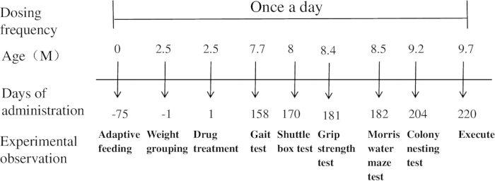
Figure 1: Schematic diagram of the experimental procedure. Please click here to view a larger version of this figure.
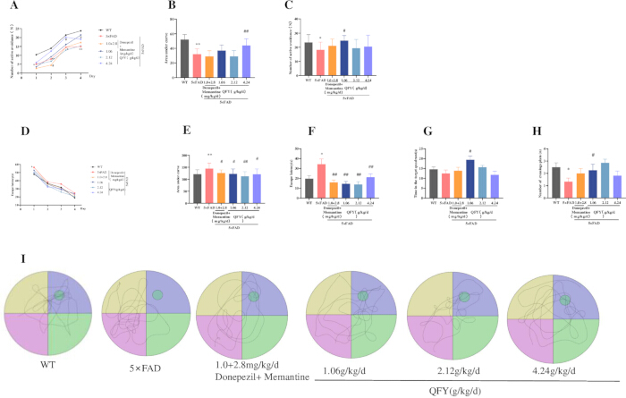
Figure 2: QFY administration improves learning and memory performance in 5xFAD mice. (A–C) Shuttle box test: (A) Number of active avoidance responses during the learning period. (B) Area under the curve (AUC) for active avoidance responses during the learning period. (C) Number of active avoidance responses during the test period. (D–I) Morris water maze test: (D) Escape latency during the learning period. (E) AUC for escape latency during the learning period. (F) Escape latency during the test period. (G) Time spent in the target quadrant. (H) Number of platform crossings. (I) Movement trajectories during the test period. Mean ± SD, n = 11–20 per group, with exact n indicated in each bar. * = p < 0.05, vs WT, Student`s t-test; # = p < 0.05, ## = p < 0.01, vs 5xFAD; One-way ANOVA followed by Dunnett's multiple comparisons test. Please click here to view a larger version of this figure.
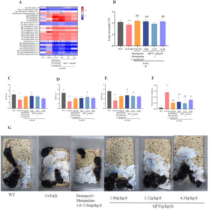
Figure 3: Effects of QFY administration on aging-associated functional measures in 5xFAD mice. (A) Gait heat map. The color scale is based on row‑wise Z‑score normalization, where red indicates values above the group mean, blue indicates values below the group mean, and white represents the mean level. (B) Grip strength test. (C–G) Colony nesting test: (C) Nesting score 6 h, (D) Nesting score at 12 h. (E) Nesting score at 24 h. (F) Untorn tissue weight. (G) Representative images of nests at 24 h. Mean ± SD, n = 3 per group, with exact n indicated in each bar. * = p < 0.05, ** = p < 0.01, vs WT, Student`s t-test; # = p < 0.05, ## = p < 0.01, vs 5x FAD, One-way ANOVA followed by Dunnett's multiple comparisons test. Please click here to view a larger version of this figure.
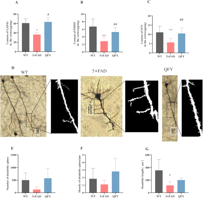
Figure 4: QFY attenuates synaptic structural alterations in the cerebral cortex of 5xFAD mice. (A) Content of GAP-43 in the cortex. (B) Content of PSD-95 in the cortex. (C) Content of SYN in the cortex. (D) Golgi-Cox staining. (E) Number of dendritic spines. (F) Density of dendritic spines. (G) Dendritic length. Mean ± SD, n = 3–14 per group, with exact n indicated in each bar. * = p < 0.05, ** = p < 0.01, vs WT, Student`s t-test; # = p < 0.05, ## = p < 0.01, vs 5xFAD, Student`s t-test. Please click here to view a larger version of this figure.
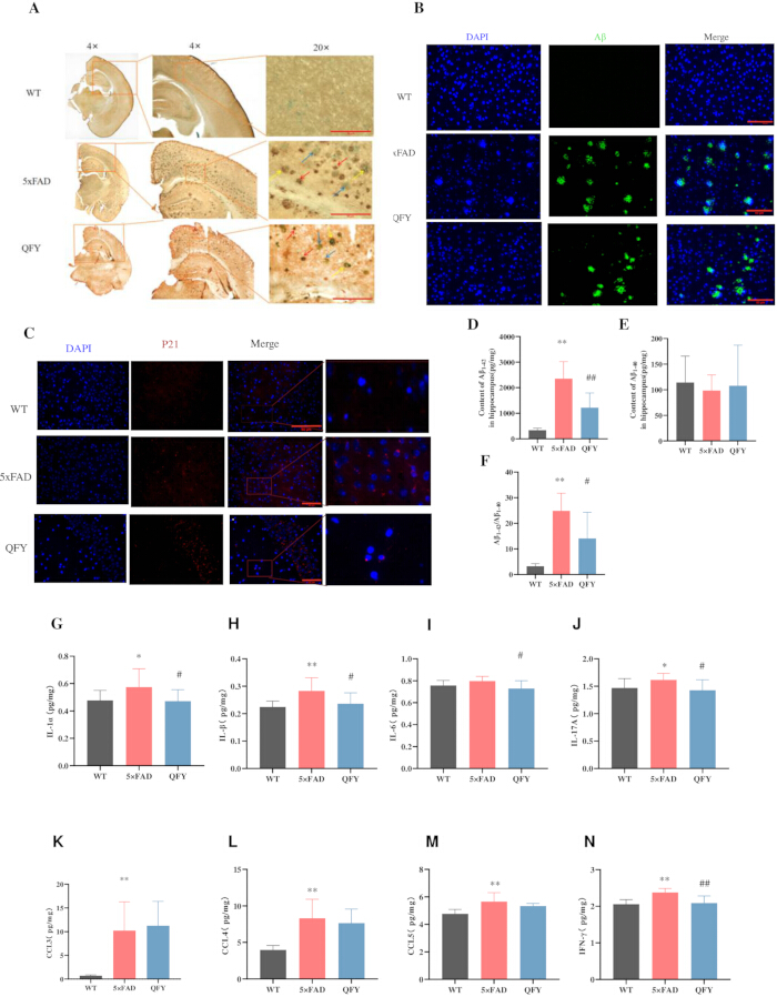
Figure 5: QFY reduced Aβ deposition and cell senescence in the brains of 5xFAD mice. (A) Combined SA-β-Gal staining and Aβ immunohistochemistry, SA-β-Gal (green, blue arrow), Aβ (brown, red arrow), SA-β-Gal + Aβ (yellow arrow), n = 3, Scale bar = 10 µm, 20x objective. (B) Immunofluorescence images showing Aβ in the cerebral cortex after treatment of QFY, n = 3, Scale bar = 50 µm, 40x objective. (C) Immunofluorescence images showing p21in the cerebral cortex after treatment of QFY. n = 1, Scale bar = 50 µm, 40x objective. (D–F) ELISA. (D) Content of Aβ1-42 in the hippocampus. (E) Content of Aβ1-40 in the hippocampus. (F) Aβ1-42/ Aβ1-40. (G–N) Luminex cytokine analysis. (G) Content of IL-1α in the cortex. (H) Content of IL-1β in the cortex. (I) Content of IL-6 in the cortex. (J) Content of IL-17A in the cortex. (K) Content of CCL3 in the cortex. (L) Content of CCL4 in the cortex. (M) Content of CCL5 in the cortex. (N) Content of IFN-γ in the cortex. Mean ± SD, n = 8–14 per group, with exact n indicated in each bar. * = p < 0.05, ** = p < 0.01, vs WT, Student’s t-test; # = p < 0.05 ## = p < 0.01, vs 5xFAD, Student’s t-test. Please click here to view a larger version of this figure.
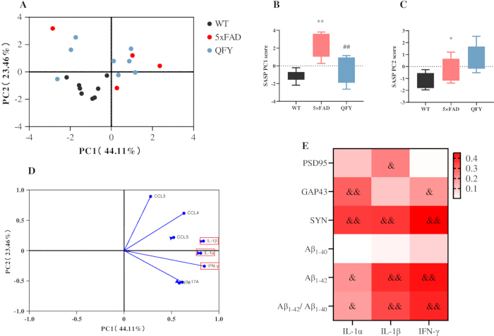
Figure 6: Principal component and correlation analyses of SASP factors, synaptic proteins, and Aβ levels in 5xFAD mice following QFY treatment. (A–D) Principal component analysis (PCA) of SASP factors. (A) PCA score plot. (B) PC1 scores. (C) PC2 scores. (D) PCA loading plot. (E) Heatmap generated from the correlation analysis between the SASP and PSD-95, GAP-43, SYN, Aβ1-40, Aβ1-42, and the ratio of Aβ1-42/Aβ1-40. Colors closer to red indicate higher R-values, while colors closer to white indicate lower R-values. Mean ± SD; n = 8 per group. * = p < 0.05, ** = p < 0.01,vs WT; ## = p < 0.01, vs 5xFAD, & = p < 0.05, && = p < 0.01, indicates a significant correlation. Pearson correlation analysis was used to assess associations between variables. Please click here to view a larger version of this figure.
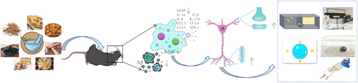
Figure 7: Proposed model of the effects of QFY on age-related cognitive decline and cellular senescence-associated phenotypes in 5xFAD mice. Please click here to view a larger version of this figure.
ディスカッション
神経シナプス可塑性は、認知機能において重要な役割を果たしています。AD患者の脳内では、Aβ沈着がさまざまな領域に広がり、それが直接的にシナプス構造の損傷、興奮性シナプスの消失、および認知機能の低下を引き起こします28。GAP-43(シナプス前膜タンパク質)29、PSD-95(シナプス後膜に位置する足場タンパク質)30、およびSYN(シナプス小胞に関連するリン酸化タンパク質)31は、シナプス構造の可塑性とADの病態形成において重要な役割を担っています32。本研究では、QFY投与により、5xFADマウスの脳において樹状突起スパインの数およびGAP-43、PSD-95、SYNのタンパク質レベルが増加し、その結果、学習・記憶能が改善し、Aβプラークの負荷が軽減され、シナプス構造が回復することが明らかになりました。
加齢はAD発症における重要なリスク要因であり、Aβ産生を促進します。加齢の特徴には、筋力の低下、歩行の不均衡、および基本的日常生活動作の低下が含まれます33,34。SA-β-Galによって特徴づけられる細胞老化は、加齢およびADなどの加齢性疾患の発症に関連していることが報告されています11,35。細胞老化を選択的に除去することで、健康寿命が延長し、加齢性疾患が遅延します36。遺伝学的または薬理学的アプローチによる老化細胞の除去は、ADモデルマウスにおいてAβ沈着を改善し、学習および記憶機能を向上させました37。DasatinibとQuercetin(D+Q)による併用療法は、オリゴデンドロサイト前駆細胞の老化を抑制し、神経炎症を緩和し、AβプラークとSA-β-Gal活性を減少させ、APP/PS1マウスにおけるAβ蓄積と認知機能障害を防止します37。Memantine治療は、SAMP8マウスの脳内におけるSA-β-Gal活性を有意に低下させ、空間学習および記憶を改善しました。同様の効果が、memantineとdonepezilの併用によっても観察されました38。本研究では、QFY治療が歩行を改善し、前肢筋力を強化し、基本的日常生活動作の能力を向上させたこと、そして特に、5xFADマウスの脳内におけるSA-β-Gal染色で示される細胞老化を緩和させることでこれらが達成されたことを見出しました。
AD患者および動物モデルのいずれにおいても、老化アストロサイト、ミクログリア、内皮細胞、およびニューロンが認められる39,40。ケモカインや炎症性サイトカインを含むSASPの分泌は、細胞老化の顕著な特徴である。過剰なSASPの分泌は慢性炎症を引き起こし、その結果として組織損傷や細胞老化を招く可能性がある41。IL-1α、IL-1β、IL-6、IL-8、IFN-γ、CCL2、CCL3、CCL4、CCL5、CXCL1などのSASPが持続的に分泌されることで、慢性炎症、正常細胞のパラクリン様老化、細胞代謝およびエピジェネティック制御の変化、そしてAβの持続的な産生が促進される42,43,44,45。AD患者およびADモデルの脳内では、IL-1α、IL-1β、IL-6、IL-17A、およびIFN-γのレベルが有意に上昇している46。研究により、D+QがIL-1αやIFN-γを含む炎症因子の分泌を抑制し、APP/PS1マウスにおけるAβプラークおよびSA-β-Gal活性を減少させることが示されている37。また、QFYがIL-1α、IL-1β、IL-6、IL-17A、およびIFN-γを含む複数のSASP因子の発現を減少させ、それによって老化に伴う炎症表現型を軽減させることが判明した。
p21はサイクリン依存性キナーゼ阻害剤のCIP/KIPファミリーに属し、サイクリン-CDK2/4複合体の近接ブレーキとして機能することで、細胞ストレス、DNA損傷、および老化が発生した際にG1/G2期停止を強制します。p21量の持続的な上昇は、安定した老化プログラムへの移行であると広く解釈されています47,48。 新たなデータにより、5xFADマウスは異常に高い脳内p21負荷を抱えており、これがD+Q併用療法によって著しく軽減されることが明らかになっています49。これと一致して、本研究では、QFYの投与がこれらの動物において皮質Aβプラークを有意に減少させ、p21発現を抑制することを見出しました。
総合的に、これらの知見は、QFY治療が5xFADマウスにおける加齢に伴う認知機能低下および細胞老化表現型の改善に関連し、同時にシナプスの構造的損傷を軽減させることを示唆している(図7)。
本研究にはいくつかの限界があることを認める必要がある。第一に、雄の5xFADマウスのみを使用したため、観察された効果に性差があるかどうかはまだ特定されていない。第二に、本データは細胞老化関連表現型の減衰と認知機能改善との間に強い相関があることを示しているが、因果関係を立証するための直接的なメカニズム介入実験は行っていない。第三に、ヒトのADに対する知見の翻訳的妥当性は、種差および臨床的な薬物動態データの不足によって制限される。これらの知見を拡張するためには、雌動物の導入、老化特異的な介入、および適切に設計された臨床試験を組み込んだ今後の研究が必要である。
開示事項
著者らに開示すべき利益相反はありません。
謝辞
本研究は、中国国家自然科学基金(82205078, 82374062)、山東省自然科学基金(ZR2026MS1293, ZR2021QH157)、山東省中医学科学技術プロジェクト(M20251730)、中国西蔵自治区科学技術プロジェクト(XZ202601ZY0167)、および山東省技術革新指導計画(中央政府指導地方科学技術発展基金)(YDZX2023137)による助成、および「クラス1新中薬としての通理腸溶性カプセルの開発および工業化研究」と題されたプロジェクトの支援を受けて実施されました。
材料
| 名前 | 会社 | カタログ番号 | コメント |
|---|---|---|---|
| 薬剤 | |||
| ドネペジル | Shanghai Yuanye Technology | ロット番号 J15HB188725 | |
| メマンチン | Shanghai Yuanye Technology | ロット J21HB189391 | |
| 気富飲 | 魯南製薬後普研究開発センター | ロット番号 2209001 | |
| 行動テスト | |||
| CatWalk XT | Noldus | CatWalkXT 10.0 ソフトウェア | |
| 握力計 | Bileadbiosci | BLD-ZL-20 | |
| モーリス水迷路 | 上海新软信息科技有限公司 | 120cm | |
| シャトルボックス | 上海新瑞安情報技術有限公司 | XR-XC404 | |
| 抗体 | |||
| 抗A抗体β1-42 抗体 | Abcam | カタログ番号 ab201061 | |
| 抗p21/CIP1/CDKN1A抗体 | Novus Biologicals | カタログ番号 NBP3-15661 | |
| アッセイキット | |||
| Aβ1-40 ELISAキット | Thermo Fisher Scientific | カタログ番号 KHB3482 | |
| Aβ1-42 ELISAキット | Thermo Fisher Scientific | カタログ番号 KHB3442 | |
| Bio-Plex Pro Mouse Cytokine 23-plex Assay kit | R&D Systems | カタログ番号 LXLBM23-1 | |
| GAP-43 ELISAキット | 江蘇省酵素免疫測定業界 | カタログ番号 MM-47506M1 | |
| Golgi-Cox染色キット | BeiJing Biolead Biology Sci & テック社 | カタログ番号 PK401 | |
| PSD-95 ELISAキット | 江蘇省酵素免疫測定業界 | Cat# MM-45953M1 | |
| 細胞老化 β-ガラクトシダーゼ染色キット | Beyotime | カタログ番号 C0602 | |
| シナプトフィジン (SYN) ELISAキット | 江蘇省酵素免疫測定業界 | カタログ番号 MM-46504M1 | |
| 試薬 & 試薬 | |||
| 抗ウサギおよび抗マウスIgG(二次抗体) | スターター | カタログ番号 S0C1001 | |
| DAB発色剤 | スターター | カタログ番号 S0C1001 | |
| ソフトウェア | |||
| CatWalk XT | Noldus Information Technology | バージョン 10.0 | |
| ImageJ | アメリカ国立衛生研究所 (NIH) | バージョン 1.53 |
参考文献
- Joe E, Ringman JM. Cognitive symptoms of Alzheimer's disease: clinical management and prevention. BMJ. 2019;367:l6217.
- Scheff SW, et al. Synaptic alterations in CA1 in mild Alzheimer disease and mild cognitive impairment. Neurology. 2007;68(18):1501-8.
- Muralidar S, et al. Role of tau protein in Alzheimer's disease: the prime pathological player. Int J Biol Macromol. 2020;163:1599-617.
- Hardy JA, Higgins GA. Alzheimer's disease: the amyloid cascade hypothesis. Science. 1992;256(5054):184-5.
- Calsolaro V, Edison P. Neuroinflammation in Alzheimer's disease: current evidence and future directions. Alzheimers Dement. 2016;12(6):719-32.
- Campisi J. Senescent cells, tumor suppression, and organismal aging: good citizens, bad neighbors. Cell. 2005;120(4):513-22.
- Yao R. An in-silico assessment suggests the potential effects of goji (fruit of Lycium barbarum L.) against aging-related diseases. Food Med Homol. 2025;2(2):9420036.
- Campisi J, d'Adda di Fagagna F. Cellular senescence: when bad things happen to good cells. Nat Rev Mol Cell Biol. 2007;8(9):729-40.
- Salminen A, Kaarniranta K, Kauppinen A. ER stress activates immunosuppressive network: implications for aging and Alzheimer's disease. J Mol Med (Berl). 2020;98(5):633-50.
- Mirabella F, et al. Prenatal interleukin 6 elevation increases glutamatergic synapse density and disrupts hippocampal connectivity in offspring. Immunity. 2021;54(11):2611-31.e8.
- Baker DJ, Petersen RC. Cellular senescence in brain aging and neurodegenerative diseases: evidence and perspectives. J Clin Invest. 2018;128(4):1208-16.
- Lucas V, Cavadas C, Aveleira CA, Hook V. Cellular senescence: from mechanisms to current biomarkers and senotherapies. Pharmacol Rev. 2023;75(4):675-713.
- Bloss EB, et al. Evidence for reduced experience-dependent dendritic spine plasticity in the aging prefrontal cortex. J Neurosci. 2011;31(21):7831-9.
- Ye XS, et al. The food and medicine homologous Chinese medicine from Leguminosae species: a comprehensive review on bioactive constituents with neuroprotective effects on nervous system. Food Med Homol. 2025;2(2):9420033.
- Dickstein DL, Weaver CM, Luebke JI, Hof PR. Dendritic spine changes associated with normal aging. Neuroscience. 2013;251:21-32.
- Wang SY, et al. Qifu-Yin attenuates AGEs-induced Alzheimer-like pathophysiological changes through the RAGE/NF-kappaB pathway. Chin J Nat Med. 2014;12(12):920-8.
- Yang X, et al. Qi-fu-yin attenuated cognitive disorders in 5xFAD mice of Alzheimer's disease animal model by regulating immunity. Front Neurol. 2023;14:1183764.
- Xiao QY, et al. Effects of Qi-Fu-Yin on aging of APP/PS1 transgenic mice by regulating the intestinal microbiome. Front Cell Infect Microbiol. 2023;12:1048513.
- Chen K, et al. Safety and toxicity of Qi-Fu Yin, a classical traditional Chinese medicine prescription: a nonclinical research. J Appl Toxicol. 2025;45(9):1811-38.
- Wang L, Qiao P, Yue L, Sun R. Is Qi Fu Yin effective in clinical treatment of dementia? A meta-analysis of 697 patients. Medicine (Baltimore). 2021;100(5):e24526.
- Li H, et al. Identification of chemical components of Qi-Fu-Yin and its prototype components and metabolites in rat plasma and cerebrospinal fluid via UPLC-Q-TOF-MS. Evid Based Complement Alternat Med. 2021;2021:1995766.
- Lei X, et al. Integrating network pharmacology and component analysis to study the potential mechanisms of Qi-Fu-Yin Decoction in treating Alzheimer's disease. Drug Des Devel Ther. 2023;17:2841-58. doi:10.2147/DDDT.S402624.
- Takeshita H, et al. Modified forelimb grip strength test detects aging-associated physiological decline in skeletal muscle function in male mice. Sci Rep. 2017;7:42323.
- Caggiano V, et al. Midbrain circuits that set locomotor speed and gait selection. Nature. 2018;553(7689):455-60.
- Ehret B, et al. Population-level coding of avoidance learning in medial prefrontal cortex. Nat Neurosci. 2024;27(9):1805-15.
- Curdt N, et al. Search strategy analysis of Tg4-42 Alzheimer mice in the Morris water maze reveals early spatial navigation deficits. Sci Rep. 2022;12:5451.
- Deacon RMJ. Assessing nest building in mice. Nat Protoc. 2006;1(3):1117-9.
- Tu S, Okamoto SI, Lipton SA, Xu H. Oligomeric Aβ-induced synaptic dysfunction in Alzheimer's disease. Mol Neurodegener. 2014;9:48.
- Benowitz LI, Routtenberg A. GAP-43: an intrinsic determinant of neuronal development and plasticity. Trends Neurosci. 1997;20(2):84-91.
- Dore K, et al. PSD-95 protects synapses from β-amyloid. Cell Rep. 2021;35(9):109194.
- Kokotos AC, et al. Synaptophysin sustains presynaptic performance by preserving vesicular synaptobrevin-II levels. J Neurochem. 2019;151(1):28-37.
- Martin SB, et al. Synaptophysin and Synaptojanin-1 in Down syndrome are differentially affected by Alzheimer's disease. J Alzheimers Dis. 2014;42(3):767-75.
- Namioka N, et al. Oxidative stress and inflammation are associated with physical frailty in patients with Alzheimer's disease. Geriatr Gerontol Int. 2016;17(6):913-8.
- Kojima G, Liljas A, Iliffe S, Walters K. Prevalence of frailty in mild to moderate Alzheimer's disease: a systematic review and meta-analysis. Curr Alzheimer Res. 2017;14(12):1256-63.
- Dimri GP, et al. A biomarker that identifies senescent human cells in culture and in aging skin in vivo. Proc Natl Acad Sci U S A. 1995;92(20):9363-7.
- Baker DJ, et al. Naturally occurring p16Ink4a-positive cells shorten healthy lifespan. Nature. 2016;530(7589):184-9.
- Zhang P, et al. Senolytic therapy alleviates Abeta-associated oligodendrocyte progenitor cell senescence and cognitive deficits in an Alzheimer's disease model. Nat Neurosci. 2019;22(5):719-28.
- Ota H, Ogawa S, Ouchi Y, Akishita M. Protective effects of NMDA receptor antagonist, memantine, against senescence of PC12 cells: a possible role of nNOS and combined effects with donepezil. Exp Gerontol. 2015;72:109-16.
- Herdy JR, et al. Increased post-mitotic senescence in aged human neurons is a pathological feature of Alzheimer's disease. Cell Stem Cell. 2022;29(12):1637-52.e6.
- Hu Y, et al. Replicative senescence dictates the emergence of disease-associated microglia and contributes to Abeta pathology. Cell Rep. 2021;35(10):109228.
- Tchkonia T, et al. Cellular senescence and the senescent secretory phenotype: therapeutic opportunities. J Clin Invest. 2013;123(3):966-72.
- Guerrero A, De Strooper B, Arancibia-Cárcamo IL. Cellular senescence at the crossroads of inflammation and Alzheimer's disease. Trends Neurosci. 2021;44(9):714-27.
- Acosta JC, et al. A complex secretory program orchestrated by the inflammasome controls paracrine senescence. Nat Cell Biol. 2013;15(8):978-90.
- Wang B, Han J, Elisseeff JH, Demaria M. The senescence-associated secretory phenotype and its physiological and pathological implications. Nat Rev Mol Cell Biol. 2024;25(12):958-78.
- Gaikwad S, Senapati S, Haque MA, Kayed R. Senescence, brain inflammation, and oligomeric tau drive cognitive decline in Alzheimer's disease: evidence from clinical and preclinical studies. Alzheimers Dement. 2024;20(1):709-27.
- Si Z, Sun L, Wang X. Evidence and perspectives of cell senescence in neurodegenerative diseases. Biomed Pharmacother. 2021;137:111327.
- Chen X, et al. Senescence-like changes induced by expression of p21Waf1/Cip1 in NIH3T3 cell line. Cell Res. 2002;12(3-4):229-33.
- Engeland K. Cell cycle regulation: p53-p21-RB signaling. Cell Death Differ. 2022;29(5):946-60.
- Choi I, et al. Autophagy enables microglia to engage amyloid plaques and prevents microglial senescence. Nat Cell Biol. 2023;25(7):963-74.