本稿では、Exonuclease Vを用いて単一のヒト卵母細胞からミトコンドリアDNAを濃縮し、Illumina互換のシーケンシングプラットフォームを用いてミトコンドリアDNA変異を高精度に検出するためのデュプレックスシーケンシングライブラリを調製するプロトコルを紹介します。
この記事について
サマリー
要約
卵母細胞には、独自のゲノムであるミトコンドリアDNA(mtDNA)を持つエネルギー産生オルガネラであるミトコンドリアが密集しています。各細胞にはmtDNAが複数コピー含まれており、そのコピー数は組織の種類によって異なります。卵母細胞は最も高いmtDNAコピー数を有しており、1細胞あたり数十万個のmtDNA分子を含んでいます。ミトコンドリアは母系のみで継承されるため、継承、老化、および疾患の研究にはmtDNA変異の正確な検出が不可欠です。複数のmtDNAコピーが存在することで、同一細胞内に野生型と変異型の分子が共存することがあり、これはヘテロプラスミーと呼ばれます。この状態では、低頻度の変異やde novo変異が1%未満の頻度で発生することがあります。従来の次世代シーケンシング(NGS)では、ライブラリ調製およびシーケンシング中に導入されるエラーと、これらの希少な変異を確実に区別するのに十分な精度が不足しています。本稿では、Exonuclease Vを用いて線状DNAを除去することで単一のヒト卵母細胞からmtDNAを濃縮し、それに続いて高精度なmtDNA解析のためのデュプレックスシーケンシングライブラリを調製するプロトコルを紹介します。このワークフローにより、個々の卵母細胞のエラー補正シーケンシングが可能となり、低頻度のmtDNA変異の信頼性の高い検出、およびヘテロプラスミーとde novo変異誘発の解析が促進されます。本プロトコルは、Illumina互換のシーケンシングプラットフォームを用いて、単一卵母細胞におけるミトコンドリアゲノム変異を調査するための再現可能なアプローチを提供します。
概要
ミトコンドリアは、エネルギー産生、アポトーシス、シグナル伝達、およびカルシウム恒常性など、必須の細胞プロセスにおいて根本的な役割を果たしています1。ミトコンドリアは独自のゲノムであるミトコンドリアDNA(mtDNA)を保持しており、これは哺乳類において長さ約16.6 kbの環状二本鎖分子です。内共生過程において、祖先的なミトコンドリア遺伝子の大部分は核ゲノムへと転移しましたが、mtDNAには、13個の酸化的にリン酸化(OXPHOS)ポリペプチド、22個の転移RNA(tRNA)、および2個のリボソームRNA(rRNA)をコードする37個の遺伝子が保持されています2。mtDNAの変異はミトコンドリアの遺伝子発現および機能を損ない、深刻な疾患を引き起こす可能性があります3。ミトコンドリアは母親の系統を通じてのみ継承されるため、卵母細胞のmtDNAは受精、胚発生、および子の健康において極めて重要な役割を担っています4。したがって、mtDNA変異とその頻度の検出および特性解析は非常に重要です。例えば、加齢や疾患に関連する生殖細胞系列の突然変異を理解することは、女性不妊症の治療戦略を開発するための重要な前提条件となります。
核DNAとは異なり、mtDNAは1細胞あたりに複数コピー存在しており、組織の種類によって体細胞では数百から数千コピー、成熟卵母細胞では100,000コピーを超えます4。このmtDNAの高コピー数は、単一細胞レベルでのミトコンドリア変異誘発を研究する上で、卵母細胞が非常に適している理由となっています。しかし、特に極めて低い頻度で存在するde novo mtDNA変異の検出は依然として困難です。これは、従来の次世代シーケンシング(NGS)のエラー率が、多くの真のバリアントの頻度を上回るためです5。したがって、ライブラリ調製、増幅、およびシーケンシング中に導入されるアーティファクトと真の変異を区別するために、エラー補正シーケンシング法が必要となります。そのようなアプローチの一つが、デュプレックスシーケンシング(DS)です6,7。DSでは、増幅前にDNA断片を、二本鎖のランダム化された12ヌクレオチド分子タグを含むアダプターにライゲーションします。これらのユニーク分子識別子(UMI)が元のDNA分子の両端にラベル付けされるため、同じテンプレート鎖に由来するシーケンシングリードを、共通のタグ配列に基づいてファミリーとしてグループ化することが可能になります。各リードファミリーから単鎖コンセンサス配列(SSCS)が生成され、その後、元のDNA二本鎖に由来する相補的なSSCSを組み合わせることで、デュプレックスコンセンサス配列(DCS)が作成されます。両方の相補的なSSCS内の大部分のリードで検出され、かつ得られたDCSで確認されたバリアントのみが真の変異とみなされるため、従来のNGSアプローチよりも数桁低いエラー率を実現できます8。この戦略は、単一細胞における低頻度のmtDNAバリアントやヘテロプラスミーの検出に特に有効です。
公表されているほとんどのデュプレックスシーケンシングプロトコルの大きな制限は、ライブラリ調製時に比較的大量の入力DNAが必要であることであり、そのためこれらの手法はシングルセルへの適用には適していません6,7,9。本プロトコルの全体的な目的は、mtDNA濃縮、ライブラリ調製、およびシーケンシングに最適化したワークフローを通じて、単一のヒト卵母細胞からのmtDNAの高精度なデュプレックスシーケンシングを可能にすることです。核内ミトコンドリアDNA断片(NUMTs)のシーケンシングを最小限に抑え、シーケンシング効率を向上させるため、本プロトコルでは、環状のmtDNAを保持しつつ線状DNAを選択的に消化するExonuclease Vベースの濃縮ステップを組み込んでいます。得られたワークフローは、単一卵母細胞における正確なmtDNA変異検出のための実用的なアプローチを提供し、ヘテロプラスミー、de novo突然変異、老化、およびミトコンドリア病を調査する研究に適しています。
プロトコル
Human oocyte collection and processing were approved by the Ethics Commission of the Johannes Kepler University Linz (Approval No. 1293/2020). Perform all procedures in accordance with institutional guidelines, including informed consent, sample anonymization, and all applicable regulations governing the use of human tissue.
NOTE: Although this protocol is described for human oocytes, adapt the species-specific primers to apply the workflow to other species. Use DNA low-binding consumables (e.g., tubes and pipette tips) throughout the protocol to minimize sample loss. Perform all steps up to the first purification in the original low-binding oocyte collection tube. Do not vortex samples, as vortexing may damage DNA strands. Instead, mix reaction components by gentle pipetting or gentle tube flicking, followed by brief centrifugation. Whenever possible, perform DNA isolation, master mix preparation, and reaction setup in dedicated polymerase chain reaction (PCR) workstations or physically separated laboratory areas to minimize contamination.
1. Preparation of Buffers and Reagents
- Prepare the Oocyte Lysis Buffer (OLB), 10 mM Tris-HCl, TE buffer, TElow buffer, and 10 mM Tris-NaCl according to Table 1 by combining the specified volumes of the stock solutions and bringing each solution to the designated final volume with molecular biology-grade water. Aliquot 1 mL of the prepared buffers, store the aliquots at 4°C, and use them within 1 year.
Table 1: Composition of buffers used throughout the protocol. Buffer compositions and final concentrations for preparation of 10 mM Tris-HCl, TE buffer, TElow buffer, 10 mM Tris-NaCl, and Oocyte Lysis Buffer (OLB). Prepare all buffers using molecular biology-grade water. Adjust the pH of Tris-HCl and EDTA stock solutions before buffer preparation, if required, according to the manufacturer's recommendations. OLB is supplemented with thermolabile proteinase K immediately before sample lysis as described in the protocol. Please click here to download this file.
2. Single Oocyte Collection
NOTE: Human oocytes used in this protocol were collected from patients scheduled for intracytoplasmic sperm injection (ICSI) at the Kinderwunsch Zentrum (fertility center) of the Kepler University Hospital, Linz, Austria. Ovarian stimulation protocols were based on the patient's predicted ovarian response, and dosages were adjusted according to individual characteristics, including age, anti-Müllerian hormone (AMH) levels, and body weight, in accordance with the recommendations of the European Society of Human Reproduction and Embryology (ESHRE)10. Pituitary suppression was achieved using either gonadotropin-releasing hormone (GnRH) agonist or GnRH antagonist protocols in combination with gonadotropin stimulation to promote follicular maturation. Follicular growth was monitored regularly by transvaginal ultrasonography before ovulation was triggered. Oocytes were then retrieved by transvaginal follicular aspiration, and the follicular fluid containing the cumulus–oocyte complexes (COCs) was collected11. Only immature or unfertilized oocytes that could not be used for the patient's ICSI treatment and would otherwise have been discarded were available for research after informed consent had been obtained. Patients with higher antral follicle counts (AFCs) were preferentially approached for oocyte donation because they were more likely to produce immature or unfertilized oocytes suitable for research. All procedures performed before the selection of oocytes for research must comply with national regulations governing in vitro fertilization and the use of human tissue. Handle all materials under sterile conditions in a laminar flow hood. Ensure that all CE-certified materials and culture media that come into direct contact with the oocytes are sterile, prewarmed to 37°C, and maintained at a pH of 7.20–7.40.
- Collect COCs from the follicular fluid and transfer them into 750 µL of GM501 Cult medium under sterile mineral oil following controlled ovarian hyperstimulation and transvaginal oocyte retrieval.
- Remove the surrounding cumulus cells from the zona pellucida (ZP) by enzymatic digestion with hyaluronidase. Incubate the oocytes in 500 µL of GM501 Hyaluronidase (80 U/mL) for 30–60 s at 37°C, followed by three washes in 750 µL of GM501 Cult medium. Carefully remove any remaining cumulus cells mechanically using denudation pipettes.
- For telophase I (TI) or metaphase II (MII) oocytes, open the ZP using a series of laser pulses adjacent to the polar body and remove the first polar body using micromanipulators. Adjust the laser settings according to the thickness of the ZP (1–3 pulses; pulse duration, 1.5–2.6 ms; spot diameter, 16–20 µm). Use the laser to remove any remaining cumulus cells, thereby minimizing contamination of the oocyte with somatic cell DNA.
- Transfer each oocyte individually into 2–3 µL of 1× phosphate-buffered saline (PBS) in a 200 µL low-binding tube. Immediately freeze the tube at −20°C. For long-term storage, transfer the frozen oocytes to −80°C.
NOTE: After thawing, perform the remaining protocol continuously without interruption until completion of the first amplification step. - To monitor potential environmental DNA contamination throughout the protocol, include a negative control consisting of 2 µL of 1× PBS without an oocyte during library preparation (Step 4 onward). Process the negative control identically to all oocyte samples. No detectable DNA should be observed in the negative control at any stage of the protocol.
3. Adapter Synthesis
NOTE: Prepare the adapters before starting library preparation. Store synthesized adapters at −80°C for up to 3 months. Prepare aliquots to avoid repeated freeze–thaw cycles, and do not refreeze adapters after thawing. All oligonucleotide sequences used in this protocol are listed in Table 2.
Table 2: Oligonucleotides and primers used throughout the protocol. The table lists all oligonucleotides used for adapter synthesis, library amplification, quantitative polymerase chain reaction (qPCR), library quantification, and mitochondrial DNA (mtDNA) enrichment estimation, together with their sequences, purification methods, supplied amounts or recommended stock concentrations, and manufacturer. The carrier oligonucleotide sequence shown is the example sequence used in this protocol. The asterisk (*) within primer sequences denotes a phosphorothioate linkage. The randomized nucleotides (N) in the mws55 adapter represent the unique molecular identifier (UMI), whereas Y denotes the degenerate pyrimidine base (C or T). Unless otherwise indicated, oligonucleotides were synthesized using standard desalting purification. Please click here to download this file.
- Prepare an aliquot of 96% ethanol (EtOH) and store it at −20°C for subsequent purification steps. Prepare 80% EtOH fresh immediately before use.
- Mix 20.5 µL each of 100 µM mws51_short and 100 µM mws55 oligonucleotides in a 200 µL low-binding tube to obtain a total volume of 41 µL (2,000 pmol of each oligonucleotide).
- Incubate the combined oligonucleotides at 95°C for 5 min in a thermocycler with the lid temperature set to 110°C. Start the timer after the sample reaches 95°C, switch off the thermocycler after 5 min, and leave the tubes inside the thermocycler for 1 h to allow slow cooling to room temperature (RT) and annealing (“annealed adapter”).
- Prepare an Extension Master Mix by combining 1× NEB Buffer 2 (5.6 µL of 10× stock), 3.5 mM deoxynucleotide triphosphates (dNTPs; 5.6 µL of 10 mM stock), 11.5 U of Klenow fragment (2.3 µL of 5 U/µL stock), and 2.5 µL of molecular biology-grade water to a final volume of 16 µL.
- Remove 1 µL of the annealed adapter prepared in Step 3.3, dilute it 1:20 in TElow buffer, label the aliquot “annealed,” and store it at 4°C for agarose gel electrophoresis.
- Add 16 µL of the Extension Master Mix to the remaining 40 µL of annealed oligonucleotides and mix thoroughly.
- Incubate the reaction at 37°C for 1 h with the thermocycler lid set to 47°C.
- Purify the extended oligonucleotides by EtOH precipitation. Add 28 µL of ammonium acetate (NH4OAc) to the 56 µL reaction mixture and mix thoroughly.
- Transfer the entire reaction to a 1.5 mL low-binding tube and add 168 µL of ice-cold 96% EtOH.
- Invert the tube several times and incubate at −20°C for 30 min to precipitate the DNA.
- Prepare 1 mL of fresh 80% EtOH and cool it to −20°C. Pre-cool the centrifuge to 4°C.
- Centrifuge at 14,000 × g for 30 min.
- Carefully remove the supernatant without disturbing the pellet. Add 1 mL of ice-cold 80% EtOH without mixing or inverting the tube.
- Centrifuge at 14,000 × g for 5 min.
- Remove all residual EtOH and air-dry the DNA pellet for 10–15 min, until no visible liquid remains and the pellet appears transparent. Do not overdry the oligonucleotides.
- Resuspend the pellet in 41 µL of molecular biology-grade water.
- Remove 1 µL, dilute it 1:20 in TElow buffer, label the aliquot “extended,” and store it at 4°C.
- Prepare a Restriction Master Mix by combining 47 µL of molecular biology-grade water, 1× CutSmart Buffer (10 µL of 10× stock), and 15 U of HpyCH4III (3 µL of 5 U/µL stock) to a final volume of 60 µL.
- Add 60 µL of the Restriction Master Mix to 40 µL of purified oligonucleotides and mix thoroughly.
- Incubate the reaction at 37°C for 16 h with the thermocycler lid set to 47°C.
- Prepare 6.5 mL of fresh 80% EtOH and cool it to −20°C. Pre-cool the centrifuge to 4°C.
- Transfer the digested adapters to a 1.5 mL low-binding tube and add 900 µL of molecular biology-grade water.
- Add 500 µL of NH4OAc and mix thoroughly.
- Divide the solution into six 250 µL aliquots in 1.5 mL low-binding tubes. Add 500 µL of ice-cold 96% EtOH to each tube.
- Invert the tubes several times and incubate at −20°C for 30 min to precipitate the DNA.
- Centrifuge at 14,000 × g for 30 min. Carefully remove the supernatant without disturbing the pellet. Add 1 mL of ice-cold 80% EtOH to each tube without mixing or inverting. Centrifuge at 14,000 × g for 5 min.
- Remove all residual EtOH and air-dry the adapter pellets.
- Resuspend each pellet in 6.7 µL of Tris-NaCl buffer and pool all six suspensions to obtain a final volume of 41 µL. Remove 1 µL, dilute it 1:10 in TElow buffer, label the aliquot “cut,” and store it at 4°C.
- Prepare aliquots of the adapter stock and store them at −80°C. Measure the adapter concentration and determine the A260/280 and A260/230 absorbance ratios of the “cut” aliquot using a spectrophotometer. The expected adapter concentration is 30–50 µM, with an A260/280 ratio of >1.7 and an A260/230 ratio of >1.9.
- Run the “annealed,” “extended,” and “cut” aliquots on a 2% agarose gel at 125 V for 45 min to verify adapter formation and complete restriction digestion (Figure 1).
NOTE: The “annealed” aliquot should contain two bands representing the annealed and unannealed fractions. The annealed adapter is expected to migrate at approximately 90 bp and consists of a 13-bp double-stranded region with a 68-nt single-stranded overhang, whereas the unannealed oligonucleotides migrate at approximately 60 bp. Following extension, the adapter consists of a 37-bp double-stranded region with a 44-nt Y-shaped overhang and is expected to migrate at approximately 110 bp. After restriction digestion, an 8-bp fragment is removed from the adapter. Three bands should be visible: a predominant band at approximately 100 bp (final adapter) and two faint bands at approximately 60 bp (remaining unannealed oligonucleotides) and 8 bp (restriction fragment).
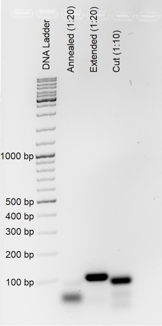
Figure 1. Representative fragment-size analysis of intermediate products and final duplex sequencing adapters. Aliquots collected during adapter synthesis after performing oligonucleotide annealing (“annealed,” diluted 1:20), fill-in extension (“extended,” diluted 1:20), and HpyCH4III restriction digestion (“cut,” diluted 1:10) were analyzed by 2% agarose gel electrophoresis to verify correct duplex sequencing adapter formation. Please click here to view a larger version of this figure.
4. Single-Oocyte Lysis
- Prepare OLB supplemented with thermolabile proteinase K (OLB+) by adding 1 µL of thermolabile proteinase K (0.120 U/µL) to 99 µL of OLB to obtain a final proteinase K concentration of 0.0012 U/µL.
- Thaw the oocytes at RT. Immediately add 4 µL of OLB+ to each oocyte. Rinse the sides of the tube several times with the lysis buffer to ensure complete transfer of the oocyte into the buffer and prevent the sample from adhering to the tube wall.
- Mix the sample thoroughly, briefly centrifuge to collect the contents at the bottom of the tube, and incubate at 37°C for 16 h in a thermocycler with the lid temperature set to 47°C.
NOTE: No independent assessment of oocyte lysis is performed before fragmentation. A 16 h incubation under the specified lysis conditions is generally sufficient for complete lysis of a single oocyte. The amount of recovered DNA may nevertheless vary because of sample loss during handling and differences in mtDNA copy number among oocytes. - Inactivate the thermolabile proteinase K by incubating the samples at 55°C for 15 min in a thermocycler with the lid temperature set to 75°C.
NOTE: Complete the subsequent workflow without interruption after lysis and proteinase K inactivation unless a later protocol step explicitly identifies a stopping point.
5. Exonuclease V Digestion
- Allow the samples to equilibrate to RT before proceeding.
- Prepare an Exonuclease V Master Mix by combining 4 µL of 25 mM magnesium chloride (MgCl₂), 1 µL of 10 mM Tris-HCl, 1 µL of 10 mM adenosine triphosphate (ATP), and 1 µL of Exonuclease V (10 U/µL stock) to obtain a final volume of 7 µL.
- Prepare a 1 mg/mL ribonuclease A (RNase A) working solution by diluting 1 µL of 10 mg/mL RNase A stock with 99 µL of molecular biology-grade water. Add approximately 0.1 µL of the diluted RNase A solution to each lysed oocyte sample.
NOTE: Use a suitable pipette for this step. When processing multiple samples simultaneously, a suitable multichannel pipette may be used, provided that precautions are taken to avoid cross-contamination between samples. Because pipetting accuracy may decrease when dispensing such small volumes with a multichannel pipette, carefully inspect all channels. Alternatively, briefly dipping the pipette tip into the diluted RNase A solution is sufficient to transfer approximately 0.1 µL. Minor deviations from this volume do not affect the performance of the protocol. Verify the approximate transferred volume beforehand using a single-channel pipette. - Add 7 µL of the Exonuclease V Master Mix to each lysed oocyte sample and mix thoroughly. Incubate the samples at 37°C for 1 h in a thermocycler with the lid temperature set to 47°C.
- Add 38 µL of TE buffer to each sample to obtain a final reaction volume of approximately 51 µL. Heat-inactivate the enzymes by incubating the samples at 70°C for 30 min in a thermocycler with the lid temperature set to 75°C.
- Proceed immediately to library preparation.
6. Fragmentation
- Shear the DNA to an average fragment size of approximately 550 base pairs (bp) using sonication.
NOTE: This protocol describes DNA fragmentation using a Covaris M220 focused-ultrasonication instrument, hereafter referred to as the sonicator; however, equivalent systems may also be used. Because the samples contain cellular debris in addition to DNA after lysis, optimize the fragmentation conditions for the specific instrument and sample type to obtain a relatively narrow fragment-size distribution centered around the intended fragment size. - Transfer the entire sample volume (approximately 51 µL) into a 50 µL focused-ultrasonication tube.
- Shear the DNA for 70 s using a Duty Factor of 10%, Peak Incident Power of 75 W, and 200 Cycles per Burst at 20°C.
- Immediately transfer each sheared sample (approximately 50 µL) from the shearing tube into a 200 µL low-binding tube, because the shearing tubes are not low-binding. The original oocyte collection tube may be reused for this purpose. Inspect the lid of the shearing tube for residual liquid and recover any remaining sample to maximize sample recovery.
- Proceed immediately to end repair and A-tailing.
7. End Repair/A-Tailing
- Prepare an End Repair/A-Tailing Master Mix containing 7 µL of End Prep Reaction Buffer and 3 µL of End Prep Enzyme Mix.
- Add 10 µL of the End Repair/A-Tailing Master Mix to each sample to obtain a final reaction volume of 60 µL. Mix thoroughly by pipetting up and down 10 times.
- Incubate the samples at 20°C for 30 min with the thermocycler lid turned off, followed by incubation at 65°C for 30 min with the lid temperature set to 75°C.
- Proceed immediately to adapter ligation.
8. Adapter Ligation
- Prepare a Ligation Master Mix containing 30 µL of Ligation Mix and 1 µL of Ligation Enhancer.
- Thaw one aliquot of the synthesized adapter and dilute it 1:4000 in Tris-NaCl buffer.
- Add 1.5 µL of the diluted adapter to each end-repaired and A-tailed DNA sample. Add 31 µL of the Ligation Master Mix and mix thoroughly.
- Incubate the samples at 20°C for 15 min. Add 1 µL of the diluted adapter to each sample to obtain a final reaction volume of 93.5 µL. Mix thoroughly and incubate at 4°C for 16 h.
- Proceed immediately to purification.
9. Purification of Adapter-Ligated DNA
- Allow the magnetic beads and TElow buffer to equilibrate to RT for at least 30 min. During this time, prepare fresh 80% EtOH. Prepare a carrier oligonucleotide solution by adding 1 µL of the carrier oligonucleotide (sequence not represented in the human genome; see Table 2) to 99 µL of TElow buffer to obtain a final concentration of 1 nM.
- Transfer 74.8 µL of magnetic beads, corresponding to a 0.8× bead-to-sample ratio, into a 1.5 mL low-binding tube. Add the entire 93.5 µL volume of adapter-ligated DNA and mix thoroughly.
- Incubate the bead–sample mixture at RT for 15 min. After 7.5 min, gently mix the suspension and briefly centrifuge the tube.
- Briefly centrifuge the tube, place it on a magnetic rack, and incubate for 5 min to allow complete bead separation. Carefully remove and discard the clear supernatant, then close the tube immediately.
- Add 400 µL of 80% EtOH, incubate for 30 s, and remove the EtOH. Add 200 µL of 80% EtOH, incubate for 30 s, and remove the EtOH.
- Remove the tube from the magnetic rack and briefly centrifuge it. Return the tube to the magnetic rack, remove any remaining EtOH, and air-dry the beads for less than 5 min.
- Add 50 µL of TElow buffer supplemented with carrier oligonucleotides. Remove the tube from the magnetic rack and thoroughly resuspend the beads by pipetting.
- Incubate the suspension at RT for 5 min, mixing occasionally. Briefly centrifuge the tube, return it to the magnetic rack, and incubate for an additional 5 min.
- During the final 5 min of the first magnetic separation, prepare a new 1.5 mL low-binding tube containing 40 µL of magnetic beads. Transfer 50 µL of the eluate into the prepared tube to perform the second purification at a 0.8× bead-to-sample ratio.
- Mix thoroughly and incubate at RT for 15 min. After 7.5 min, gently mix the suspension and briefly centrifuge the tube.
- Place the tube on the magnetic rack and incubate for 5 min to allow complete bead separation. Carefully remove and discard the clear supernatant, then close the tube immediately.
- Add 400 µL of 80% EtOH, incubate for 30 s, and remove the EtOH. Add 200 µL of 80% EtOH, incubate for 30 s, and remove the EtOH.
- Remove the tube from the magnetic rack and briefly centrifuge it. Return the tube to the magnetic rack, remove any remaining EtOH, and air-dry the beads for less than 5 min.
- Add 15.5 µL of Tris-HCl to each sample. Remove the tube from the magnetic rack and thoroughly resuspend the beads by pipetting.
- Incubate the suspension at RT for 5 min, mixing occasionally. Briefly centrifuge the tube, return it to the magnetic rack, and incubate for an additional 5 min.
- Transfer 15 µL of the eluate to a new 200 µL low-binding tube. Remove 1 µL of the eluate and dilute it 1:10 in Tris-HCl for attomole quantitative PCR (qPCR) and mitochondrial DNA (mtDNA) enrichment qPCR analyses.
10. Attomole Estimation
NOTE: Determine the approximate amount of adapter-ligated DNA to adjust the DNA input and the number of cycles used in the subsequent amplification and indexing PCRs. Because the sample quantity is limited, direct concentration measurements may not be reliable; therefore, estimate DNA quantity by qPCR. Analyze the amplification products by agarose gel electrophoresis to assess fragment-size distribution and detect residual adapter dimers. Cq values may vary depending on the reagents and real-time PCR instrument used. Adjustments to Cq values based on adapter-dimer detection, as well as downstream DNA input amounts and PCR cycle numbers, are based primarily on empirical observations and may require optimization for individual library preparations.
- Prepare an attomole qPCR Master Mix containing 5 µL of 2× KAPA HiFi HotStart Reaction Mix (hereafter referred to as the 2× high-fidelity PCR mix), 1 µL of the Dual-NEBNext Universal PCR Primer for Illumina (10 µM; hereafter referred to as the universal library primer), 1 µL of the mws20 primer (10 µM), 0.5 µL of 20× EvaGreen, and 0.5 µL of molecular biology-grade water per reaction.
- Add 8 µL of the Master Mix to each designated well of a qPCR-compatible 96-well plate. Add 2 µL of the 1:10 diluted sample to obtain a final reaction volume of 10 µL.
- Seal the plate and briefly centrifuge it before loading it into the real-time PCR instrument.
- Perform qPCR using the following thermal cycling conditions: 45 s at 98°C, followed by 45 cycles of 15 s at 98°C, 30 s at 65°C, and 45 s at 72°C.
- Determine the quantification cycle (Cq) by setting the fluorescence threshold to 1,000 relative fluorescence units (RFU).
NOTE: Select a fluorescence threshold that is compatible with the real-time PCR instrument and use the same threshold in all experiments to permit comparison of Cq values. - Separate the qPCR amplification products on a 1.5% agarose gel at 125 V for 40 min.
- Inspect the gel for residual adapter dimers. Adapter dimers migrate at approximately 130 bp (Figure 2).
- If adapter dimers are visible, adjust the Cq value using the appropriate attomole Cq correction factor (ACF) shown in Figure 2. Select the ACF according to the intensity of the adapter-dimer band observed in the agarose gel, using the representative examples in Figure 2 as a reference. An ACF-corrected attomole Cq value of 22–26 is optimal; however, values <29 are generally acceptable.
NOTE: Instead of correcting the Cq value, an additional purification may be performed when necessary; however, this may result in substantial library loss. Samples with strong adapter-dimer bands are typically associated with high attomole qPCR Cq values, indicating low DNA input. Samples with Cq values ≥29 generally produce large family sizes and low mitochondrial DNA sequencing depths (<100×) and may therefore be excluded from sequencing. Because Cq values depend on the real-time PCR instrument and assay conditions, establish laboratory-specific cutoff values when implementing this protocol.
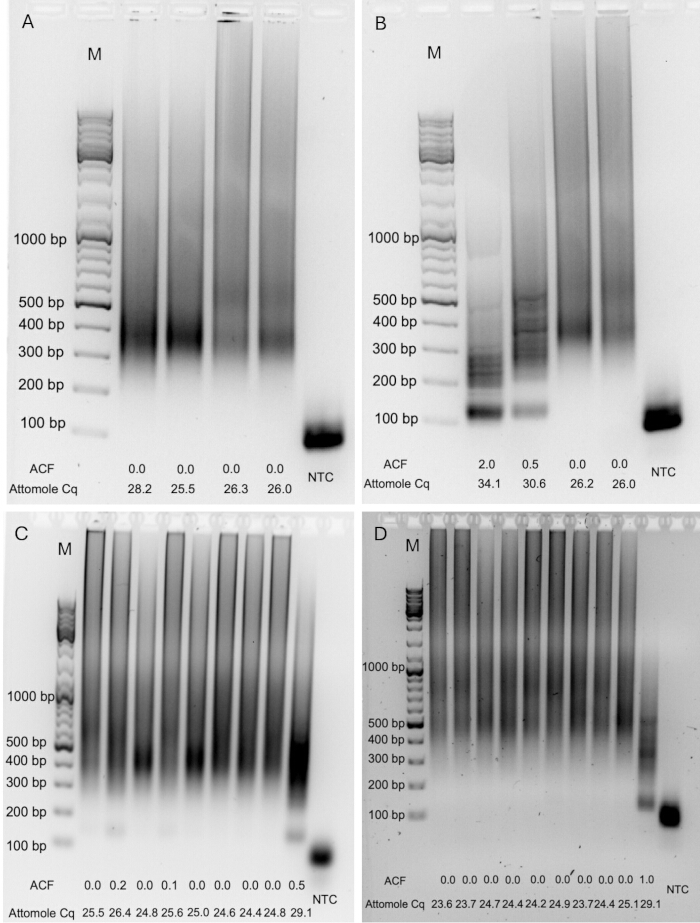
Figure 2. Representative attomole quantitative PCR (qPCR) products used to estimate library input and identify adapter/primer dimers. Amplified attomole qPCR products were analyzed by 1.5% agarose gel electrophoresis to assess fragment-size distribution and detect residual adapter/primer dimers. (A–D) Representative examples showing libraries with different amounts of adapter-ligated DNA and varying levels of adapter/primer dimers. The attomole Cq correction factor (ACF) and the corresponding uncorrected quantification cycle (Cq) are shown for each sample. M, DNA size marker; NTC, non-template control. Please click here to view a larger version of this figure.
11. Mitochondrial DNA Enrichment Estimation (Optional)
NOTE: Estimate mitochondrial DNA (mtDNA) enrichment by qPCR using primers targeting the mitochondrial ND6 gene and nuclear Alu elements. Calculate the difference in Cq values between the nuclear and mitochondrial targets to estimate mtDNA enrichment. Use the absolute Cq value of the mitochondrial target to estimate the amount of mtDNA in the sample. Cq values may vary depending on the reagents, consumables, and real-time PCR instrument used. Adjust the primer sequences for other species. Refer to previous publications for mouse- and macaque-specific primers12,13. This step is optional and provides a rough estimate of mtDNA enrichment before sequencing.
- Prepare separate Master Mixes for the mitochondrial and nuclear targets. For each reaction, combine 5 µL of 2× PowerUp SYBR Green Mix, 0.4 µL of each primer in the respective primer pair (10 µM each), and 2.1 µL of molecular biology-grade water.
- Add 8 µL of the appropriate Master Mix to each designated well of a qPCR-compatible 96-well plate. Add 2 µL of the 1:10 diluted sample to obtain a final reaction volume of 10 µL.
- Seal the plate and briefly centrifuge it before loading it into the real-time PCR instrument.
- Perform qPCR using the following thermal cycling conditions: 2 min at 95°C, followed by 45 cycles of 15 s at 95°C, 20 s at 56°C, and 30 s at 72°C.
- Determine the Cq by setting the fluorescence threshold to 100 RFU.
NOTE: Select a fluorescence threshold that is compatible with the real-time PCR instrument and use the same threshold in all experiments to permit comparison of Cq values. Correlate Cq values with sequencing-derived enrichment efficiencies after sequencing and use these values as references for subsequent library preparations. - Calculate the enrichment-associated ΔCq by subtracting the nuclear-target Cq from the mitochondrial-target Cq (CqmtDNA − CqnDNA). Once initial sequencing data are available, generate a laboratory-specific standard curve to estimate the proportion of sequencing reads derived from mtDNA.
NOTE: If sequencing data are not yet available, Equation 1 may be used to obtain a rough estimate of the expected percentage of mtDNA-derived sequencing reads using an example standard curve generated from seven oocyte libraries (ΔCq = −0.8, 16.83%; ΔCq = −2.1, 33.62%; ΔCq = −3.0, 45.57%; ΔCq = −3.9, 60.34%; ΔCq = −5.0, 77.98%; ΔCq = −6.2, 89.25%; ΔCq = −8.4, 95.09%). A ΔCq value <−1 should be achieved to ensure efficient removal of nuclear DNA. Lower enrichment efficiencies can be compensated for by allocating additional sequencing reads to obtain sufficient mtDNA sequencing depth; however, this may increase nuclear mitochondrial DNA segment (NUMT) contamination. Because the relationship between ΔCq and mtDNA content depends on reagents, consumables, instrumentation, and experimental conditions, Equation 1 should be considered an example calibration and laboratory-specific standard curves should be established whenever possible.
Estimated mtDNA (%) = −11.006 × ΔCq + 13.776 (1)
12. Library Amplification
NOTE: Perform the first amplification in two consecutive PCR steps. Perform the first PCR using a single primer to generate linear amplification. Subsequently, add the second primer to enable exponential amplification during the second PCR.
- Determine the DNA input for the first amplification PCR using the results of the attomole qPCR.
- For single-oocyte libraries, use samples with an attomole qPCR Cq value typically greater than 22. If a sample has a lower Cq value, dilute it to a Cq value of approximately 22 according to Table 3. Libraries with Cq values of ≥29 typically produce large family sizes and shallow mtDNA sequencing depths and are therefore not recommended for continued library preparation or sequencing.
- Prepare an amplification Master Mix containing 20 µL of 2× high-fidelity PCR mix and 4 µL of the mws20 primer (10 µM) per reaction.
- Add 24 µL of the Amplification Master Mix to 14 µL of the diluted sample.
- Perform the linear amplification PCR using the following thermal cycling conditions: 45 s at 98°C, followed by 12 cycles of 15 s at 98°C, 30 s at 60°C, and 45 s at 72°C, followed by 2 min at 72°C.
- Add 4 µL of the universal library primer (10 µM) and mix thoroughly.
- Perform the exponential amplification PCR using the following thermal cycling conditions: 45 s at 98°C, followed by 9 cycles of 15 s at 98°C, 30 s at 65°C, and 45 s at 72°C, followed by a 2 min extension at 72°C.
NOTE: After completion of the first amplification, the samples may be stored at 4°C and the protocol resumed later if necessary. Perform all preceding steps, from oocyte lysis through the first amplification, without interruption. - Add 10 µL of molecular biology-grade water to each sample.
- Purify the amplified DNA using 40 µL of magnetic beads (0.8× the sample volume), following the second magnetic-bead purification procedure described in Steps 9.9–9.15. Wash the beads twice with 200 µL of 80% EtOH, elute the DNA in 15.5 µL of Tris-HCl, and transfer 15 µL of the eluate to a new 200 µL low-binding tube.
NOTE: This is a suitable stopping point. Store the purified DNA at 4°C or proceed immediately to indexing.
Table 3: Dilution factors and corresponding quantitative polymerase chain reaction (qPCR) quantification cycle (Cq) correction factors used to standardize DNA input for the first library amplification polymerase chain reaction (PCR). Samples with attomole qPCR Cq values below the target value were diluted with molecular biology-grade water before the first amplification PCR. The corrected Cq value was obtained by adding the appropriate dilution correction factor to the experimentally measured Cq value and was subsequently used to determine the indexing PCR cycle number and the target sequencing depth (Table 4). The sample and water volumes produce a final input volume of 14 µL for the first amplification PCR. The asterisk (*) indicates that the listed dilution correction factor should be added to the experimentally measured attomole qPCR Cq value to obtain the corrected Cq. The correction factors were determined empirically for this workflow. Please click here to download this file.
13. Indexing
- Determine the number of indexing PCR cycles using the dimer- and dilution-corrected attomole qPCR Cq value according to Table 4.
- Prepare an indexing master mix containing 25 µL of 2× high-fidelity PCR mix and 10 µL of a 10 µM Unique Dual Index Primer Pair for each sample.
- Add 35 µL of the indexing master mix to 15 µL of each amplified sample.
- Perform the indexing PCR using the following thermal cycling conditions: 45 s at 98°C, followed by the appropriate number of cycles consisting of 15 s at 98°C, 30 s at 65°C, and 45 s at 72°C, followed by a final extension at 72°C for 2 min.
- Purify the indexed libraries using 40 µL of magnetic beads (0.8× sample volume). Wash the beads twice with 80% EtOH and elute the DNA in 21 µL of TElow buffer.
- Transfer the eluate to a DNA low-binding tube.
- Measure the DNA concentration using a Qubit High Sensitivity DNA Assay or an equivalent fluorometric assay.
- Store the libraries at 4°C before sequencing or at −80°C for long-term storage.
NOTE: This is a safe stopping point.
Table 4: Indexing polymerase chain reaction (PCR) cycle numbers and target sequencing depth determined from the corrected attomole quantitative polymerase chain reaction (qPCR) quantification cycle (Cq). The corrected Cq value incorporates the experimentally measured attomole qPCR Cq together with any applicable adapter-dimer correction factor (Figure 2) and dilution correction factor (Table 3). The corrected Cq value was used to determine both the number of indexing PCR cycles and the recommended number of paired-end sequencing reads allocated to each library for pooling. The target sequencing read allocations serve as an initial guideline and may require laboratory-specific optimization depending on the real-time PCR instrument, sequencing platform, multiplexing strategy, and experimental requirements. Please click here to download this file.
14. Quality Control
- Assess library quality, fragment-size distribution, and the presence of residual adapter or primer dimers using a Bioanalyzer, TapeStation, or an equivalent nucleic acid fragment analysis instrument. Fragment sizes should range from approximately 300 to 1,000 bp. Small residual peaks (<5% of the sample fluorescence intensity [RFU] in the Bioanalyzer trace) were generally acceptable (Figure 3A–D). Residual adapter or primer dimers appeared as distinct peaks at approximately 70–150 bp (Figure 3E–H). The final library concentration should be at least 5 ng/µL. Refer to Supplementary File 1 (Troubleshooting Guide) if any of these quality criteria are not met.
- (Optional) If adapter or primer dimers are detected (Figure 3E–H), adjust the library volume to 50 µL with molecular biology-grade water, add 40 µL of magnetic beads (0.8× sample volume), and perform an additional purification. Wash the beads twice with 80% EtOH and elute the purified library in 21.5 µL of TElow buffer.
- (Optional) Repeat the quality-control analysis (Step 14.1) to confirm complete removal of adapter and primer dimers.
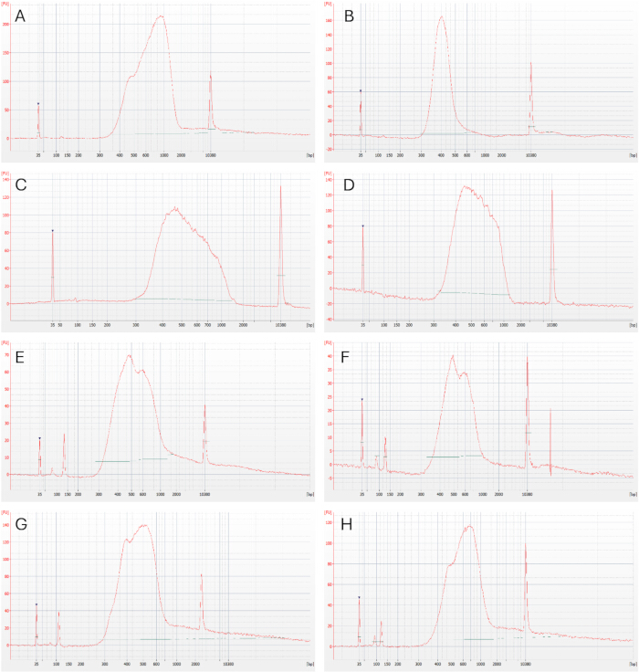
Figure 3. Representative fragment analysis of indexed sequencing libraries. Representative electropherograms generated by Bioanalyzer fragment analysis showing library quality after indexing PCR. (A–D) Libraries with the expected fragment-size distribution (approximately 300-1000 bp) and no detectable adapter/primer dimers, suitable for sequencing without additional purification. (E–H) Libraries containing residual adapter and/or primer dimers that require an additional magnetic-bead purification before sequencing. Peaks at approximately 35 bp and 10,380 bp correspond to the lower and upper internal markers, respectively. Please click here to view a larger version of this figure.
15. Pooling and Sequencing
- Measure the concentration of each indexed library using a qPCR-based library quantification assay compatible with Illumina-prepared libraries, such as the Collibri Library Quantification Kit, according to the manufacturer’s protocol. Dilute each library 1:100,000 in the supplied library dilution buffer and analyze each sample in at least duplicate; triplicate measurements are recommended. Analyze the standards in triplicate.
- Calculate the mean quantification cycle (Cq) value for each sample and standard. Generate a standard curve from the mean Cq values of the standards and calculate the concentration of each library according to the manufacturer’s instructions.
- Pool the libraries according to the intended number of paired-end reads determined from the adapter-dimer- and dilution-corrected attomole qPCR Cq value obtained in Step 10 and the allocations provided in Table 4.
NOTE: The pooling ratio depends on the corrected Cq value of each oocyte library and the corresponding target number of paired-end reads. The values in Table 4 are approximate and may vary among real-time PCR instruments. Establish laboratory-specific values for optimal performance. - Calculate the relative pooling fraction for each library by dividing its target number of paired-end reads by the sum of the target reads assigned to all libraries. Determine the required amount of each library by multiplying this fraction by the total molar amount of the final pool, then calculate the corresponding library volume from its measured molar concentration. Combine the calculated volumes to generate the final pool.
- Sequence the pooled libraries using an Illumina sequencing platform or another platform compatible with Illumina adapter sequences. Use a paired-end configuration with a minimum read length of 2 × 150 bp. Longer reads, such as 2 × 250 or 2 × 300 bp, are recommended because they improve the ability to identify and filter reads derived from shorter nuclear mitochondrial DNA segments (NUMTs).
- Sequence the pooled libraries, for example, on an Illumina NovaSeq 6000 platform using two-channel sequencing-by-synthesis chemistry with an SP Reagent Kit v1.5 (500 cycles) and a paired-end configuration of 2 × 250 bp including a 5% PhiX spike-in. Load the library and perform sequencing according to the manufacturer’s instructions. Representative sequencing performance should achieve ≥75% of bases with a quality score of ≥Q30 and ≥60% of clusters passing filter.
16. Bioinformatic Analysis
NOTE: The following workflow describes data analysis in Galaxy using the Du Novo analysis tools8,14,15. Analysis may also be performed using a local installation of Du Novo or other software developed for duplex sequencing data.
- Upload the demultiplexed paired-end FASTQ files, generated using BCL Convert or an equivalent demultiplexing tool, to a local Galaxy installation or a publicly available Galaxy instance14.
- Assess sequencing-read quality using FastQC (Galaxy version 0.72+galaxy1). Inspect, at minimum, the Per Base Sequence Quality, Per Sequence GC Content, and Adapter Content modules.
- Generate single-strand consensus sequences (SSCSs) and DCSs from the demultiplexed paired-end FASTQ files using the Du Novo pipeline (Galaxy version 3.0.2). Use a minimum family size of three reads for SSCS formation and call a consensus nucleotide when it is present in at least 70% of the reads15. Enable barcode error correction with up to three mismatches. Refer to the Galaxy Training Network Du Novo tutorial for detailed instructions on use of the pipeline16.
- Use Sequence Content Trimmer (Galaxy version 0.2.3) to remove bases represented by “NRYSWKMBDHV” and discard reads shorter than 10 bp.
- Trim the first 10 nucleotides from the 5′ end of each DCS using FASTQ Trimmer (Galaxy version 1.1.5) to reduce end-repair-associated bias. Align the trimmed DCS reads to the human reference genome, such as GRCh38.p14 containing the revised Cambridge Reference Sequence (rCRS; NC_012920.1), using BWA-MEM (Galaxy version 0.7.17.1).
NOTE: Other human genome assemblies, including T2T-CHM13v2.0 or newer assemblies, may be used. - Filter the BAM files using BAMTools Filter BAM datasets on a variety of attributes (Galaxy version 2.5.2+galaxy1). Retain reads with a mapping quality >20 that map to chrM, represent primary alignments, are paired, are properly paired, and have a mapped mate. These criteria reduce potential NUMT-derived alignments17.
- Left-align the reads using Bam Left Align (Galaxy version 1.3.1). Clip overlapping regions of paired DCS reads using BAMUtil clipOverlap (Galaxy version 1.0.15+galaxy1).
- Call single-nucleotide variants (SNVs) and insertions/deletions (indels) using Call variants with LoFreq (Galaxy version 2.1.5+galaxy2) with default settings.
- Exclude paired-end DCS reads containing more than two variants, when present, to reduce potential NUMT contamination. Samples with efficient mtDNA enrichment are typically free of detectable NUMT-derived reads; however, insufficient depletion of nuclear DNA may result in NUMT-derived sequences.
- Inspect each library for potential cross-sample contamination using donor-specific mtDNA sequence differences, including fixed variants and high-frequency heteroplasmies. If cross-sample contamination is detected, apply the precautions described in Supplementary File 1 (Troubleshooting Guide). If the fragment-size distribution exceeds the recommended range or the average fragment size is >900 bp, perform a double size-selective purification as described previously18.
- Perform downstream analyses of the detected variants according to the objectives of the study.
- Only include samples with a mean mitochondrial DCS sequencing depth of ≥100× for downstream analysis. No fixed minimum thresholds were applied for paired-end read count or SSCS or DCS yield.
結果
胚盤胞(GV)、第一減数分裂中期(MI)、第二減数分裂中期(MII)、前核期(0PN)、およびTI卵子を含む、異なる成熟段階を代表するヒト卵子からデュプレックスシーケンシングライブラリを調製した。正常に調製されたライブラリは、Illumina NovaSeq 6000プラットフォームを用い、SPフローセルを使用して2 × 250 bpのペアエンドリードでシーケンシングを行った。データ解析はGalaxyにて実施し、リードファミリーのグルーピングおよびコンセンサスリードの生成にはDu Novoパイプラインを使用した8,14,15。正常に処理されたライブラリの代表的なシーケンシング指標を補足表1にまとめ、ライブラリ調製中に最適ではない結果が得られシーケンシングから除外したサンプルを補足表2にまとめた。
ライブラリ調製の間、アダプターが結合したDNA量、mtDNA濃縮度、ライブラリ濃度、およびフラグメントサイズ分布を評価するために、いくつかの品質管理ステップが実施されました。最初の品質管理ステップはアトモレqPCRアッセイであり、これを用いてアダプター結合DNA量を推定しました(Figure 4A,B)。qPCR増幅産物のアガロースゲル電気泳動によりアダプターダイマーまたはプライマーダイマーが検出された場合、算出された定量サイクル(Cq)値を調整しました(Figure 2A–D)。アダプターダイマーまたはプライマーダイマーは通常、Cq値が高いサンプルで観察され、これはアダプター結合DNA量が少ないことを示しています。本プロトコルに記載された条件を用いた場合、調整後のアトモレqPCR Cq値の平均は25.8でした(Supplementary Table 1)。補正後のCq値に基づき、最初の増幅PCRへのDNA投入量をTable 3に示す希釈スキームに従って調整しました。補正後のCq値が≥22のサンプルは希釈せずに使用し、補正後のCq値が<22のサンプルは増幅前に希釈しました。この調整は、リードファミリーサイズを最適化し、必要なシーケンシングリード数を削減するために行われました。補正後のCq値が>28のサンプルは、一般的にリードファミリーサイズが大きくなり(平均ファミリーサイズ 21.8)、その結果、DCSが少なくなり、mtDNAの平均シーケンシング深度は<200×となりました(Supplementary Table 1)。補正後のCq値を22–28に調整した結果、平均ファミリーサイズは7.91となり、これは以前にデュプレックスシーケンシングに最適であると報告された約6というファミリーサイズに近くなります6,7。とはいえ、単一卵母細胞のような極めて低投入量のサンプルでは、シーケンシング深度を高めることができるため、大きなファミリーサイズが有利な場合があります。
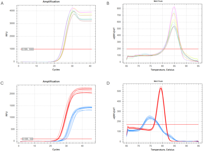
図4ライブラリの定量およびミトコンドリアDNA(mtDNA)濃縮度の推定に使用される定量PCR(qPCR)アッセイ。 精製されたアダプターライゲーション済みDNAを1:10に希釈し、qPCR解析に供した。(A) アダプターライゲーション済みDNA量を推定するために用いたアトモルqPCRアッセイの増幅曲線。蛍光しきい値は1,000相対蛍光単位(RFU)に設定した。(B) アトモルqPCRアッセイの融解曲線分析。(C) mtDNA濃縮qPCRアッセイの増幅曲線。蛍光しきい値は100 RFUに設定した。ミトコンドリアNADH脱水素酵素サブユニット6(ND6)ターゲットの増幅を赤色で、核のAlu反復配列ターゲットの増幅を青色で示す。(D) ミトコンドリアおよび核のqPCR産物の融解曲線分析。 この図の拡大版を表示するには、ここをクリックしてください。
ライブラリのプーリング前に、すべてのライブラリをBioanalyzerまたはTapeStationで分析し、断片サイズの分布を評価して、残留アダプターまたはプライマーダイマーを検出しました。最適なライブラリと、残留アダプターまたはプライマーダイマーを含むライブラリの代表例をFigure 3A–Hに示します。アダプターまたはプライマーダイマーは約70–150 bpにピークとして現れました(Figure 3E–H)。これらが検出された場合、シーケンシング前に磁気ビーズによる追加精製を行いました。サンプルの損失を最小限に抑えるため、意図的にサイズセレクションを省略したため、シーケンシングしたライブラリのインサートサイズの中央値は275 bpでした(Supplementary Table 1)。
mtDNAの濃縮度は、ミトコンドリアおよび核の標的遺伝子座を用いたqPCRによって推定され、これによりミトコンドリアゲノムにマッピングされると予想されるシーケンシングリードの割合を予測した(図 4C,D)。平均Cq値は、ミトコンドリア標的で25.2、核標的で29.7であり(付随表 1)、これは平均ΔCq(CqmtDNA − CqnDNA)の−4.5に相当する。平均して、ペアエンドシーケンシングリードの67.5%がミトコンドリアリファレンスゲノムにマッピングされた。ライブラリのプーリングに際しては、mtDNA濃縮度の推定値と補正後のアトモルqPCR Cq値の両方を考慮した。サンプル間でバランスの取れたシーケンシング出力を促進するため、補正後のCq値が高く、mtDNA濃縮効率が高いライブラリには、表 4に示すシーケンシング割り当てスキームに従って、比例的に少ないシーケンシングリードを割り当てた。
補正後のアトモールqPCR Cq値に基づき、1ライブラリあたり平均400万ペアエンドリードのターゲット割り当てを行った(表4)。クラスター生成の効率が悪かったため、シーケンシング後の1ライブラリあたりの平均ペアエンドリード数は130万リードでしたが、このシーケンシング収量は、その後のデュプレックスシーケンシング解析を行うのに十分な量でした(付随表1DCSをミトコンドリア参照ゲノムにマッピングした結果、シーケンシング深度の中央値は567であった。× ミトコンドリアゲノム全体にわたって(図5A,B,D; 補足表1)。ライブラリあたり平均で344,554個のSSCSおよび96,322個のDCSが生成された(補足表1)。得られたライブラリの平均リードファミリーサイズは8.32であり、一般的に、補正後のアトモルqPCR Cq値が高いライブラリほど、ファミリーサイズが大きくなる傾向が見られた(図5C,E; 付随表 1)。これらの結果は、最適なクラスター密度で実施された先行研究で得られた結果と一致しており、その研究におけるmtDNAシーケンス深度の中央値は1,440であった。× 達成された19.
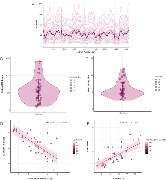
図5. 単一卵母細胞デュプレックスシーケンシングライブラリのシーケンシング性能。 (A) ミトコンドリアゲノム全域におけるデュプレックスコンセンサス配列(DCS)深度。細線は個々のライブラリを、太線は全ライブラリの平均深度を示す。(B) 個々のライブラリにおける中央値DCS深度の分布。(C) 個々のライブラリにおける平均DCSファミリーサイズの分布。(B)および(C)において、各点は1つのライブラリを表し、点の色はアトモルqPCR定量サイクル(Cq)を示す。(D) mtDNA濃縮推定値(ΔCq)と、ミトコンドリアリファレンスゲノムにアラインしたシーケンシングリードの割合とのピアソン相関。(E) アトモルqPCR Cq値と平均DCSファミリーサイズのピアソン相関。(D)および(E)において、各点は1つのライブラリを表し、点の色はアトモルqPCR Cq値を、点の大きさはそのライブラリで得られたペアエンドリードの総数を表す。n = 39ライブラリ。こちらのリンクをクリックして、この図の拡大版を表示してください。
付随表 1. 最適なヒト卵母細胞デュプレックスシーケンシングライブラリのシーケンシングおよびライブラリ品質指標。 この表は、品質管理に合格し、その後の解析に含まれたライブラリの、シーケンシング前のライブラリ品質指標、定量ポリメラーゼ連鎖反応(qPCR)測定値、シーケンシング性能、およびデュプレックスシーケンシング解析結果をまとめたものである。報告されたパラメータには、卵母細胞の発達段階、ミトコンドリアDNA(mtDNA)濃縮qPCR結果、アトモルqPCR測定値、アダプターダイマー補正係数(ACF)、インデックスPCRサイクル数、予定ペアエンドシーケンシング深度、ライブラリ濃度、シーケンシング収量、mtDNA濃縮度、シングルストランドコンセンサス配列(SSCS)およびデュプレックスコンセンサス配列(DCS)のリード数、ファミリーサイズ統計、シーケンシング深度、およびインサートサイズ分布が含まれる。サンプル識別子は解析前に匿名化された。予定ペアエンドリード数は、補正済みアトモルqPCR定量サイクル(Cq)値に基づいて割り当てられた(表 4)。 こちらをクリックしてファイルをダウンロードしてください。
補足表2. 後続のデュプレックスシーケンシング解析から除外された、品質不十分なヒト卵母細胞ライブラリーのシーケンシング前品質指標。 この表は、後続のシーケンシング解析への組み込み基準を満たさなかったライブラリーの、シーケンシング前の品質管理結果をまとめたものである。報告されているパラメータには、サンプル識別子、卵母細胞の発達段階、ミトコンドリアDNA (mtDNA) 濃縮定量PCR (qPCR) 結果、アダプターダイマー補正係数 (ACF)、補正後アトモルqPCR定量サイクル (Cq)、インデックスPCRサイクル数、および Qubit High Sensitivity DNA Assay を用いて測定したライブラリー濃度が含まれる。ライブラリーは、プロトコルに記載された品質管理基準に従って除外された。 こちらのリンクからファイルをダウンロードしてください。
補足ファイル 1. 単一卵母細胞ミトコンドリアDNAデュプレックスシーケンシングライブラリ調製のためのトラブルシューティングガイド。 この補足ファイルでは、サンプルの損失防止、アダプターの調製と品質評価、磁気ビーズ精製、ライブラリの品質管理、シーケンシング性能、サンプル間の交差汚染、および核内ミトコンドリアDNAセグメント(NUMT)汚染を含む、ワークフローの重要なステップに関するトラブルシューティングの推奨事項を提供します。本ガイドはメインプロトコルを補完するものであり、ライブラリの品質指標やシーケンシング性能が推奨範囲外となった場合に参照してください。 こちらのリンクをクリックしてファイルをダウンロードしてください。
ディスカッション
卵母細胞で同定されるmtDNA変異は、de novo変異、世代間のヘテロプラスミーの変動、ならびに加齢および疾患が女性生殖細胞系に及ぼす影響の組み合わせを反映しています。本プロトコールでは、単一の卵母細胞からmtDNAを濃縮し、それに続いてデュプレックスシーケンシングライブラリの調製およびシーケンシングを行う手法について記述します。これにより、mtDNA変異の極めて高精度な検出と解析が可能になります。卵母細胞には大量のmtDNAコピー数が存在しますが、本ワークフローでは単一細胞から完全なシーケンシングライブラリを調製する必要があります。したがって、補足ファイル1(トラブルシューティングガイド)に詳述されている通り、手順全体を通して慎重なサンプルハンドリングが不可欠です。慎重にサンプルを扱ったとしても、初期のハンドリングステップにおける卵母細胞の消失や、ライブラリ調製中のサンプル消失により、ライブラリ調製の約5%が失敗する可能性があります。
ライブラリ調製中および調製後の品質管理は、アダプターライゲーションの成功の確認、mtDNA濃縮度の評価、増幅のためのDNA投入量の最適化、残留アダプターまたはプライマーダイマーの検出、および追加の精製や増幅ステップが必要かどうかの判断を行うために不可欠である。ライブラリ調製全体を通して同一のリアルタイムqPCR機器を使用することで、反応条件が一定に保たれ、後続の品質管理やプロトコルの最適化に用いられる測定値の比較可能性、再現性、および信頼性が向上する。本プロトコルに記載されているライブラリ増幅およびインデックス付加のためのPCRサイクル数は、異なる機器、酵素、または試薬を使用する場合、調整が必要となる可能性がある。最初の増幅ステップは、単一のプライマーを用いた線形増幅フェーズと、それに続く2番目のプライマー添加後の指数関数的増幅フェーズに分かれている。この戦略により、最初の増幅サイクルで導入されたエラーが、本物のDNA変異と区別が困難なレベルまで増幅される可能性を最小限に抑えることができる。推奨されるサイクル数は、デュプレックスシーケンシング解析に最適なリードファミリーサイズを得るために選定されているが、投入されるDNAの量と質に応じて最適化が必要な場合がある。DNA投入量が少ないサンプルでは追加の増幅サイクルが必要となる場合があり、一方でDNA投入量が多いサンプルでは、過剰増幅を避けライブラリの複雑性を維持するために、サイクル数を減らす必要がある場合がある。
本プロトコルではヒトの単一卵母細胞を用いて実演していますが、このワークフローはヒトのサンプルに限定されるものではありません。我々は以前、マウスおよびマカクにおける生殖細胞系列の変異誘発を調査するために同様のアプローチを適用しており12,13、種特異的なプライマー配列および、必要に応じてその他の実験パラメータを最適化することで、デュプレックスシーケンシングのライブラリ調製が複数種の卵母細胞に適用可能であることを示しています。デュプレックスシーケンシングは広範な適用性を持ちますが、ここで述べるmtDNA濃縮およびライブラリ調製のワークフローは、単一の体細胞には容易に転用できません。なぜなら、体細胞のmtDNAコピー数は通常、卵母細胞よりも数桁低く、ライブラリ調製に利用可能な入力DNA量が制限されるためです。それにもかかわらず、ライブラリ調製前に最適化されたmtDNA濃縮を組み合わせれば、バルクの体細胞サンプルからデュプレックスシーケンシングライブラリを生成することが可能です。デュプレックスシーケンシングのもう一つの制限は、DNA断片化が必要なことであり、これにより、特に断片末端付近でアーティファクトによるエラーが導入される可能性があります。その結果、断片末端に位置する塩基は通常、後続の解析から除外され、挿入および欠失の検出と特性評価も断片化プロセスの影響を受ける可能性があります。ロングリードシーケンシング技術、特にOxford Nanopore Technologies社によって開発された技術の最近の進歩により、将来的には断片化なしでミトコンドリアDNA分子全体の高精度なシーケンシングが可能になるかもしれません。しかし、信頼性の高い変異検出に必要な精度を達成するには、現状では非増幅のネイティブDNA分子の解析に依存しています。単一の卵母細胞に存在するDNA量はこれらのアプローチには不十分であるため、現時点ではこのサンプルタイプへの適用は現実的ではありません。
ロングレンジPCRや従来の大量並列シーケンシング20,21,22を含む先行研究で用いられた手法と比較して、本プロトコルにはいくつかの利点があります。増幅ステップごとに、低頻度変異の検出を妨げるアーティファクト変異が導入されるリスクがあり、多くの場合、マイナーアレル頻度の閾値を少なくとも1%に設定する必要があります。対照的に、デュプレックスシーケンシングによって達成される大幅に低いエラー率は、変異コールの精度を向上させ、従来のシーケンシング手法を上回ります5。その結果、本手法は希少なmtDNA変異を検出および定量するためのより信頼性の高い枠組みを提供します。このワークフローを用いて、我々は以前、ほとんどの体細胞組織とは異なり、卵母細胞は加齢に伴うmtDNA変異の蓄積を概ね回避していることを示しました19。この明らかな保護効果が、子宮内膜症など卵巣の微小環境を変化させ卵母細胞の質を低下させる疾患状態で維持されるかどうかは不明であり、さらなる調査が必要です。
開示事項
著者らは、競合する利益がないことを宣言します。
謝辞
卵子の採取にご協力いただいたケプラー大学病院リンツの体外受精センターのチームに心より感謝いたします。また、Galaxy上でデュプレックスシーケンシング解析インスタンスを開発し、データ解析にご指導いただいたN. Stoler氏とA. Nekrutenko氏に感謝いたします。本研究は、オーストリア科学基金(FWF)のSchrödinger Fellowship(DOI: 10.55776/J4096; BA)およびFWF Stand-Alone Project(DOI: 10.55776/P36928; BA)の支援を受けました。KDMは、米国国立衛生研究所(NIH)の助成金R01GM116044、およびペンシルベニア州立大学エバリー科学大学のWillaman Chair Endowment Fundから一部支援を受けました。
材料
| 名前 | 会社 | カタログ番号 | コメント |
|---|---|---|---|
| 96 Unique Dual Index Primer Pairs | New England Biolabs | E6440S/L | インデックスPCR用のユニークデュアルインデックスプライマーペア |
| Absolute ethanol, molecular biology grade, 96% | Fisher BioReagents | 15518181 | 分子生物学グレードの水を用いて、新鮮な80%エタノールを調製する |
| Agarose, low electroendosmosis | Biozym | 840000 | 1.5%および2%アガロースゲルの作製用 |
| Ammonium acetate solution, 5 M | Thermo Fisher Scientific | J60688.AD | エタノール沈殿に使用 |
| ATP, 10 mM | New England Biolabs | P0756S/L | Exonuclease V消化に使用 |
| Automated electrophoresis instrument | Agilent Technologies | G2992AA | 生産終了となったAgilent 2100 Bioanalyzerを使用した。4150 TapeStationまたは同等の核酸断片解析装置を使用可能。 |
| Bioanalyzer High Sensitivity DNA Kit | Agilent Technologies | 5067-4626 | ライブラリのサイズ分布およびアダプター/プライマーダイマーの評価用 |
| Carrier oligonucleotide | Integrated DNA Technologies (IDT) | Custom synthesis | ヒトゲノムに相補的でない配列。例として表2に配列を示す。100 nMのストック溶液を調製し、最終濃度1 nMまで希釈する。 |
| CFX96 Touch Real-Time PCR Detection System | Bio-Rad | 12011319 | 本研究で使用した装置は生産終了となっている。Cq値が変動する可能性があるが、CFX Opus 96または同等のリアルタイムPCR装置を使用可能。 |
| Collibri Library Quantification Kit | Invitrogen | A38524100 | プーリング前のインデックス済みライブラリの定量用 |
| Cumulus-cell removal medium containing hyaluronidase (80 U/mL) | Gynemed | 4 HY 0010 | GM501 Hyaluronidase |
| DNA low-binding PCR tube, 200 µL | Corning | PCR-02-L-C | Axygen Maximum Recovery tube |
| DNA low-binding tube, 0.5 mL | Biozym | 710136 | |
| DNA low-binding tube, 1.5 mL | Biozym | 710176 | |
| DNA Polymerase I, Large (Klenow) Fragment (5 U/µL) | New England Biolabs | M0210S/L | アダプター伸長時に使用 |
| dNTP Mix (equimolar dATP, dCTP, dGTP, and dTTP), 10 mM | New England Biolabs | N0447S/L | アダプター伸長時に使用 |
| EDTA, 0.5 M (pH 8.0) | Fisher BioReagents | 10628203 | TEおよびTElowバッファーの成分 |
| EvaGreen dye, 20× | Biotium | 31077-T | アトモルqPCRに使用 |
| Exonuclease V (RecBCD), 10 U/µL | New England Biolabs | M0345S/L | 線状DNAの消化に使用 |
| Fixed-height comb | Bio-Rad | 1704446EDU | アガロースゲル電気泳動用 |
| Focused-ultrasonication instrument | Covaris | 500295 | M220 集中超音波破砕装置 |
| Focused-ultrasonication tube, 50 µL | Covaris | 520166 | microTUBE-50 AFA Fiber Screw-Cap |
| Gel-loading dye, 6× | Thermo Fisher Scientific | R1161 | TriTrack DNA Loading Dye |
| GM501 Mineral Oil | Gynemed | 4 MO 0100 | 卵細胞回収培地の上に重ねて蒸発を防ぎ、卵細胞の取り扱い中に温度、浸透圧、pHなどの安定した無菌培養条件を維持するために使用 |
| High-fidelity PCR Mix, 2× | KAPA Biosystems | KK2602 | KAPA HiFi HotStart ReadyMix |
| High-sensitivity fluorometric DNA assay kit | Invitrogen | Q32854 | Qubit dsDNA High Sensitivity Assay Kit |
| HpyCH4III restriction enzyme (5 U/µL) | New England Biolabs | R0618S/L | アダプターの制限酵素消化に使用 |
| Laboratory centrifuge, refrigerated | Eppendorf | 5406000313 | 4 °Cで14,000 × gに対応していること |
| Laser system for zona pellucida opening | Vitrolife | 19310/0146 | 極体除去に使用 |
| Library preparation kit | New England Biolabs | E7645S/L | NEBNext Ultra II DNA Library Prep Kit(末端修復/A-tailingおよびライゲーション試薬を含む) |
| Magnesium chloride solution, 25 mM | New England Biolabs | B9021S/L | Exonuclease V消化に使用 |
| Magnetic bead purification reagent | Beckman Coulter | A63881 | AMPure XP Beads |
| Magnetic rack | Invitrogen | 12-321-D | 磁気ビーズ分離用 |
| Micromanipulator | Luigs & Neumann GmbH | SM II/2 | 極体除去に使用 |
| Microscope | Olympus | IX51 | 極体除去に使用 |
| Molecular biology-grade water | Thermo Fisher Scientific | 327290010 | 試薬の調製および希釈に使用 |
| Oocyte culture medium | Gynemed | 4 GM 501H-20 | GM501 Cult medium |
| PCR plate, 96-well | Biozym | AF4TI-0960-C | 透明ウェル、透明フレーム、ロープロファイル、スカート付き |
| PCR plate seal | Bio-Rad | MSB1001B | リアルタイムPCR装置に対応 |
| Phosphate-buffered saline (PBS), 20× | Cell Signaling Technology | 9808S | 分子生物学グレードの水で1×に希釈 |
| Pipettes for manipulation and transfer of cumulus–oocyte complexes (COCs) and oocytes | CooperSurgical | MXL3-150 (150 µm) | 卵丘細胞の機械的除去、およびCOCおよび卵細胞の移送に使用。他のピペットサイズについては、メーカーのウェブサイトを参照すること。 |
| Power supply for gel electrophoresis | Bio-Rad | 1645050 | PowerPac Basic Power Supply |
| PowerUp SYBR Green Master Mix, 2× | Applied Biosystems | 15350929 | ミトコンドリアおよび核のqPCRアッセイに使用 |
| Qubit Fluorometer | Invitrogen | Q33238 | 生産終了となったQubit 1 Fluorometerを使用した。最新バージョンまたは同等の蛍光核酸定量装置を使用可能。 |
| RNase A, 10 mg/mL | Thermo Fisher Scientific | 10753721 | 使用前に1 mg/mLに希釈 |
| Spectrophotometer | Thermo Fisher Scientific | NDULTRAGL | 生産終了となったNanoDrop OneCを使用した。最新バージョンまたは同等の微量分光光度計を使用可能。 |
| SYBR Safe DNA Gel Stain | Invitrogen | S33102 | アガロースゲル電気泳動用 |
| Thermal cycler | Bio-Rad | 1861096 | T100 Thermal Cycler |
| Thermolabile Proteinase K (0.120 U/µL) | New England Biolabs | P8111S/L | 単一卵細胞の溶解に使用 |
| Tris-acetate-EDTA (TAE) buffer, 50× | Fisher Scientific | BP1332-1 | アガロースゲル電気泳動用に1×に希釈 |
| Tris-HCl (pH 8.0), 1 M | Fisher BioReagents | 10336763 | 試薬の調製および希釈に使用 |
| Tween 20 | Fisher BioReagents | 11417160 | 卵細胞溶解バッファーの成分 |
| Universal DNA ladder | Thermo Fisher Scientific | SM0333 | GeneRuler Ready-to-Use DNA Ladder (100–1,000 bp) |
| Wide Mini-Sub Cell GT Cell | Bio-Rad | 1704468EDU | アガロースゲル電気泳動用 |
| Wide Mini-Sub Cell GT UV-Transparent Gel Tray (15 × 7 cm) | Bio-Rad | 1704426 | アガロースゲル電気泳動用 |
参考文献
- Dyall SD, Brown MT, Johnson PJ. Ancient invasions: from endosymbionts to organelles. Science. 2004;304:253-257. doi:10.1126/science.1094884.
- Pakendorf B, Stoneking M. Mitochondrial DNA and human evolution. Annu Rev Genomics Hum Genet. 2005;6:165-183. doi:10.1146/annurev.genom.6.080604.162249.
- Ng YS, Turnbull DM. Mitochondrial disease: genetics and management. J Neurol. 2016;263:179-191. doi:10.1007/s00415-015-7884-3.
- Shoubridge EA, Wai T. Mitochondrial DNA and the mammalian oocyte. In: St John JC, editor. Current Topics in Developmental Biology. Vol. 77: The Mitochondrion in the Germline and Early Development. San Diego (CA): Academic Press; 2007. p. 87-111. doi:10.1016/S0070-2153(06)77004-1.
- Fox EJ, Reid-Bayliss KS, Emond MJ, Loeb LA. Accuracy of next generation sequencing platforms. Next Gener Seq Appl. 2014;1:1000106. doi:10.4172/jngsa.1000106.
- Kennedy SR, et al. Detecting ultralow-frequency mutations by Duplex Sequencing. Nat Protoc. 2014;9:2586-2606. doi:10.1038/nprot.2014.170.
- Schmitt MW, et al. Detection of ultra-rare mutations by next-generation sequencing. Proc Natl Acad Sci U S A. 2012;109:14508-14513. doi:10.1073/pnas.1208715109.
- Stoler N, et al. Family reunion via error correction: an efficient analysis of duplex sequencing data. BMC Bioinformatics. 2020;21:96. doi:10.1186/s12859-020-3419-8.
- Abascal F, et al. Somatic mutation landscapes at single-molecule resolution. Nature. 2021;593:405-410. doi:10.1038/s41586-021-03477-4.
- The ESHRE Guideline Group on Ovarian Stimulation, Ata B, Bosch E, Broer S, Griesinger G, Grynberg M, et al. ESHRE guideline: ovarian stimulation for IVF/ICSI: an update in 2025. Hum Reprod. 2026;41:498-514. doi:10.1093/humrep/deag018.
- The ESHRE Working Group on Ultrasound in ART, et al. Recommendations for good practice in ultrasound: oocyte pick up. Hum Reprod Open. 2019;2019:hoz025. doi:10.1093/hropen/hoz025.
- Arbeithuber B, et al. Age-related accumulation of de novo mitochondrial mutations in mammalian oocytes and somatic tissues. PLoS Biol. 2020;18:e3000745. doi:10.1371/journal.pbio.3000745.
- Arbeithuber B, et al. Advanced age increases frequencies of de novo mitochondrial mutations in macaque oocytes and somatic tissues. Proc Natl Acad Sci U S A. 2022;119:e2118740119. doi:10.1073/pnas.2118740119.
- The Galaxy Community. The Galaxy platform for accessible, reproducible, and collaborative data analyses: 2024 update. Nucleic Acids Res. 2024;52:W83-W94. doi:10.1093/nar/gkae410.
- Stoler N, Arbeithuber B, Guiblet W, Makova KD, Nekrutenko A. Streamlined analysis of duplex sequencing data with Du Novo. Genome Biol. 2016;17:180. doi:10.1186/s13059-016-1039-4.
- Nekrutenko A, Stoler N. Calling very rare variants. Galaxy Training Network. Available from: https://training.galaxyproject.org/training-material/topics/variant-analysis/tutorials/dunovo/tutorial.html. Accessed July 25, 2026.
- Barnett DW, et al. BamTools: a C++ API and toolkit for analyzing and managing BAM files. Bioinformatics. 2011;27:1691-1692. doi:10.1093/bioinformatics/btr174.
- Illumina. Double sided size selection and bead clean up. Illumina Knowledge Base. Available from: https://knowledge.illumina.com/library-preparation/general-library-prep/library-preparation-general-library-prep-reference_material-list/000006157. Accessed July 25, 2026.
- Arbeithuber B, et al. Allele frequency selection and no age-related increase in human oocyte mitochondrial mutations. Sci Adv. 2025;11:eadw4954. doi:10.1126/sciadv.adw4954.
- Mertens J, et al. Mitochondrial DNA variants segregate during human preimplantation development into genetically different cell lineages that are maintained postnatally. Hum Mol Genet. 2022;31:3629-3642. doi:10.1093/hmg/ddac059.
- Mertens J, et al. Children born after assisted reproduction more commonly carry a mitochondrial genotype associating with low birthweight. Nat Commun. 2024;15:1232. doi:10.1038/s41467-024-45446-1.
- Van Der Kelen A, et al. The interplay between mitochondrial DNA genotypes, female infertility, ovarian response, and mutagenesis in oocytes. Hum Reprod Open. 2025;2025:hoae074. doi:10.1093/hropen/hoae074.
再版と許可
この記事は公開されました
動画は近日公開