The method integrates exhaustive swimming exercise, intramuscular administration of mesenchymal stem cells and boric acid, and physiological, biochemical, and histopathological assessments of muscle recovery. The protocol provides a reproducible experimental framework for investigating cellular, metabolic, and regenerative interventions in a rat model of exercise-induced skeletal muscle injury and repair.
研究記事
Effects of Intramuscular Mesenchymal Stem Cell and Boric Acid Administration on Muscle Damage and Recovery in an Exhaustive Swimming Rat Model
この記事について
サマリー
要約
Exhaustive exercise induces damage to skeletal muscle, reducing athletic performance and delaying recovery. Effective experimental strategies are needed to mitigate this damage and accelerate tissue regeneration. This study aimed to address limitations in the therapeutic efficacy of bone marrow-derived mesenchymal stromal/stem cell-like cells (MSCs) associated with limited cell viability by evaluating boric acid (BA) as an antioxidant and metabolic modulator to enhance cell viability and muscle repair.
Twenty-five female Wistar albino rats underwent a 15 day exhaustive swimming protocol and received intramuscular injections of MSCs (1 × 10⁶ cells), BA (10 µg), or their combination, according to the experimental group allocation. Bone marrow obtained from five additional donor rats was used exclusively for MSC isolation and culture. Daily exhaustion time, serum biomarkers lactate, lactate dehydrogenase (LDH), creatine kinase (CK), and gastrocnemius histology were evaluated. Intramuscular BA improved endurance and reduced biochemical markers of muscle injury, whereas MSC application promoted structural muscle regeneration, as evidenced by centrally located nuclei and improved muscle fiber integrity.
Combined MSCs + BA treatment improved several biochemical and histological outcomes compared with exercise alone. Serum lactate concentrations remained elevated in treated groups 24 h after the final exercise session, indicating altered post-exercise metabolic responses. Overall, intramuscular BA modulated metabolic responses, whereas administration of bone marrow-derived stromal/stem cell-like cells was associated with structural regeneration in this rat model of exercise-induced skeletal muscle injury, providing a reproducible platform for evaluating regenerative interventions.
概要
Mesenchymal stem cells (MSCs) are widely studied for their ability to support tissue repair and modulate immune and inflammatory responses. By migrating to sites of tissue injury and secreting bioactive factors, MSCs contribute to tissue repair and immune regulation. Experimental and clinical studies have demonstrated their therapeutic potential in wound healing and the treatment of neurological, hepatic, renal, skeletal, and cardiovascular disorders1. Despite these promising effects, their clinical application remains limited by low cell yield, donor-dependent variability, and genetic alterations associated with in vitro expansion, highlighting the need for standardized production protocols2.
Exercise may disrupt physiological homeostasis, resulting in muscle damage, oxidative imbalance, and endocrine alterations that can negatively affect recovery and performance3. Strenuous exercise may also induce skeletal muscle damage, as evidenced by increased plasma creatine kinase (CK) and lactate dehydrogenase (LDH) activities4. As a result, exercise-induced skeletal muscle injury has become a widely used preclinical model for investigating mechanisms of fatigue, tissue damage, and recovery5. Exhaustive swimming is a well-established experimental model that induces systemic fatigue, oxidative stress, and skeletal muscle injury and is widely used to evaluate recovery interventions in rodents6,7.
Regenerative medicine has emerged as a promising strategy for enhancing recovery after exercise. Mesenchymal stem cells support muscle regeneration through differentiation and growth factor secretion8. Intramuscular injection, which is widely used in clinical practice, increases drug absorption and bioavailability more than oral or other parenteral routes. In the present study, intramuscular administration was selected to deliver therapeutic agents directly into the gastrocnemius muscle, the primary muscle affected by the exhaustive swimming protocol, to maximize local tissue exposure while minimizing systemic dilution. Intramuscular administration may improve local bioavailability and facilitate muscle repair9.
Transplantation of rat bone marrow-derived MSCs into skeletal muscle has been reported to support muscle repair10. To improve the therapeutic potential of MSCs in cell-based applications, several approaches have been investigated, including the use of growth factors, hormones, cytokines, vitamins, and pharmaceutical compounds11. Nevertheless, limited cell survival, proliferation, and engraftment remain important challenges12. Boron is a trace mineral of interest in sports research because of its potential effects on hormone regulation, bone health, cognitive function, and athletic performance13. Boric acid (BA), a water-soluble form of boron, has also gained attention because of its antioxidative and anti-inflammatory properties, vital role in bone formation and maintenance, and role in musculoskeletal health14,15,16. Therefore, interventions capable of modulating these responses may contribute to improved recovery and endurance performance17.
Experimental evidence18,19 suggests that BA may influence cellular proliferation, differentiation, and apoptosis, indicating potential therapeutic value in tissue repair. Boric acid was selected in the present study because it is a simple, water-soluble boron-containing compound that can be readily prepared at controlled concentrations for both in vitro MSC culture and intramuscular administration. This property allowed dose optimization and compatibility testing with cultured MSCs under controlled experimental conditions. Therefore, BA was considered suitable for evaluating whether a boron-based treatment environment could support MSC viability and contribute to skeletal muscle recovery in this experimental model. The present study aimed to investigate the effects of MSCs and BA, alone and in combination, on exercise-induced skeletal muscle injury and recovery in a rat model.
Lactate metabolism is a key indicator of exercise-induced physiological stress, with lactate accumulation and increased LDH activity during exhaustive exercise impairing contractility20,21. Lactate is commonly used as a biochemical marker to monitor exercise intensity, metabolic stress, and fatigue-related responses in athletes22. Despite the reported regenerative effects of MSCs and the cytoprotective properties of boric acid, studies evaluating their combined effects on exercise-induced skeletal muscle injury remain limited. Therefore, this study aimed to establish a reproducible protocol for evaluating the effects of intramuscularly administered bone marrow-derived MSCs and boric acid, individually and in combination, on functional performance, serum biochemical markers, and histopathological recovery following exhaustive swimming-induced skeletal muscle injury in rats.
プロトコル
All experimental procedures were conducted in accordance with the ARRIVE guidelines and the national regulations for the care and use of laboratory animals in Türkiye23. The study protocol was reviewed and approved by the Kirsehir Ahi Evran University Animal Experiments Local Ethics Committee/Institutional Animal Care and Use Committee (Approval No: 68429034/08; Approval Date: 07 March 2024). All efforts were made to minimize animal pain, suffering, and distress, and to reduce the number of animals used in accordance with the 3R principles. The reagents and the equipment used are listed in the Table of Materials.
Experimental animals, housing, grouping, and treatment
Twelve-week-old female Wistar Albino rats were used. Rats were housed at 22 ± 2 °C under a 12/12 h light/dark cycle, with food and water provided ad libitum. Animals were randomly assigned to five experimental groups (n = 5) using a computer-generated randomization sequence. The experimental groups included a non-exercised control group, an exercise-only group, a BA-treated exercise group, an MSCs + BA before-exercise group, and an MSCs + BA after-exercise group. Five additional donor rats were used for MSC isolation. All interventions were performed over a period of 3 weeks. To minimize circadian variability, all procedures were performed between 09:00 h and 11:00 h.
The experimental groups were as follows: Group I, Control (C), a non-exercised group receiving an intramuscular injection of sterile 0.9% saline (1 mL) into the left gastrocnemius muscle; Group II, Exercise (Ex); Group III, Boric acid + Exercise (BA + Ex); Group IV, MSCs + BA administered prior to exercise (MSCs + BA + Ex); and Group V, MSCs + BA administered after exercise (Ex + MSCs + BA).
The timing of BA and MSCs administration was designed to distinguish between prophylactic and early post-injury therapeutic effects in the exercise-induced skeletal muscle injury model. The primary aim was to evaluate whether BA, cultured bone marrow-derived adherent mesenchymal stromal/stem-like cells, or their combination could attenuate muscle damage when administered before exhaustive exercise. Therefore, the groups receiving BA, MSCs, or a combination of MSCs + BA before exercise were considered the prophylactic/preconditioning intervention groups.
In contrast, the group receiving MSCs + BA after exhaustive exercise was included to evaluate the early therapeutic/regenerative potential of the combined treatment following exercise-induced muscle injury. Thus, the study design allowed comparison between preventive administration before injury induction and early post-injury administration after exercise. The selected time points were chosen to model two clinically relevant strategies: pre-exercise protection against muscle injury and post-exercise support of tissue repair.
Exercise protocol
Rats were subjected to a 3 week, 15 day exhaustion-based swimming protocol24,25,26. Exercise was performed in an 80 × 40 cm tank with water maintained at 25 °C. During the adaptation period, the water depth was gradually increased: 10 cm on day 1, 20 cm on days 2–3, and 30 cm on days 4–5. After adaptation, swimming was performed in a water depth of 50 cm. Sessions continued until complete exhaustion, defined as loss of coordinated movement or immobility for 2–3 s. Mean exhaustion time for each group was recorded27.
The experimental protocol consisted of three periods: adaptation (P1), exhaustive swimming with treatment administration (P2), and post-intervention exhaustive swimming (P3). During Period 1, rats underwent adaptation from days 1–5. During Period 2, rats swam to exhaustion daily from days 5–10, and BA or MSCs + BA were administered at the end of this period in the prophylactic/preconditioning groups. During Period 3, rats again swam to exhaustion daily from days 10–15 to complete the protocol. In the post-exercise treatment group, MSCs + BA were administered on day 15. Sample collection was performed on day 16. The exact timing of each intervention relative to the swimming protocol is shown in Figure 1.
Exhaustion time (ET) measured on Days 5, 10, and 15 was analyzed using a repeated-measures ANOVA with Period as the within-subject factor and Group as the between-subject factor. Because Mauchly's test indicated a violation of the sphericity assumption, the Greenhouse-Geisser correction was applied. Bonferroni-adjusted pairwise comparisons were used for post hoc analyses. Statistical significance was accepted at p < 0.05.
Cultivation of bone marrow-derived stromal/stem-like cells (operationally defined as MSCs)
Bone marrow obtained from five donor rats was pooled to obtain a sufficient number of cells for transplantation. After the bone ends were cut to expose the marrow cavity, each femur and tibia was flushed with sterile complete DMEM containing 10% FBS, 100 µg/mL penicillin–streptomycin, and L-glutamine using a sterile syringe. The collected cell suspension was centrifuged at 500 × g for 5 min, and the resulting pellet was resuspended in complete medium. Cells were then seeded into T75 culture flasks and maintained at 37 °C in 5% CO₂. During medium changes performed every 3 days, non-adherent cells were removed. When cultures reached approximately 80% confluence, adherent cells were detached with trypsin-EDTA, counted with a hemocytometer, and assessed for viability with trypan blue (Figure 2)12,28.
Preparation of boric acid for intramuscular injection
Boric acid (BA) was first dissolved in phosphate-buffered saline (PBS) to prepare a 1 mg/mL stock solution. Immediately before administration, 10 µL of the BA stock solution, corresponding to 10 µg of BA, was added to a suspension containing 1 × 10⁶ bone marrow-derived mesenchymal stromal/stem-like cells in DMEM. The final injectable suspension was adjusted to a total volume of 1 mL, yielding a BA concentration of 10 µg/mL. Thus, each rat received 10 µg (0.01 mg) of BA, along with 1 × 10⁶ cells, in a total injection volume of 1 mL. The preparation was administered slowly into the right gastrocnemius muscle using a sterile 30 G × 13 mm syringe.
The BA stock solution and the final injectable suspension were prepared under aseptic conditions immediately before administration. The BA solution was not subjected to sterile membrane filtration. The BA dose was standardized according to the composition of the injectable MSC preparation rather than body weight; therefore, a body-weight-normalized BA dose (mg/kg) was not calculated.
Intramuscular injection of MSCs
BM-MSCs (1 × 10⁶ cells/1 mL) combined with BA (10 µg) were injected intramuscularly into the left gastrocnemius muscle at the end of the second exercise period. A total injection volume of 1 mL was administered intramuscularly into the left gastrocnemius muscle of each rat. Swimming resumed the following day. Injections were performed slowly using a 30 G × 13 mm syringe. After 6 days, rats were anesthetized and euthanized by exsanguination, and blood was collected into serum separator tubes containing a clot activator. Both gastrocnemius muscles were subsequently harvested, and the left gastrocnemius muscle was fixed in 10% neutral buffered formalin for histological analysis, whereas the right gastrocnemius muscle was stored at −80 °C for potential subsequent analyses. The BA dose used for injection was previously optimized based on cell viability in culture.
Since no previous studies have reported an intramuscular dose of boric acid (BA) for skeletal muscle administration in combination with mesenchymal stem cells (MSCs), the optimal BA concentration was determined in vitro prior to the animal experiments. Bone marrow-derived MSCs were cultured and exposed to increasing concentrations of BA (0, 5, 10, 20, 40, and 80 µg/mL) for 24 h and 48 h under standard culture conditions (37 °C, 5% CO₂).
Cell viability and proliferation were evaluated using a viability assay, and cellular morphology was monitored by phase-contrast microscopy. The optimal concentration was defined as the highest BA concentration that preserved MSC viability, maintained normal spindle-shaped morphology, and did not significantly reduce cell proliferation compared with untreated control cells. Based on these criteria, 10 µg/mL was selected as the optimal concentration and used for subsequent intramuscular administration (Figure 3).
Histological analysis
Gastrocnemius muscle tissues from all experimental groups were fixed in 10% neutral buffered formalin, processed routinely, and embedded in paraffin (Figure 4). Serial 5 µm sections were prepared, stained with hematoxylin and eosin (H&E), and examined using a light microscope. Histological damage was assessed using a predefined histological scoring system based on the presence and severity of leukocyte infiltration, separation between muscle fibers, disruption of muscle fiber integrity, and Z-band irregularities. For each animal, 10 randomly selected, non-overlapping microscopic fields were evaluated separately. Microscopic fields containing sectioning artifacts, tissue folds, or other technical distortions were excluded from the assessment. The histological evaluation was performed by a single experienced histologist who was blinded to the experimental group allocation during microscopic examination and scoring, thereby minimizing potential observer-related bias.
Each of the 10 microscopic fields was evaluated according to the predefined histological scoring criteria. The scores obtained from the 10 fields were averaged to obtain a representative histological damage score for each animal, and the resulting animal-level scores were used for subsequent statistical analysis. As the histological assessment was performed by a single blinded observer, interobserver agreement could not be assessed29.
Euthanasia, blood collection, and tissue harvesting
At 24 h after the final intervention, animals were anesthetized with intraperitoneal ketamine (75 mg/kg) and xylazine (10 mg/kg). Adequate anesthesia was confirmed by the absence of corneal and pedal withdrawal reflexes. Euthanasia was then performed by exsanguination followed by thoracotomy. Blood was collected into serum separator tubes containing a clot activator, allowed to clot for 30 min at room temperature, and centrifuged at 1,200 × g for 10 min. The resulting serum was transferred into 1.5 mL microcentrifuge tubes and stored at −80 °C until analysis. Thereafter, both gastrocnemius muscles were isolated; the left gastrocnemius muscle was fixed in 10% neutral buffered formalin for histological analysis, whereas the right gastrocnemius muscle was stored at −80 °C for potential subsequent analyses30,31,32.
Lactate, LDH, and CK analysis
Serum lactate, lactate dehydrogenase (LDH), and creatine kinase (CK) levels were measured in serum samples collected 24 h after the final exercise session. The serum samples were obtained from blood collected into serum separator tubes containing a clot activator and were analyzed using commercial assay kits on an automated biochemistry analyzer33.
Statistical analysis
Data normality was evaluated using the Shapiro–Wilk test, and variance homogeneity was assessed using Levene’s test. Results are presented as mean ± SD. For two-group comparisons, normally distributed data were analyzed using Student’s t-test, whereas non-normally distributed data were analyzed using the Mann–Whitney U-test. For comparisons among multiple groups, one-way ANOVA was performed, followed by Tukey’s post hoc test when variances were homogeneous or Dunnett’s T3 post hoc test when variances were unequal. Statistical significance was defined as p < 0.05.
The main planned comparisons were performed between the Exercise group and each treatment group to evaluate the protective or therapeutic effects of the interventions against exercise-induced skeletal muscle damage. Additional comparisons were performed between the Control group and the treatment groups, and between single-treatment and combined-treatment groups, where appropriate.
結果
Body weight and temperature parameters
At the end of the experimental period (P3), the overall mean body weight across groups was 145.0 g, with the Exercise (Ex) group exhibiting the highest mean body weight (156.8 ± 9.52 g). Compared to the Ex group, BA administration during exercise (BA + Ex) resulted in a significant reduction in body weight (135.6 ± 13.76 g, p < 0.05), whereas the combined MSCs + BA + Ex group (140.8 ± 15.83 g, p > 0.05) maintained body weight at levels comparable to the Control group (Table 1). Additionally, body temperatures ranged from 37.3 °C to 39.0 °C, with the highest mean value in the Ex group (37.94 ± 0.89 °C) and the lowest in the BA + Ex group (37.38 ± 0.43 °C) at the end of the study (p > 0.05) (Table 2).
Exhaustion time parameters
Exhaustion time was analyzed using a repeated-measures ANOVA with Periods (Days 5, 10, and 15) as the within-subject factor and Group as the between-subject factor. Mauchly's test indicated that the assumption of sphericity was violated (W = 0.187, p < 0.001); therefore, Greenhouse-Geisser correction was applied. A significant main effect of Period was observed (Greenhouse-Geisser: F = 168.043, p < 0.001), indicating that exhaustion time changed significantly across the experimental periods. In addition, a significant Period × Group interaction was detected (Greenhouse-Geisser: F = 10.349, p < 0.001), indicating that longitudinal changes in exhaustion time differed significantly among the treatment groups.
Exhaustion time increased across all periods (P1–P3), indicating improved endurance. The Ex group had the lowest values, whereas BA treatment significantly enhanced endurance and reduced fatigue. The MSCs + BA combination occasionally showed a greater effect, although it did not consistently yield the highest values (Table 3).
Lactate level parameters
Serum lactate levels measured 24 h after exhaustive swimming differed significantly among the groups (p < 0.05). The exercise-only group (Ex) exhibited lower lactate levels (p < 0.05) compared with the control group. In contrast, lactate levels were significantly higher in the BA + Ex (p < 0.05), MSCs + BA + Ex, and Ex + MSCs + BA groups (p > 0.05) compared with the Ex group (Figure 5).
Lactate dehydrogenase (LDH) level parameters
BA and MSCs administration reduced LDH (U/L) levels 24 h after exhaustive swimming (Figure 6). The Ex group showed elevated LDH levels, indicating exercise-induced muscle damage, whereas the BA application showed lower levels. The reduction in LDH was statistically significant for the MSCs +BA + Ex group compared with the Ex group (p < 0.05), with trends toward lower levels in other treatment combinations.
Creatine kinase (CK) level parameters
Mean serum creatine kinase (CK) levels (U/L) were evaluated across the experimental groups (C, Ex, BA+Ex, MSCs+BA+Ex, and Ex+MSCs+BA) (p < 0.05). Serum CK activity was markedly reduced in the prophylactic treatment groups, with the lowest values observed in the MSCs+BA+Ex group, followed by the BA+Ex group (p < 0.05), compared to the Control and Exercise groups. In contrast, post-exercise administration (Ex+MSCs+BA) exhibited higher mean CK levels (p < 0.05) with greater variability (Figure 7).
Stem cell culture
Cultured mesenchymal stem cells successfully adhered to the culture surface and exhibited the characteristic spindle-shaped fibroblast-like morphology during expansion. Representative phase-contrast images of the cultured MSCs are shown in Figure 2.
Optimal boric acid dose for MSC viability
Mesenchymal stem cells (MSCs) were cultured under standard conditions (37 °C, 5% CO₂) to determine the optimal boric acid (BA) concentration that maintained cell viability. A BA concentration of 10 µg/mL maintained MSC viability in culture (Figure 3).
Histological analysis results
Histological evaluation of the gastrocnemius–soleus muscle sections stained with hematoxylin and eosin revealed distinct structural differences among the experimental groups (Figure 4). Histological damage was evaluated according to the predefined scoring criteria described in the Protocol section, including leukocyte infiltration, separation between muscle fibers, disruption of muscle fiber integrity, and Z-band irregularities. For each animal, 10 randomly selected, non-overlapping microscopic fields were evaluated, and the mean score obtained from these fields was used as the representative histological damage score for each animal.
The control group exhibited normal muscle architecture, characterized by intact muscle fibers, peripherally located oval-shaped nuclei, normal sarcoplasmic appearance, and preserved striation patterns. No necrotic muscle fibers or inflammatory cell infiltration were observed in this group.
In contrast, the exercise-only group demonstrated pronounced histopathological alterations, including widened inter-fiber spaces, disrupted muscle fiber integrity, irregular nuclear localization, pale cytoplasm, and disorganization of the normal striation pattern. Leukocyte infiltration and overall histological damage scores were significantly higher in the exercise group compared with the control and other exercise-treated groups (p < 0.05).
Muscle sections from the BA + Exercise and MSCs + BA + Exercise groups showed reduced structural damage compared with the exercise-only group and exhibited histological features similar to those observed in the control group. Centrally located nuclei, which may reflect regenerative activity in skeletal muscle, were observed predominantly in the MSC-treated groups, particularly in the combined MSCs + BA group.
Overall, these findings indicate that MSCs and/or BA administration attenuated the histopathological alterations associated with exhaustive exercise and was associated with improved preservation of muscle tissue architecture and morphological features suggestive of regenerative activity.
Data availability:
The datasets used and analyzed during the current study are provided within Supplemental Table S1.
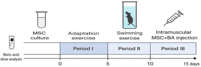
Figure 1. Experimental timeline of the study. The graphical abstract summarizes the experimental workflow. Before the adaptation period, bone marrow-derived stromal/stem cell-like cells were isolated and cultured, and the boric acid dose study was performed. During Period I, rats were acclimatized to the experimental conditions and the swimming procedure. During Period II, the exhaustive swimming protocol was performed to induce exercise-related skeletal muscle injury. At the end of Period II (Day 10), the prophylactic/preconditioning groups received an intramuscular injection of BA or MSCs + BA, according to the experimental group allocation. The post-exercise treatment group received MSCs + BA intramuscularly on Day 15 following completion of the exhaustive exercise protocol. Abbreviations: MSCs = bone marrow-derived mesenchymal stromal/stem cell-like cells; BA = boric acid. Please click here to view a larger version of this figure.
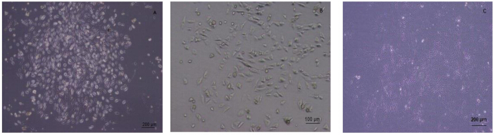
Figure 2. Representative phase-contrast images of cultured mesenchymal stem cells. Phase-contrast images show cultured mesenchymal stem cells at different magnifications. (A) Colony formation. (B) Spindle-shaped fibroblast-like morphology. (C) Low-magnification overview of the culture. Abbreviation: MSCs = mesenchymal stem cells. Please click here to view a larger version of this figure.
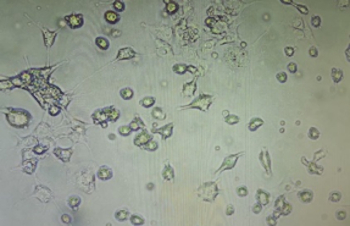
Figure 3. Representative microscopic images of bone marrow-derived stromal/stem cell-like cells following boric acid exposure. The morphology of bone marrow-derived stromal/stem cell-like cells was examined by microscopy following exposure to boric acid (BA) under standard culture conditions. The representative image illustrates the cellular morphology observed during the preliminary in vitro assessment. Based on the overall cellular response, including cell viability, proliferation, and preservation of normal fibroblast-like morphology, 10 µg/mL BA was selected for subsequent intramuscular administration in the animal experiments. Please click here to view a larger version of this figure.
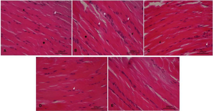
Figure 4. Histological evaluation of gastrocnemius muscle tissue. Representative hematoxylin and eosin-stained sections of gastrocnemius muscle tissue are shown for the following groups: (A) Control, (B) Exercise, (C) BA + Exercise, (D) MSCs + BA + Exercise, and (E) Exercise + MSCs + BA. White arrows indicate nuclei; black arrows indicate separations between muscle fibers; white stars indicate increased connective tissue; black stars indicate peripheral core structures. Scale bar = 100 µm. Abbreviations: MSCs = mesenchymal stem cells; BA = boric acid. Please click here to view a larger version of this figure.
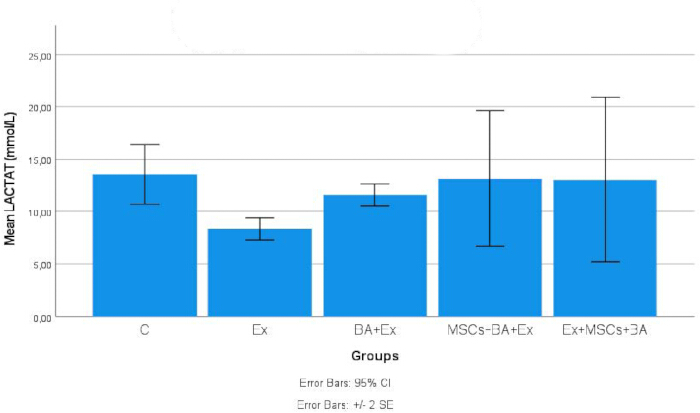
Figure 5. Comparison of serum lactate levels among experimental groups. Data are expressed as mean ± 95% confidence interval (95% CI) (n = 3 per group). The y-axis represents lactate concentration in mmol/L. Statistical comparisons between pairwise groups were performed using independent-samples t-test. *p = 0.025 vs. C group; #p = 0.011 vs. Ex group. Abbreviations: C = Control; Ex = Exercise; BA + Ex = Boric Acid + Exercise; MSCs + BA + Ex = Mesenchymal Stem Cells + Boric Acid + Exercise; Ex + MSCs + BA = Exercise + Mesenchymal Stem Cells + Boric Acid; CI = confidence interval. Please click here to view a larger version of this figure.
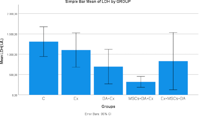
Figure 6. Comparison of serum lactate dehydrogenase levels among experimental groups. Data are expressed as mean ± 95% confidence interval (95% CI) (n = 3 per group). The y-axis represents LDH activity in U/L. Statistical analyses were performed using independent-samples t-test and confirmed by Mann-Whitney U test. Statistical markers are indicated in the figure. Abbreviations: C = Control; Ex = Exercise; BA + Ex = Boric Acid + Exercise; MSCs + BA + Ex = Mesenchymal Stem Cells + Boric Acid + Exercise; Ex + MSCs + BA = Exercise + Mesenchymal Stem Cells + Boric Acid; LDH = lactate dehydrogenase; CI = confidence interval. Please click here to view a larger version of this figure.
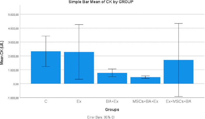
Figure 7. Comparison of serum creatine kinase levels among experimental groups. Data are expressed as mean ± 95% confidence interval (95% CI) (n = 3 per group). The y-axis represents CK activity in U/L. Pairwise comparisons were performed using independent-samples t-test and confirmed by Mann-Whitney U test. Statistical markers are indicated in the figure. Abbreviations: C = Control; Ex = Exercise; BA + Ex = Boric Acid + Exercise; MSCs + BA + Ex = Mesenchymal Stem Cells + Boric Acid + Exercise; Ex + MSCs + BA = Exercise + Mesenchymal Stem Cells + Boric Acid; CK = creatine kinase; CI = confidence interval. Please click here to view a larger version of this figure.
| groups | C | Ex | BA+Ex | MSCs+BA+Ex | Ex+MSCs+BA |
| Body-Weight(gr) P1 | 128 ± 7,90 | 140,2 ± 9,36 | 122,6 ± 11,13 | 137,4 ± 6,50 | 135,0 ± 2,83 |
| Body-Weight(gr) P2 | 139,0 ± 10,39 | 144,4 ± 11,8 | 130,8 ± 12,60 | 138,2 ± 14,96 | 140,8 ± 15,83 |
| Body-Weight(gr) P3 | 143,6 ± 10,38 | 156,8 ± 9,52 | 135,6 ± 13,76 | 140,8 ± 15,83 | 148,20 ± 12,70 |
Table 1: Body weight parameters. Body weight values measured during the experimental periods are shown for the experimental groups. Data are presented as mean ± standard deviation. Abbreviations: BW = body weight; C = Control; Ex = Exercise; BA = boric acid; MSCs = mesenchymal stem cells; P1 = Period 1; P2 = Period 2; P3 = Period 3; SD = standard deviation.
| groups | C | Ex | BA+Ex | MSCs+BA+Ex | Ex+MSCs+BA |
| BT (oC) P1 | 37,0 ± 0,29 | 36,28 ± 0,71 | 36,16 ±1,29 | 37,78 ± 0,39 | 37,78 ± 0,38 |
| BT (oC) P2 | 37,6 ± 0,40 | 38,34 ± 0,18 | 38,2 ± 0,71 | 38,6 ± 0,54 | 37,58 ± 0,29 |
| BT (oC) P3 | 37,46 ± 0,45 | 37,94 ± 0,89 | 37,38 ± 0,43 | 37,58 ± 0,28 | 37,64 ± 0,15 |
Table 2: Body temperature parameters. Body temperature values measured during the experimental periods are shown for the experimental groups. Data are presented as mean ± standard deviation. Abbreviation: BT = body temperature. Abbreviations: BT = body temperature; C = Control; Ex = Exercise; BA = boric acid; MSCs = mesenchymal stem cells; P1 = Period 1; P2 = Period 2; P3 = Period 3; SD = standard deviation.
| groups | Ex | Ex+BA | MSCs+BA+Ex | Ex+MSCs+BA |
| Exhaustion Time (min) P1 | 17,20 ± 0,84 | 9,80 ± 0,84 | − | 12,20 ± 0,84 |
| Exhaustion Time (min) P2 | 29,40 ± 7,67 | 58,40 ± 10,74 | 12,60 ± 1,67 | 17,40 ± 1,64 |
| Exhaustion Time (min) P3 | 68,20 ± 16,51 | 145,60 ± 19,48 | 76,00 ± 38,81 | 77,80 ± 28,36 |
Table 3: Repeated measurements of exhaustion time across experimental periods. Exhaustive swimming time was measured repeatedly in the same animals during three experimental periods (P1, P2, and P3). Data are presented as mean ± standard deviation and expressed in minutes. Repeated measurements were analyzed using a General Linear Model for repeated measures. Significant effects of time, group, and the time × group interaction were observed (all p < 0.001). Abbreviations: P1 = Period 1; P2 = Period 2; P3 = Period 3. Abbreviations: ET = exhaustion time; C = Control; Ex = Exercise; BA = boric acid; MSCs = mesenchymal stem cells; GLM = General Linear Model; SD = standard deviation.
Supplemental Table S1. Raw dataset used for quantitative analyses. This file contains the raw data used for body weight, body temperature, exhaustion time, and serum biochemical analyses. Data are organized by experimental group and animal number. Body weight, body temperature, and exhaustion time are shown across the experimental periods P1, P2, and P3. Serum lactate, lactate dehydrogenase, and creatine kinase values are provided for the animals included in biochemical analyses. Blank cells indicate that no value was available for that animal/parameter. Abbreviations: BW = body weight; BT = body temperature; ET = exhaustion time; LDH = lactate dehydrogenase; CK = creatine kinase; C = Control; Ex = Exercise; BA = boric acid; MSCs = mesenchymal stem cells; P1 = Period 1; P2 = Period 2; P3 = Period 3. Please click here to download this file.
ディスカッション
The present study established a reproducible experimental model of exercise-induced skeletal muscle injury and demonstrated that combining functional, biochemical, and histological assessments provides a comprehensive evaluation of muscle recovery. The protocol distinguished the contributions of boric acid (BA) and mesenchymal stem cells (MSCs) and indicated that treatment efficacy depends on the timing of administration.
Functional assessment demonstrated that BA exerted the most pronounced effect on endurance performance. General Linear Model (GLM) repeated-measures analysis demonstrated significant effects of time, experimental group, and the time × group interaction (p < 0.001). Rats receiving BA before exercise exhibited longer exhaustive swimming times than the Exercise group during the repeated swimming protocol, indicating improved resistance to fatigue. Previous studies have suggested that boron-containing compounds influence muscle differentiation, oxidative balance, and tissue repair34. However, angiogenesis and muscle maturation pathways were not directly measured here; thus, the improved endurance in BA-treated rats reflects a functional outcome potentially associated with reduced exercise-induced stress rather than direct evidence of vascular or structural maturation.
Biochemical analyses demonstrated that treatment timing influenced the response to muscle injury. CK levels typically rise within 6–24 h post-exercise, with clearance and structural recovery spanning 24–72 h35,36. Pretreatment with MSCs combined with BA (MSCs + BA + Ex) significantly reduced serum CK levels compared to the Exercise group, indicating attenuated sarcolemmal disruption. In contrast, postexercise administration (Ex + MSCs + BA) did not produce comparable biochemical reductions, suggesting that these treatments were more effective when administered prophylactically rather than therapeutically post-injury. LDH activity was lower in the treatment groups than in the Exercise group. The reduction reached statistical significance in the MSCs + BA pretreatment group, whereas BA alone showed a numerical reduction that did not reach statistical significance. These findings suggest that the combined pretreatment may provide greater protection against exercise-induced muscle damage than BA alone. Because LDH is a well-established biochemical marker of muscle cell membrane damage, these observations suggest that prophylactic BA administration prior to exhaustive exercise may suggest a potential protective trend. Whether this tendency relates to antioxidant or cytoprotective mechanisms requires further investigation, as the direct molecular pathways were not evaluated in the present study37,38.
Serum lactate measured 24 h post-exercise reflects recovery-phase dynamics rather than acute fatigue. Lactate levels in the Exercise group approached baseline by 24 h, showing only a borderline difference from Controls. Similarly, near-control lactate levels in the combined MSC + BA groups indicate preserved or restored lactate homeostasis post-injury (p > 0.05). While lactate threshold correlates positively with endurance capacity and delayed fatigue39, lactate metabolism is complex. Because lactate transporters, glycolytic enzyme activity, mitochondrial function, tissue oxygenation, and signaling pathways were not directly assessed, these findings cannot serve as definitive evidence of enhanced metabolic efficiency or reprogramming. Alternative mechanisms, such as altered clearance, transient metabolic stress, or altered glycolytic flux, must be considered. The contribution of MSCs to lactate dynamics may also be indirect and time-dependent; intramuscularly administered MSCs can persist for up to seven days, promoting protein turnover and satellite cell activation8, which may temporarily increase local metabolic demand during tissue repair.
Histological analysis of the gastrocnemius–soleus muscle complex supported these biochemical findings. The exercise-only group exhibited classic signs of exercise-induced injury: widened inter-fiber spaces, disrupted fiber integrity, irregular nuclear localization, pale cytoplasm, and striation disorganization—yielding significantly higher damage scores and leukocyte infiltration compared to controls (p < 0.05). Conversely, tissues from the MSC, BA, and combined MSC + BA groups exhibited structural preservation resembling controls. Centrally located nuclei in MSC-containing groups indicate active myofiber repair and satellite cell activation, aligning with evidence that persistent intramuscular MSCs stimulate regenerative signaling8.
Selecting an optimal intramuscular BA dose was essential. To our knowledge, this is the first study to combine intramuscular bone marrow-derived mesenchymal stromal/stem-like cells and BA in a muscle injury model. Dose selection (10 µg/mL) was based on preliminary in vitro optimization to ensure a non-cytotoxic concentration that preserves cell viability, proliferation, and morphology. Consistent with this strategy, BA administration attenuated histopathological damage (fiber separation) and supported lower serum CK and LDH levels. Though BA can modulate myogenic differentiation, oxidative stress, and membrane stability in a dose-dependent manner40,41,42,43, these specific pathways were not directly evaluated in this study.
Collectively, these findings suggest a complementary interaction: transplanted MSC-like cells contribute primarily to structural repair, whereas BA optimizes the local biochemical environment and endurance performance. However, because surface marker profiling and trilineage differentiation were not performed, the cultured plastic-adherent cells should be formally recognized as bone marrow-derived mesenchymal stromal cell-like cells rather than fully characterized MSCs.
Strengths and limitations
A major strength of this protocol is the integration of functional, biochemical, and histological metrics within a single model to simultaneously evaluate function, cellular damage, and tissue repair. Several limitations remain: recovery was evaluated at a single post-exercise time point, specific molecular mechanisms (oxidative stress, mitochondrial function, satellite cell activation) were not directly quantified, and findings in an experimental rat model require validation before clinical translation.
A limitation of the present study is that the pH and osmolality of the final BA-containing injectable preparation were not measured. In addition, no specific endotoxin testing or quantitative particulate analysis was performed. Therefore, although the preparation was made under aseptic conditions immediately before administration, these physicochemical and quality-control parameters could not be independently verified.
In conclusion, this study establishes a reproducible rat model for evaluating exercise-induced skeletal muscle injury. Boric acid administration primarily improved functional endurance performance, whereas MSC treatment predominantly supported structural muscle regeneration. Furthermore, pre-exercise intervention yielded superior outcomes compared to post-exercise delivery. These findings warrant further mechanistic and long-term preclinical evaluation prior to clinical translation.
開示事項
The authors have no conflicts of interest to declare.
During the preparation of this work, the authors used the ChatGPT model to improve the language. After using this tool, the authors reviewed and edited the content as needed and take full responsibility for the publication's content.
謝辞
The authors would like to thank the students of Kirsehir Ahi Evran University Faculty of Medicine for their participation and commitment to this study: Aysenur Sahin, Dileksu Turgut, Egemen Yıldız, Hilal Ozdemir, Mehmet AliCagır, Meryem Tekkol, Muhammed Eren Acıkgoz, Muhammed Köksal Özel, Nagihan Kutlu, Omer Metindogan, Sultan Kose, and to Davut Yolcu for his technical support for performing the biomarker analyses (Baran Medikal/ Otto Scientific). Special thanks to Kirsehir Ahi Evran University for their support and resources. This research was supported by Kirsehir Ahi Evran University Scientific Research Projects Directorate with Project number TIP.A3.24.012.
材料
| 名前 | 会社 | カタログ番号 | コメント |
|---|---|---|---|
| Boric Acid | BioShop Canada Inc | 4H88611 | Used for BA treatment preparation |
| Centrifuge | Hettich | 320R | Used for serum separation and cell processing |
| Centrifuge Tubes | ISOLAB | CTPPS24050001 | Used for serum separation and cell processing |
| Clot activator tubes | Vacuette | A24093F8 | Used for blood collection and serum preparation |
| Conical Tube | SPL Life Sciences | 50015 | Used for the isolation of mesenchymal stem cells. |
| CO2 incubator | Heal Force | HF90 | Used for Cell culture at 37 °C and 5% CO2 |
| Creatine Kinase | Otto Scientific | OTTOBC136 | Used for serum creatine kinase (CK) measurement. |
| Deep Freezer -80°C (Model DF590) | Nüve | DF590 | Used for sample storage at -80 °C |
| Digital Microscope Camera (Axiocam 208 color) | Carl Zeiss | model | Used to Image acquisition for histopathology |
| Dulbecco’s modified Eagle medium | Capricorn Scientific, USA | model | Used as Basal medium for BMSC/MSC-like cell culture |
| Dulbecco's PBS | Capricorn Scientific, USA | PBS-1A | Used for washing cells and preparing suspensions. |
| Electronic Balance | SHIMADZU | ATX224 | Used for weighing reagents |
| Eosin Y | Merck / Sigma-Aldrich | 1159350025 | Used as Eosin component for H&E staining |
| Eppendorf 1000 µL | microcult | 251810 | Used for MSCs transplantation |
| Eppendorf 200 μL | microcult | 251410 | Used for BA treatment preparation |
| Ethanol Absolute (99.5%) | Tekkim Kimya | TK.200655.05001 | Used to prepare graded ethanol series for tissue dehydration |
| Fetal Bovine Serum | Biological Industries | 1937209 | Used as Supplement for cell culture medium |
| Formaldehyde solution | Merck / Sigma-Aldrich | 1040022500 | Used for tissue fixation |
| Harris Hematoxylin Solution | BESTLAB | BS-002 | Used for H&E staining (hematoxylin component) |
| Hemocytometer | Neubauer | model | Used for cell counting |
| Injector | Genject | 1222301 | Used for MSCs isolation |
| Ketamine hydrochloride | Pharmaceutical grade(commercial source not specified) | N/A | Used for intraperitoneal anesthesia |
| Lactate Assay kit | Randox | Lec3980 | Used for serum lactate measurement |
| Laminar flow cabinet | Metisafe | model | Used for aseptic cell culture procedures |
| LDH Assay kit | Otto Scientific | OttoBC129 | Used for serum LDH measurement |
| L-glutamine | Capricorn Scientific | model | Used as Supplement for cell culture medium |
| Light microscope | OLYMPUS DP22 | TV0.5XC-3 | Used for cell morphology evaluation |
| Light Microscope (Primo Star) | Carl Zeiss | Primo Star | Used for Histopathological imaging |
| penicillin-streptomycin | Capricorn Scientific, USA | PS-B | Used as antibiotic supplement for cell culture |
| Rotary Microtome (Autocut) | Leica Biosystems | RM Autocut | Used for Cutting 5 μm paraffin sections |
| Slide Warmer | Leica Biosystems | HI1210 | Used for Drying histology slides |
| Syringe- 30G ×1/2” 0,3mm × 13mm | BD MicrolanceTM 3 | model | Used for intramuscular injection |
| Trypsin / EDTA | Capricorn Scientific, USA | TRY-38 | Used for detaching adherent cells. |
| Xylazine hydrochloride | Pharmaceutical grade(commercial source not specified) | N/A | Used for intraperitoneal anesthesia |
| Xylene (mixture of isomers) | Tekkim Kimya | TK.090270.05003 | Used for tissue clearing before paraffin embedding |
参考文献
- Margiana R et al. Clinical application of mesenchymal stem cell in regenerative medicine: a narrative review. Stem Cell Res Ther. 2022;13:366.
- Chu DT et al. An update on the progress of isolation, culture, storage, and clinical application of human bone marrow mesenchymal stem/stromal cells. Int J Mol Sci. 2020;21(3):708.
- Bilden A et al. Biochemical and histopathological effects of hirudotherapy in a rat model of chronic exercise-induced physiological stress. J Vis Exp. 2026;(231):e70494.
- Zhang Y et al. The contrasting effects of two distinct exercise training modalities on exhaustive exercise-induced muscle damage in mice may be associated with alterations in the gut microbiota. Int J Mol Sci. 2024;25(14):7837.
- Schwiete C et al. Overlaps of skeletal muscle fatigue and skeletal muscle damage: the muscle injury continuum. Sports Med Open. 2025;11(1):73.
- Thirumalai T, Viviyan TS, Elumalai EK, David E. Intense and exhaustive exercise induce oxidative stress in skeletal muscle. Asian Pac J Trop Dis. 2011;1(1):63-6.
- de Araujo et al. Low-intensity swimming training does not protect the skeletal muscle against exhaustive exercise-induced injuries in rats. Rev Bras Med Esporte. 2011;17(3).
- Takegaki J et al. Intramuscular injection of mesenchymal stem cells activates anabolic and catabolic systems in mouse skeletal muscle. Sci Rep. 2021;11(1):21224.
- Ayinde O, Hayward RS, Ross JDC. The effect of intramuscular injection technique on injection associated pain: a systematic review and meta-analysis. PLoS One. 2021;16(5):e0250883.
- Kamei N, Adachi N, Ochi M. Magnetic cell delivery for the regeneration of musculoskeletal and neural tissues. Regen Ther. 2018;9:116-9.
- Al-Azab M, Idiiatullina E, Safi M, Hezam K. Enhancers of mesenchymal stem cell stemness and therapeutic potency. Biomed Pharmacother. 2023;162:114356.
- Kestane S, Bilden A, Kahraman M, Cicek M. Leech saliva extract increases mesenchymal stem cells proliferation and viability: in vitro study. Indian J Exp Biol. 2025;63(7):605-11.
- Pizzorno L. Nothing boring about boron. Integr Med (Encinitas). 2015;14(4):35-48.
- Moussa FH, Akbas E, Yuzbasioglu D, Unal F. Boric acid prevents MMC- and H₂O₂-related DNA damage: evidence from cytogenetic and comet assays. Biol Trace Elem Res. 2026;204(2):677-96.
- Bita A et al. New insights into boron essentiality in humans and animals. Int J Mol Sci. 2022;23(16):9147.
- Sedighi-Pirsaraei N et al. Boron in wound healing: a comprehensive investigation of its diverse mechanisms. Front Bioeng Biotechnol. 2024;12:1475584.
- Sharaif B, Al Busafi M, Al Kitani M, Nasr Aldeen N. The effect of boron supplementation on free testosterone activity in the blood during muscle strength training of volleyball players. Ovidius Univ Ann Ser Phys Educ Sport Sci Mov Health. 2020;20(1):54-9.
- Nielsen FH. Update on human health effects of boron. J Trace Elem Med Biol. 2014;28(4):383-7.
- Varol M, Subaşı B, Koldemir-Gündüz M, Yerlikaya A. Boron's role in B16-F10 melanoma: cytotoxic, antioxidant, apoptotic, and anti-inflammatory effects. Biol Trace Elem Res. 2026;204(3):1319-28.
- Yu L, Dong X, Li H, Mi L. The value of blood lactate and lactate clearance rate in evaluating the prognosis of athletes with heat illness of varying degrees after high-intensity exercise. BMC Sports Sci Med Rehabil. 2025;17(1):7.
- Huang T et al. Novel insights into athlete physical recovery concerning lactate metabolism, lactate clearance and fatigue monitoring: a comprehensive review. Front Physiol. 2025;16:1459717.
- Soler-López A, Moreno-Villanueva A, Gómez-Carmona CD, Pino-Ortega J. The role of biomarkers in monitoring chronic fatigue among male professional team athletes: a systematic review. Sensors (Basel). 2024;24(21):6862.
- Kilkenny C et al. Improving bioscience research reporting: the ARRIVE guidelines for reporting animal research. PLoS Biol. 2010;8(6):e1000412.
- Ridwan R, Razak HRA, Adenan MI, Saad WMM. Supplementation of 100% flesh watermelon [Citrullus lanatus (Thunb.) Matsum. and Nakai] juice improves swimming performance in rats. Prev Nutr Food Sci. 2019;24(1):41-8.
- Ozocak O, Kestane S, Duran HH, Coksevim B. The curative effect of swimming exercise on electrophysiological parameters after sciatic nerve injury. Turk J Sport Sci. 2023;6(2):37-50.
- Yan K et al. Establishment and identification of an animal model of long-term exercise-induced fatigue. Front Endocrinol (Lausanne). 2022;13:915937.
- Reis CHO et al. Effects of physical training on the metabolic profile of rats exposed to chronic restraint stress. Clinics (Sao Paulo). 2024;79:100411.
- Tseng HC, Wu MR, Lee CH, Hsiao JK. Differentiation capacity of bone marrow-derived rat mesenchymal stem cells from DsRed and Cre transgenic Cre/loxP models. Cells. 2022;11(17):2769.
- Sanz-Nogués C et al. Development and validation of a multiparametric semiquantitative scoring system for the histopathological assessment of ischaemia severity in skeletal muscle. J Tissue Eng Regen Med. 2023;2023:5592455.
- Kalinich JF, Vergara VB, Hoffman JF. Oxidative damage in metal fragment-embedded Sprague-Dawley rat gastrocnemius muscle. Curr Res Toxicol. 2022;3:100083.
- Procópio IM et al. Histomorphometric comparison of the corpus cavernosum of rats submitted to euthanasia with ketamine and xylazine or isoflurane. Acta Cir Bras. 2021;36(11):e361103.
- Metwally MAF et al. Cardiac puncture as a survival and multiple blood collection method in laboratory rats. J Basic Appl Zool. 2026;87:32.
- de Souza RF et al. Ultra-endurance associated with moderate exercise in rats induces cerebellar oxidative stress and impairs reactive GFAP isoform profile. Front Mol Neurosci. 2020;13:157.
- Apdik H et al. Dose-dependent effect of boric acid on myogenic differentiation of human adipose-derived stem cells (hADSCs). Biol Trace Elem Res. 2015;165(2):123-30.
- Brancaccio P, Maffulli N, Limongelli FM. Creatine kinase monitoring in sport medicine. Br Med Bull. 2007;81-82:209-30.
- Peake JM et al. Muscle damage and inflammation during recovery from exercise. J Appl Physiol (1985). 2017;122(3):559-70.
- Banfi G, Colombini A, Lombardi G, Lubkowska A. Metabolic markers in sports medicine. Adv Clin Chem. 2012;56:1-54.
- Yılmaz B et al. Boron-enhanced mitochondrial repair: DeepA-I tissue regeneration. Scientifica (Cairo). 2025;2025:5343930.
- Sung DJ, So WY, Choi DH, Jeong TT. Blood lactate levels after all-out exercise depend on body fat percentage in Korean college students. Iran J Public Health. 2016;45(6):817-9.
- Powers SK, Jackson MJ. Exercise-induced oxidative stress: cellular mechanisms and impact on muscle force production. Physiol Rev. 2008;88(4):1243-76.
- Kozakowska M, Pietraszek-Gremplewicz K, Jozkowicz A, Dulak J. The role of oxidative stress in skeletal muscle injury and regeneration: focus on antioxidant enzymes. J Muscle Res Cell Motil. 2015;36(6):377-93.
- Turkez H et al. Effects of some boron compounds on peripheral human blood. Z Naturforsch C J Biosci. 2007;62(11-12):889-96.
- Ince S et al. The effects of dietary boric acid and borax supplementation on lipid peroxidation, antioxidant activity, and DNA damage in rats. J Trace Elem Med Biol. 2010;24(3):161-4.