This video demonstrates the procedure of co-culturing amoebae with mutant bacterial strains to evaluate their intracellular survival and identify virulence markers through differences in colony counts.
이 콘텐츠를 보려면 JoVE 구독이 필요합니다. 로그인하거나 오늘 무료 평가판을 시작하세요.
방법 논문
Detecting Bacterial Virulence Using Amoeba as a Host System
153 조회수
⸱
2026년 7월 1일
이 논문에서
초록
프로토콜
- Co-culture of amoebae-Mycobacterium abscessus Tn mutant: first screen
NOTE: This takes 3–4 months for the screening of 6,000 mutants.- Spread 5 x 10⁴ amoebae(Acanthamoeba castellanii) /well to a 96-well plate in 100 µL of AC (Acanthamoeba castellanii) buffer. Incubate for 1 h at 32 °C without shaking. Allow the floating amoeba trophozoïtes time to adhere to the wells. While incubating, thaw bacteria on ice for 1 h.
- Add 5 µL of the thawed bacteria cultures with an electronic multichannel pipette. Infect for 1....
액세스가 제한되었습니다. 이 콘텐츠를 보려면 로그인하거나 체험판을 시작하세요.
결과
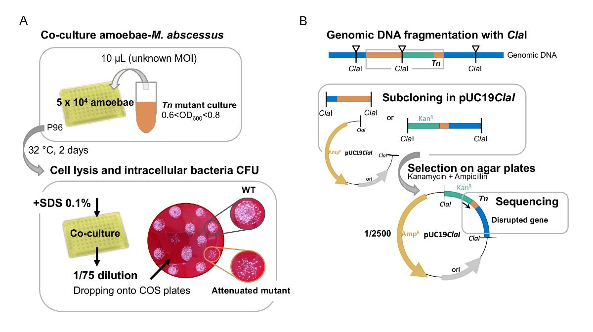
Figure 1: A. First visual screen of M. abscessus Tn mutants in amoeba. (A) large-scale screen of Tn mutants on amoebae is performed in.......
액세스가 제한되었습니다. 이 콘텐츠를 보려면 로그인하거나 체험판을 시작하세요.
재료
| 이름 | 회사 | 카탈로그 번호 | 댓글 |
|---|---|---|---|
| 24-well plates | Thermofisher | 11874235 | |
| 96-well plates | Thermofisher | 10687551 | |
| QuBit fluorometer | Thermofisher | Q33226 | |
| Acanthamoeba castellanii | ATCC | 30010 | strain |
| Amikacin | Mylan | 150927-A | powder |
| CaCl2 | Sigma-Aldrich | C1016 | >93% granular anhydrous |
| Columbia agar | Biomerieux | 43041 | 90 mm |
| D-Glucose | Sigma-Aldrich | G8270 | powder |
| DMEM | Thermofisher | 11500596 | medium |
| DNase and RNase free water | Invitrogen | 10977-035 | solution |
| Escherichia coli | Clinical isolate | personal stock | bacteria |
| Fetal Calf Serum | Gibco | 10270 | serum |
| Müller-Hinton agar | Biorad | 3563901 | powder |
| SDS 20% | Biorad | 1610418 | solution |
