마이크로 - 온 - 칩 플랫폼은 포토 리소그래피 리플 로우 레지스트 기술, 소프트 리소그래피, 그리고 미세 유체의 조합에 의해 개발되었다. endothelialized 마이크로 플랫폼은 생체 내 미세 혈관에서의 3 차원 (3D) 구조를 모방 제어 지속적인 혈류의 흐름에서 실행하고, 고품질의 실시간 영상을 허용하고 미세 혈관 연구에 적용 할 수 있습니다.
이 콘텐츠를 보려면 JoVE 구독이 필요합니다. 로그인하거나 오늘 무료 평가판을 시작하세요.
이 논문에서
요약
초록
생체 내 동물 연구는 더 많은 시간이 소요되는, 비싼, 그리고 관찰과 정량화가 매우 어려운됩니다 때문에 노력은 미세 혈관의 연구를위한 생체 분석의 개발에 초점을 맞추고있다. 세 가지 차원 (3D) 형상에 대해 생체 내 미세 혈관의 대표 및 지속적인 유체 흐름을 제공 할 때 그러나, 생체 미세 혈관 분석에서 기존의 제한이 있습니다. 포토 리소그래피 리플 로우 레지스트 기술, 소프트 리소그래피, 그리고 미세 유체의 조합을 사용하여, 우리는 멀티 깊이 원형 단면 endothelialized 개발 한 제어 연속 관류에 따라 생체 내 미세 혈관에서의 3D 형상을 모방하고 실행 마이크로 - 온 - 칩 흐름. 긍정적 인 리플 로우 레지스트는 반원형 단면 미세 네트워크와 마스터 몰드를 제조하는 데 사용되었다. REPL 개의 폴리 디메틸 실록산 (PDMS) 마이크로의 정렬 및 본딩마스터 금형 icated, 원통형 마이크로 네트워크를 만들었습니다. 마이크로의 직경은 잘 제어 할 수 있습니다. 또한, 칩 내부에 씨앗을 품고 기본 인간 제대 정맥 내피 세포 (HUVECs)는 세포 4 일 이주 사이의 기간 동안 지속적인 제어 관류에 따라 마이크로의 내부 표면을 지어 것으로 나타났다.
서론
미세 혈관은 혈액 순환 시스템의 일부로서, 혈액과 조직 사이의 상호 작용을 중재 신진 대사 활동을 지원, 조직의 미세 환경을 정의하고, 많은 건강과 병적 상태에 중요한 역할을한다. 체외에서 기능 미세 혈관의 재현부는 복잡한 혈관 현상의 연구 플랫폼을 제공 할 수 있습니다. 그러나, 내피 세포 마이 그 레이션 분석, 내피 관 형성 분석과 쥐 및 쥐 대동맥 링 분석 등의 생체 미세 혈관 분석, 종래의 입체 (3D) 형상과 연속 흐름 제어에 대해 생체 내 미세 혈관의를 다시 할 수 없습니다 1-8. 동물 모델과 같은 각막 신생 혈관 분석, 병아리 융모 막 혈관 분석 및 마트 리젤 플러그 분석 등의 생체 분석,에서를 사용하여 미세 혈관의 연구는 더 많은 시간이 소요되는 비용 높은 관찰과 quantifications에 대해 도전하고 있습니다윤리적 문제 1, 9-13 올립니다.
하지 않았을 등 쉽게 엄격하게 제어 생물학적 조건과 역동적 인 유체 환경과 같은 연구 14, 동물 생체에 관련된 높은 실험 비용과 복잡성을 축소 및 단축하면서 micromanufacturing 및 미세 유체 칩 기술의 발전은 생명 과학에 대한 통찰력의 다양한 활성화 기존의 거시적 기술로 할 수 있었다.
여기, 우리는 endothelialized를 구성하는 방법을 제시 마이크로 - 온 - 칩 생체 내 미세 혈관에서의 3D 형상을 모방하고 포토 리소그래피 리플 로우 레지스트 기술, 소프트 리소그래피, 그리고 미세 유체의 조합을 사용하여 제어 지속적인 혈류의 흐름에서 실행됩니다.
액세스가 제한되었습니다. 이 콘텐츠를 보려면 로그인하거나 체험판을 시작하세요.
프로토콜
1. 포토 레지스트 마스터 금형의 석판 제작
다음 프로토콜은 30 ~ 60 μm의 사이 직경의 미세을 제작하는 과정을 보여줍니다. 작은 직경 (이하 30 μm의), 포토 레지스트의 단일 스핀 코팅 마이크로를 얻으려면이 필요합니다.
- 사용하기 전에 클린 24 시간에 4 ° C에서 냉장고에서 리플 로우 레지스트를 전송하고 실내 온도를 따뜻하게 할 수 있습니다.
- 실리콘 웨이퍼를 청소하고 탈수 할 수 있도록 150 ° C에서 한 시간 동안 구워. 탈수는 실리콘 기판에 포토 레지스트 접착 도움을 줄 것입니다.
- 스핀 코팅 포토 레지스트의 첫 번째 레이어는 다음과 같은 조리법을 사용하여 :
| 단계 | 시간 (초) | 속도 (rpm) | 가속 |
| 1 | 2 | 300 | 최고 |
| 2 | 10 | 0 | |
| 3 | 3 | 300 | 최고 |
| 4 | 60 | 600 | 최고 |
| 5 | 10 | 500 | 최고 |
| 6 | 10 | 600 | 최고 |
- 그런 다음 90 초 동안 110 ° C의 온도 열판에 웨이퍼를 굽는다. 부드러운 빵을 한 후 포토 레지스트의 두께는 20 ~ 30 μm의 수 있습니다.
- 코트에게 첫 번째 레이어에 사용되는 것과 동일한 조리법에 따라 포토 레지스트의 두 번째 레이어를 회전.
- 부드러운 빵을 90 초 동안 110 ° C의 온도 열판에 배치하여 다시 웨이퍼. 이 부드러운 빵을 한 후 포토 레지스트의 두께는 40 ~ 60 μm의 수 있습니다.
- photor를 노출하여 긍정적 인 마스터 패턴을 생성영화 마스크를 통해 14,500 엠제이 / cm 2의 노광량으로 UV 빛을 ESIST.
- 탈 (DI) 물 (1시 2분 (V / V))의 개발을 희석. 패턴이 완전히 개발 될 때까지 용액에 반복적으로 웨이퍼를 씻어. 다음 DI 물과 웨이퍼를 세척하고 질소 가스를 사용하여 말립니다.
- 리플 로우 : 4 분, 용매 증발을 방지하기 위해 유리 페트리 접시 덮개를위한 120 ° C의 온도 열판에 배치 웨이퍼. 열판에서 웨이퍼를 제거하고 실온까지 냉각 할 수 있습니다. 리플 로우 후의 레지스트 두께는 50 μm의 수 있습니다.
2. PDMS 마이크로 채널 네트워크 소프트 리소그래피 제작
- 10:1 중량 비율 (기본 : 경화제)에서 폴리 디메틸 실록산 (PDMS) 솔루션을 준비하고 행성 원심 믹서를 사용하여 철저하게 섞는다.
- 리플 로우 포토 레지스트 마스터 금형에 PDMS 솔루션을 캐스팅합니다. 가스를 15 분 동안 데시 케이 터에서 주조 PDMS를 놓고. 필요한 경우 남아있는 거품을 제거하기 위해 질소 가스를 사용합니다.
- 그것을 치료 할 수 있도록 3 시간 동안 60 ° C의 온도에서 오븐에 PDMS를 굽는다. 그런 다음 마스터 금형 치료 PDMS 층을 제거합니다.
- 채널 네트워크에 구멍을 펀치로 유입 / 유출 구멍을 만들 날카롭게 주먹을 사용합니다. 질소 가스를 사용하여 PDMS의 표면을 청소합니다.
- 4.5 × 10 -2 Torr의 및 3.5 피트 / 분의 산소 유량의 압력에서 플라즈마 클리너 내부에 30 초 동안 산소 플라즈마 두 PDMS 레이어를 처리합니다. 그런 다음 광학 현미경으로 수동으로 PDMS의 표면을 맞 춥니 다. 물 한 방울 정렬의 더 나은 제어에 필요한 경우에 사용합니다.
- 60 ° C 영구적 인 결합을 달성하기 위해 30 분 동안 오븐에서 장치를 굽는다.
3. 칩 HUVEC 문화 및 시드
- 문화는 L-글루타민과 문화 매체와 HUVECs 10 % 태아 소 혈청 (FBS)와 보충. 일에 대한2와 5 사이에 전자 실험 구절을 사용 하였다.
- HUVECs가 합류되면, 37 ℃에서 2-6 분 동안 세포를 계산하고 최초의 HEPES이 (HEPES-BSS) 생리 식염수를 버퍼로 세포를 세척하여 그들을 수확 후 트립신 / EDTA (에틸렌 다이아 민 테트라 초산)과 부화와 세포 치료 트립신이 완료된 후, 트립신을 중화 솔루션과 트립신 / EDTA (에틸렌 다이아 민 테트라 초산)을 중화. 원심 분리를 수집하기 위해 8 % 덱스 트란과 문화 매체에있는 세포를 일시 중지합니다. 덱스 트란은 더 나은 세포 파종 및 첨부 돕기 위해 중간 점도를 높이기 위해 사용되었다.
- 5 4.5 × 10 -2 Torr의의 작동 압력 분 및 3.5의 산소 유량 피트 3 / 분 산소 플라즈마 장치를 취급합니다. 다음 DI 워터로 장치를로드하고 멸균 층류 생물 안전 후드에서 8 시간 동안 UV 빛으로 취급합니다.
- 세포가 준비되어 하루 전에 1X 인산염 장치를 세척 피브로넥틴 (100 ㎍ / ㎖, DIL와 코트 다음 (1X PBS) 완충 식염수1X PBS로 uted), 4 ° C에서 하룻밤 냉장고에 품어.
- 피브로넥틴 코팅 후 1X PBS 다음 배지와 부하 장치를 씻는다. 15 분 37 ° C의 온도에서 장치를 품어.
- 3-4 X 10 6 세포 / ML의 세포 농도와 8 % 덱스 트란 문화 매체에 HUVEC 세포를로드합니다. 장치의 하나 입구에서 세포의 20 μL 방울을 배치하고 미세 유체 채널에 세포를 소개를 기울이십시오. 15-20 분 후 세포는 채널의 측면 벽에 부착하기 시작합니다. 장치에게 전지보다 균일 한 분포를 생성 할 때마다 15 분을 돌립니다. 필요한 경우, 추가 하중을 수행 할 수 있습니다.
4. 장기 관류 설정
정적 문화의 5-6 시간 후, 첨부 HUVECs 완전히 확산하기 시작합니다. 10 μL / 시간의 지속적인 흐름으로 원격 제어 주사기 펌프 시스템을 사용하여 관류를 설정합니다. 관류는 F를 조정할 수 있습니다또는 더 높은 유량,는 2 주 4 일 사이 기간 동안 지속될 수있다.
5. 세포 염색 및 현미경 특성
- 세포가 장치 안에 합류에 도달하면, 첫째, 철저하게 매체를 제거 1X PBS로 장치를 세척한다. 다음 희석제 (4 μL-1 ML)로 희석 붉은 염료로 장치를로드합니다. 셀 로딩 절차와 비슷 염료를 넣습니다. 상온에서 5 분 동안 어둠 속에서 장치를 배양 한 후 염색을 중지 배지로 장치를 세척한다. 염료의 긴 배양은 세포 독성과 disadhesion가 발생할 수 있습니다.
- 1X PBS (ML 당 2 방울)에 희석 파란색 염료로 장치를로드합니다. 상온에서 5 분 동안 어둠 속에서 부화 후 철저하게 1X PBS로 장치를 세척한다.
- 거꾸로 광학 현미경으로 세포 염색을 검사합니다. 염색이 좋으면 (1X PBS로 희석 3.5 % 파라 포름 알데히드) 고정 매체로 마이크로를로드 한 후 잠수함중간과 완전히 알루미늄 호일로 커버를 고정에있는 장치. 4의 온도에 냉장고에 장치를 보관 ° C 건조하고 photobleaching에에서 장치를 방지 할 수 있습니다. 고정 장치는 이제 공 촛점 현미경 스캐닝 레이저로 할 수있는 공 촛점 이미징을위한 준비가되어 있습니다.
액세스가 제한되었습니다. 이 콘텐츠를 보려면 로그인하거나 체험판을 시작하세요.
결과
멀티 깊이 마이크로 네트워크를 제조하는 우리의 접근 방식은 미세 크로스 섹션에게 15 반올림하는 생체 미세 혈관에서의 복잡한 3D 형상을 모방. 또한, 부모 분기 채널의 직경과 딸 채널은 대략 전체 채널 저항이 낮고 유속이 네트워크 16-18 전역에 균일 있도록 필요한 수준에서의 유체 흐름을 유지하기 위해 머레이의 법칙을 준수하십시오. 반원형 포토 레지스트 마스터 금형의 제조 및 원형 단면 PDMS 마이크로 채널 네트워크에 대한 프로세스 및 결과는 영화 1, 그림 2는 각각 영화 2에서 설명 하였다. PDMS 마이크로 채널 네트워크의 기하학적 특징은 특징과 그림 2에 나타내었다. 우리의 결과는 포토 레지스트 리플 로우 기술은 다중 깊이 분기 채널 네트워크를 만들 수 있다는 것을 보여 전NA 더 편리 포토 레지스트의 리플 로우 기술로 접근, 약 머레이의 법칙을 순종 혈...
액세스가 제한되었습니다. 이 콘텐츠를 보려면 로그인하거나 체험판을 시작하세요.
토론
1. 마스터 금형 제작
혈관의 형태 학적위한 설계 원칙 중 하나는 네트워크를 통해 혈관 직경의 분포는 최소의 에너지 고려에 의해 지배되는 진술 머레이의 법칙 16로 알려져 있습니다. 또한 분기점에서 부모 혈관의 직경 큐브 딸 혈관의 직경 큐브의 합에 해당된다는 진술 ( 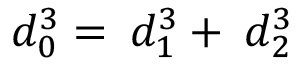 또한) 19 Poiseuille의 법칙은 다음과 같이 혈관의 대부분에서 전단 응력의 크기를 추정하는 데 사용되었습니다
액세스가 제한되었습니다. 이 콘텐츠를 보려면 로그인하거나 체험판을 시작하세요.
공개 사항
저자는 그들이 더 경쟁 재정적 이익이 없다는 것을 선언합니다.
감사의 글
이 연구는 부분적으로 국립 과학 재단 (NSF 1,227,359)는 각각 국립 과학 재단 (EPS-1003907), 국립 과학 재단 (1007978) 후원 WVU 사전 사무실 및 WVU PSCoR에 의해 자금 WVU EPSCoR 프로그램에 의해 지원되었다. 미세 작업은 WVU 공유 연구 시설 (클린 룸 설비)과 웨스트 버지니아 대학의 칩 연구소 (마이크로 칩 연구소)에 미세 유체 통합 세포 연구로 이루어졌다. 공 촛점 이미징 WVU 현미경 이미징 시설에서 이루어졌다.
액세스가 제한되었습니다. 이 콘텐츠를 보려면 로그인하거나 체험판을 시작하세요.
재료
| 이름 | 회사 | 카탈로그 번호 | 댓글 |
|---|---|---|---|
| Reagent/Material | |||
| Reflow Photoresist | AZ Electronic Materials | AZP4620 | |
| Developer | AZ Electronic Materials | AZ 400K | |
| PDMS | Dow Corning Corporation | Sylgard 184 | |
| MCDB 131 Culture Medium | Invitrogen | 10372-019 | |
| NacBlue 핵 염색 | Invitrogen | H1399 | |
| PKH Red Stain | Sigma | MINI26 및 PKH26GL | |
| Fibronectin | Gibco | PHE0023 | |
| L-Glutamine | Sigma G7513 | Buffered | |
| Saline Invitrogen | 14040-133 | ||
| HEPES Buffered Saline | Solution Lonza | CC-5024 | |
| 트립신/EDTA | 인비트로젠 | 25300-062 | |
| 트립신 중화액 | Lonza | CC-5002 | |
| PDMS 경화제 | 다우코닝 코퍼레이션 | 실가드 184 | |
| 1차 인간 배꼽 정맥 내피 세포 | Lonza | CC-2517 | |
| 소 태아 혈청 | Lonza | 14-501F | |
| 희석제 C | Sigma | CGLDIL | |
| Hoechst33342 | Invitrogen, Molecular Probes | R37605 | |
| 덱스트란 | Sigma | 95771 | |
| 3.5% 파라포름알데히드 | 전자 현미경 과학 | 15710-S | |
| Equipment | |||
| Spinner | Laurell Technologies Corporation | WS-400BZ-6NPP/ LITE | |
| 데시케이터 | BelArt 제품 | 999320237 | |
| 도립 현미경 | Nikon | Eclipse Ti | |
| 주사기 펌프 시스템 | 하버드 장치 | PHD Ultra | |
| Laminar Biosafety Hood | Thermo Scientific | 1300 시리즈 A2 | |
| 행성 원심 믹서 | Thinky | ARE-310 | |
| Isotemp 오븐 | Fisher Scientific | 13-246-516GAQ | |
| 광학 현미경 | Zeiss | Invertoskop 40C | |
| 플라즈마 클리너 | Harrick Plasma | PDC-32G | |
| 핫플레이트 | Barnstead/Thermolyne Cimarec | SP131635 | |
| 레이저 스캐닝 컨포칼 현미경 | Zeiss | LSM 510 |
참고문헌
- Adair, T. H. Angiogenesis: Integrated systems physiology: from molecule to function to disease. , 1st edition, Biota Publishing. (2011).
- Goodwin, A. M. In vitro assays of angiogenesis for assessment of angiogenic and anti-angiogenic agents. Microvasc. Res. 74, 172-183 (2007).
- Smith, E. J., Staton, C. A. Tubule formation assays. Angiogenesis assays: A critical appraisal of current techniques. , John Wiley & Sons Ltd. West Sussex, UK. 65-87 (2006).
- Nakatsu, M. N., Davis, J. J., Hughes, C. C. W. Optimized fibrin gel bead assay for the study of angiogenesis. J. Vis. Exp. (3), e186(2007).
- Nicosia, R. F., Ottinetti, A. Growth of microvessels in serum-free matrix culture of rat aorta. A quantitative assay of angiogenesis in vitro. Lab. Invest. 63, 115-122 (1990).
- Aplin, A. C., Fogel, E., Zorzi, P., Nicosia, R. F. The aortic ring model of angiogenesis. Methods Enzymol. 443, 119-136 (2008).
- Nicosia, R. F. The aortic ring model of angiogenesis: A quarter century of search and discovery. J. Cell. Mol. Med. 13, 4113-4136 (2009).
- Griffith, L. G., Swart, M. A. Capturing complex 3D tissue physiology in vitro. Nat. Rev. Mol. Cell Biol. 7, 211-224 (2006).
- Folkman, J. History of angiogenesis. Angiogenesis: An integrative approach from science to medicine. , Springer. (2008).
- Auerbach, R., Lewis, R., Shinners, B., Kubai, L., Akhtar, N. Angiogenesis assays: A critical overview. Clin. Chem. 49, 32-40 (2003).
- Auerbach, R., Akhtar, N., Lewis, R. L., Shinners, B. L. Angiogenesis assays: Problems and pitfalls. Cancer Metastasis Rev. 19, 167-172 (2000).
- Staton, C. A., Reed, M. W., Brown, N. J. A critical analysis of current in vitro and in vivo angiogenesis assays. Int. J. Exp. Pathol. 90, 195-221 (2009).
- Staton, C. A., Stribbling, S. M., et al. Current methods for assaying angiogenesis in vitro and in vivo. Int. J. Exp. Pathol. 85, 233-248 (2004).
- Moraes, C., Mehta, G., Lesher-Perez, S. C., Takayama, S. Organs-on-a-Chip: A focus on compartmentalized microdevices. Ann. Biomed. Eng. 40 (6), 1211-1227 (2012).
- Huang, Z., Li, X., Martins-Green, M., Liu, Y. Microfabrication cylindrical microfluidic channel networks for microvascular research. Biomedical Microdevices. 14 (5), 873-883 (2012).
- Murray, C. D. The physiological principle of minimum work applied to the angle of branching of arteries. J. Gen. Physiol. 9 (6), 835-841 (1926).
- Zamir, M., Medeiros, J. A. Arterial branching in man and monkey. J. Gen. Physiol. 79, 353-360 (1982).
- Gafiychuk, V. V., Lubashevsky, I. A. On the principles of the vascular network branching. J. Theor. Biol. 212, 1-9 (2001).
- Sherman, T. F. On connecting large vessels to small. The meaning of Murray's law. J. Gen. Physiol. 78 (4), 431-453 (1981).
- Kamiya, A., Bukhari, R., Togawa, T. Adaptive regulation of wall shear stress optimizing vascular tree function. Bull Math Biol. 46 (1), 127-137 (1984).
- LaBarbera, M. Principles of design of fluid transport systems in zoology. Science. 249, 992-1000 (1990).
- Emerson, D. R., Cieslicki, K., Gu, X., Barber, R. W. Biomimetic design of microfluidic manifolds based on a generalized Murray's law. Lab Chip. 6, 447-454 (2006).
- Lu, H., Koo, L. Y., et al. Microfluidic shear devices for quantitative analysis of cell adhesion. Anal. Chem. 76, 5257-5264 (2004).
- Shevkoplyas, S. S., Gifford, S. C., Yoshida, T., Bitensky, M. W. Prototype of an in vitro model of the microcirculation. Microvasc. Res. 65, 132-136 (2003).
- Kaihara, S., Borenstein, J., et al. Silicon micromachining to tissue engineer branched vascular channels for liver fabrication. Tissue Eng. 6, 105-117 (2000).
- Fisher, A. B., Chien, S., Barakat, A. I., Nerem, R. M. Endothelial cellular response to altered shear stress. Am. J. Physiol. Lung Cell. Mol. Physiol. 281 (3), L529-L533 (2001).
- Nerem, R. M., Alexander, R. W., et al. The study of the influence of flow on vascular endothelial biology. Am. J. Med. Sci. 316 (3), 169-175 (1998).
- Daly, D., Stevens, R. F., Hutley, M. C., Davies, N. The manufacture of microlenses by melting photoresist. Meas. Sci. Technol. 1 (8), 759-766 (1990).
- Schilling, A., Merz, R., Ossmann, C., Herzig, H. P. Surface profiles of reflow microlenses under the influence of surface tension and gravity. Opt. Eng. 39 (8), 2171-2176 (2000).
- Young, B., Heath, J. W. Wheater's functional histology: A text and colour atlas. , 4th edition, Churchill Livingstone. (2000).
- O'Neill, F. T., Sheridan, J. T. Photoresist reflow method of microlens production. Part 1: Background and experiments. Optik Int. J. Light Electron Opt. 113, 391-404 (2002).
- de Gennes, P. G. Wetting: statics and dynamics. Rev. Mod. Phys. 57, 827-863 (1985).
- Elias, H. G. An Introduction to Polymer Science. , VCH Publishers. New York. (1997).
- Voinov, O. V. Dynamic edge angles of wetting upon spreading of a drop over a solid surface. J. Appl. Mech. Tech. Phys. 40, 86-92 (1999).
- Daly, D., Stevens, R. F., Hutley, M. C., Davies, N. The manufacture of microlenses by melting photoresist. Meas. Sci. Technol. 1, 759(1990).
- Jay, T. R., Stern, M. B. Preshaping photoresist for refractive microlens fabrication. Opt. Eng. 33, 3552-3555 (1994).
- Nerem, R. M., Alexander, R. W., et al. The Study of the influence of flow on vascular endothelial biology. Am. J. Med. Sci. 316 (3), 169-175 (1998).
- Chien, S., Li, S., Shyy, Y. J. Effects of mechanical forces on signal transduction and gene expression in endothelial cells. Hypertension. 31, 162-169 (1998).
- Li, Y. S., Haga, J. H., Chien, S. Molecular basis of the effects of shear stress on vascular endothelial cells. J. Biomech. 38, 1949-1971 (2005).
- Lee, E. J., Vunjak-Novakovic, G., Wang, Y., Niklason, L. E. A biocompatible endothelial cell delivery system for in vitro tissue engineering. Cell Transplant. 18, 731-743 (2009).
- Lee, E. J., Niklason, L. E. A novel flow bioreactor for in vitro microvascularization. Tissue Eng. Part C Methods. 16, 1191-1200 (2010).
- Chau, L., Doran, M., Cooper-White, J. A novel multishear microdevice for studying cell mechanics. Lab Chip. 9, 1897-1902 (2009).
- Meeson, A., Palmer, M., Calfon, M., Lang, R. A relationship between apoptosis and flow during programmed capillary regression is revealed by vital analysis. Development. 122, 3929-3938 (1996).
- Van Royen, N. J., Piek, J., Schaper, W., Bode, C., Buschmann, I. Arteriogenesis: mechanisms and modulation of collateral artery development. J. Nucl. Cardiol. 8, 687-693 (2001).
- Schaper, W. Therapeutic arteriogenesis has arrived. Circulation. 104 (17), 1994-1995 (2001).
- Tarbell, J. M. Shear stress and the endothelial transport barrier. Cardiovas. Res. 87 (2), 320-330 (2010).
- Potter, C. M., Lundberg, M. H., et al. Role of shear stress in endothelial cell morphology and expression of cyclooxygenase isoforms. Arterioscler. Thromb. Vasc Biol. 31, 384-391 (2011).
- Montesano, R. In vitro rapid organization of endothelial cells into capillary-like networks is promoted by collagen matrices. J. Cell Biol. 97, 1648-1652 (1983).
- Darland, D. C., D'Amore, P. A. TGF beta is required for the formation of capillary-like structures in three-dimensional cocultures of 10T1/2 and endothelial cells. Angiogenesis. 4 (1), 11-20 (2001).
- Lawley, T. J., Kubota, Y. Induction to morphologic differentiation of endothelial cells in culture. J. Invest. Dermatol. 93, 59S-61S (1989).
- Kanzawa, S., Endo, H., Shioya, N. Improved in vitro angiogenesis model by collagen density reduction and the use of type III collagen. Ann. Plast. Surg. 30, 244-251 (1993).
- Davis, G. E., Bayless, K. J., Mavila, A. Molecular basis of endothelial cell morphogenesis in three-dimensional extracellular matrices. Anat. Rec. 268, 252-275 (2002).
- Velazquez, O. C., Snyder, R., Liu, Z., Fairman, R. M., Herlyn, M. Fibroblast-dependent differentiation of human microvascular endothelial cells into capillary-like 3-dimensional networks. FASEB J. 16, 1316-1318 (2002).
- Donovan, D., Brown, N. J., Bishop, E. T. Comparison of three in vitro human "angiogenesis" assays with capillaries formed in vivo. Angiogenesis. 4, 113-121 (2001).
- Tang, D. G., Conti, C. J. Endothelial cell development, vasculogenesis, angiogenesis, and tumor neovascularization: an update. Semin. Thromb. Hemost. 30, 109-117 (2004).
- Takayama, S., McDonald, J. C., et al. Patterning cells and their environments using multiple laminar fluid flows in capillary networks. Proc. Natl. Acad. Sci. U.S.A. 96, 5545-5548 (1999).
- Cho, B. S., Schuster, T. G., et al. Passively driven integrated microfluidic system for separation of motile sperm. Anal. Chem. 75, 1671-1675 (2003).
- Parsa, H., Upadhyay, R., Sia, S. K. Uncovering the behaviors of individual cells within a multicellular microvascular community. Proc. Natl. Acad. Sci. U.S.A. 108 (12), 5133-5138 (2011).
액세스가 제한되었습니다. 이 콘텐츠를 보려면 로그인하거나 체험판을 시작하세요.
재인쇄 및 허가
