우리는 심장 수술을 받은 환자에서 표준 transesophageal 심장 초음파 기술을 사용 하 여 소용돌이 형성 시간, 왼쪽된 심 실 충전 효율의 색인을 측정 프로토콜을 설명 합니다. 우리는 다른 심장 병 리과 환자의 여러 그룹에서 소용돌이 형성 시간 분석에이 기술을 적용.
이 콘텐츠를 보려면 JoVE 구독이 필요합니다. 로그인하거나 오늘 무료 평가판을 시작하세요.
방법 논문
심장 수술 중 심장 Transesophageal 초음파를 사용 하 여와 동 형성 시간의 비 침범 성 결정
이 논문에서
요약
초록
트랜스 mitral 혈 생성 3 차원 회전체 액체의 강화의 효율 왼쪽 심 실 (LV) 작성 하는 소용돌이 링 알려진 연속 선형 jet와 비교. 소용돌이 링 개발은 가장 자주 소용돌이 형성 시간 (VFT), 크기가 없는 매개 변수는 엄밀한 관에서 액체 방출에 따라 정량. 우리의 그룹은 심장 수술 하는 동안 효율성을 채우는 LV에 영향을 주는 요인에 관심을. 이 보고서에서 우리는 표준 2 차원 (2D) 및 도플러를 사용 하는 방법을 설명 transesophageal 심장 초음파 (티) noninvasively 파생 변수 VFT 계산 하는 데 필요한. 우리는 트랜스 mitral 초기 LV 작성 및 심 방 systole 혈액 흐름 속도 파형 중반 식도 4 챔버 티 뷰에서 측정 속도 시간 전체에서 심 방 충전 분수 (β)을 계산 합니다. LV 유출 트랙의 직경의 제품 중반-식도 긴 축 티 뷰에서 측정 및 유출 트랙을 통해 혈액의 흐름의 속도 시간 전체 펄스 파를 사용 하 여 깊은 transgastric 보기에 결정으로 치기 양 (SV) 계산 도플러입니다. 마지막으로, 승 모 판 직경 (D) 주요 하 고 작은 축 길이 직교 중반 식도 bicommissural와 긴 축 이미징 비행기에 각각 측정의 평균으로 결정 됩니다. VFT는 다음 4 × (1-β) × SV로 계산 / (πD3). VFT 다른 심장 이상 가진 환자의 여러 그룹 분석에이 기술을 사용 했습니다. 우리는이 기술 및 그것의 잠재적인 한계의 우리의 응용 프로그램 토론 하 고 또한 우리의 결과 날짜를 검토. VFT 티를 사용 하 여의 비 침범 성 측정은 심장 수술 마 취 환자에서 간단 합니다. 기술은 심장 담당 및 외과 병 적인 조건 및 LV에 외과 개입의 영향 평가를 실시간으로 효율성을 작성 수 있습니다.
서론
유체 역학은 왼쪽된 심 실 (LV) 작성의 중요 한 아직 자주 무시 결정 이다. 3 차원 회전체 액체, 액체 통과 구멍1,2,3때마다 소용돌이 반지로 알려진이 생성. 이 소용돌이 링 연속 선형 제트4와 비교 하는 유체 수송의 효율성을 향상 시킵니다. 초기 LV 작성 동안 승 모 판 통해 혈액의 운동5,6,,78 를 소용돌이 링 원인과 유체 운동량을 보존 하 여 챔버로의 전파를 용이 하 게 하 고 운동 에너지9. 이러한 작업 강화 LV 효율4,10,11,,1213작성. 링 LV apex14,15,,1617 혈액 흐름 정체의 억제 뿐만 아니라 앞쪽 mitral 전단지7, 아래 우선적으로 흐름 안내 18, 꼭대기 혈전 형성의 위험을 감소 하 고 LV 유출의 작성을 용이 하 게 하는 효과 각각19, 추적 합니다. 17심장 초음파, 도플러 벡터 흐름 매핑6,20,21, 자기 공명 영상7, 그리고 입자 이미징 velocimetry9,22 대조 ,,2324 모양 및 정상 및 병 적인 조건 하에서 트랜스 mitral 소용돌이 링의 동작을 보여 주기 위해 사용 되었습니다. 왼쪽된 심 방 LV 압력 기울기, 확장기 mitral 환상 여행, 심장, 그리고 속도 LV 휴식의 범위 동안 달성 최소 LV 압력의 정도 기간, 크기, 흐름, 강도와의 위치의 4 개의 주요 결정 요인 트랜스 mitral 반지2,12,25,26,27,,2829.
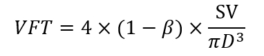
소용돌이 링 개발은 가장 자주는 크기가 없는 매개 변수 (와 동 형성 시간; 계량 VFT)는 엄밀한 관3, VFT 시간 평균 유체 속도의 제품 및 오리 피스 직경으로 나눈 방출의 기간으로 정의 됩니다 액체 방출 기반으로 합니다. 소용돌이 링의 최적의 크기 VFT 4 체 외에 제트기를 후행 하기 때문에 고 에너지 한계는 더 큰 크기3,4달성에서 그것을 방지 이루어집니다. 승 모 판 VFT는 임상 심장 transthoracic 초음파8,,3031를 사용 하 여 직선 근사 줄 되었습니다. 트랜스 mitral 혈액 흐름 속도 및 승 모 판 직경 (D)의 분석을 바탕으로, 그것은 쉽게 일 수 있다 표시8 그 VFT = 4 × (1-β) × EF × α3, 어디 β = 심 방 충전 분수, EF = LV 방출 분수, 그리고 α = EDV1/3/D, 어디 EDV = 끝 확장기 볼륨입니다. 방출 분수는 치기 양 (SV)의 비율 및 EDV, VFT을 단순화 하는이 방정식을 수 있도록 4 × (1-β) × SV = / (πD3). VFT 치수 (볼륨/볼륨) 이므로이 인덱스 체중 또는 신체 표면적8에 대 한 조정 없이 다양 한 크기의 환자 사이의 직접 비교 가능 3.3과 건강 한 과목8, 그리고 결과에 5.5 사이의 최적의 VFT 범위는 유체 역학 모델3,32에서 얻은 그와 일치 합니다. VFT는 ≤ 2.0 환자의 우울된 LV 수축 기 기능, 또한 이론적인 예측8에서 지원 되는 결과 수를 표시 했다. VFT 절감 독립적으로 예측 하지 사망률30심장 마비 가진 환자에 있는 사망률. 높은 LV afterload33, Alzheimer의 질병34, 비정상적인 심장 확장 기능19, 그리고 네이티브 승 모 판 족35 로 교체도 VFT 감소 표시 되었습니다. VFT의 측정 혈액 흐름 정체의 또는 혈전 증 환자에서 급성 심근 경색36,37식별도 유용할 수 있습니다.
우리의 그룹은 심장 수술38,39,,4041동안 효율성을 채우는 LV에 영향을 주는 요인에 관심을. 우리가 noninvasively VFT 계산 하는 데 필요한 변수를 파생 표준 2 차원 및 도플러 transesophageal 심장 초음파 (티)를 사용 합니다. 이 보고서에서 우리는 구체적에서이 방법론을 설명 하 고 날짜를 우리의 연구 결과 검토.
액세스가 제한되었습니다. 이 콘텐츠를 보려면 로그인하거나 체험판을 시작하세요.
프로토콜
클레멘트 제이 Zablocki 노 병 사변 의료 센터의 제도적 검토 보드 프로토콜을 승인 했다. 서 면된 동의 침략 적인 심장 모니터링 및 티는 정기적으로 우리의 기관에 심장 수술을 받은 모든 환자에 사용 하기 때문에 기권 했다. 티 그 반복 메디아 sternotomy 또는 응급 수술 및 심 방 또는 심 실 tachyarrhythmias와 그에 대 한 상대 또는 절대 금기 환자 참여에서 제외 했다.
1입니다. 마 취
- 수술 전에 의식 진정에 대 한 정 맥 midazolam (1 ~ 3 mg)와 펜타닐 (50 ~ 150 µ g) 각 환자를 제공 합니다.
- 취 (피하 1 %lidocaine)를 사용 하 여 정 맥 및 요 골 동맥 카 테 터의 삽입에 대 한. pinprick와 취의 품질을 테스트 합니다.
- 그 환자 받는 보충 산소 비 강 정 (2 ~ 4 L/min)를 사용 하 여 확인 합니다.
- 중앙 정 맥 또는 폐 동맥 카 테 터를 오른쪽 이나 왼쪽 적절 한 임상 표시를 기반으로 하는 내부 경 정 맥 초음파 지도 통해 무 균 조건 하에서 취 (피하 1 %lidocaine)을 사용 하 여 배치 합니다.
- 정 맥 펜타닐 (5 mcg/kg), propofol (1 ~ 2 mg/kg), rocuronium (0.1 mg/kg)를 사용 하 여 마 취를 유도. Isoflurane (끝 갯벌 농도 1%의) 공기 산소 혼합물, 펜타닐 (1 ~ 2 µ g/k g/h), 흡입 마 취를 사용 하 여 rocuronium (0.05 mg/kg) 신경 근육 학 모니터링을 사용 하 여 효과를 적정 유지 관리 합니다.
- 위는 구강-위 튜브를 사용 하 여 흡입.
- 환자의 hypopharynx에 초음파 젤리를 배치 합니다. 턱을 anteriorly 들어올린 hypopharygeus 근육의 저항을 극복 하기 위해 부드러운 압력으로 식도에 티 조사 사전.
2. Transesophageal 심장 초음파
- 다음과 같은 미국 사회의 심장 초음파/사회 각 환자에서 심혈 관 사들이 지침42 의 포괄적인 티 검사를 수행 합니다.
- 펄스 파 도플러 샘플 볼륨 비행기 (그림 1) 이미징 중반 식도 4 챔버 티에 기록 트랜스 mitral 혈액 흐름 속도를 mitral 전단지의 끝 사이 놓습니다.
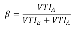
- 초기 LV 작성 및 트랜스 mitral 혈액 흐름 속도의 심 방 systole 혈액 흐름 파형을 확인 하 고 그들의 해당 최대 속도 및 속도 시간 전체 측정 (VTIE 와 VTIA, 각각)를 사용 하는 심장 초음파 장비 통합된 소프트웨어 패키지 (그림 1).
- 심 방 충전 분수 (β) 총 LV 작성 하 심의 비로 계산:
- 중간 systole (그림 2A) 동안 중반 식도 대동맥 밸브 긴 축 티 보기에에서 대동맥 밸브 바로 아래 LV 유출 관의 최대 직경을 측정 합니다.
- Π/4 제품 및 직경의 제곱 원형 형상을 가정 LV 유출 관의 면적을 계산 (단계 위의 2.5 참조).
- 깊은 transgastric 긴 축 티 보기 고 어디는 직경 (단계 위의 2.5 참조) 측정 했다; 동일한 수준에서 혈액 흐름 속도 봉투 (그림 2B)를 기록 하는 원심 LV 유출 관에 펄스 파 도플러 샘플 볼륨을 배치 VTI를 심장 초음파 장비 소프트웨어 패키지를 사용 하 여이 파형의 영역을 통합 합니다.
- 곱하기 결과 속도-시간 적분 (VTI) LV 유출 트랙 혈액 흐름 속도 파형 (그림 2B) 유출의 영역으로의 추적 (단계 2.6 참조) 치기 양 (SV)를 합니다.
- 중반-식도 bicommissural의 비디오 클립을 기록 하 고 긴 축 티 이미징 비행기, 각각42LV. 각 기록에 여러 심장 주기를 포함 해야 합니다.
- 시각적으로 검사 (단계 위의 2.9 참조) 비디오 클립의 슬로우 모션 이미지 후는 ECG T-파 승 모 판 전단의 최대 개방을 선택 하.
- Mitral 전단지 (그림 3A 와 3B) 사이의 거리를 측정 심장 초음파 장비 "캘리퍼스" 기능을 사용 하 여.
- 주요 (transcommissural 앞쪽-측면-후부-중간)과 부 (이전 후부) 길이 평균으로 승 모 판 직경 (D)를 계산 합니다.
- VFT는 수식을 사용 하 여 계산 합니다.
- 끝 만료에 3 중에서 모든 양적 echocardiographic 측정을 수행 합니다.
3. 실험 설계
- 결정 VFT, LV 확장기 기능과 hemodynamics 인덱스 정상 조건 30 분 전과 관상동맥에서 정상적인 수술 LV 방출 분수 10 환자에서 심폐 바이패스 (CPB) 후 15, 30, 및 60 분 CPB 뚜렷이 VFT39감소 가설을 테스트 하는 수술.
- 대동맥 밸브 협 착 증에 의해 생산 하는 LV 압력 과부하 비 심한 대동맥 협 착 증 및 8의 다른 그룹에 관측 비교 (8 환자 관상동맥 밸브 보충의 한 그룹)에서 검사 하 여 VFT 감소 가설 테스트 환자 정상 LV 벽 두께 관상 동맥 수술40을 받고. 정상 상태 조건 CPB 전에 30 분 동안 VFT, LV 확장기 기능, hemodynamics, 및 끝 확장기 후부 벽 두께 측정 합니다.
- 비정상적인 확장기 혈 LV를 입력 트랜스 mitral LV 대동맥 밸브 협 착 증 및 보통 대동맥 불충분 regurgitant 밸브 없는 대동맥 협 착 증 환자 8 대 8 환자에 효율성 작성에 영향을 미치는 가설 테스트 38. 측정 VFT 및 다른 매개 변수 (3.2 단계) 위에서 설명한 대로.
- 고급 나이 가설 테스트 LV 작성 VFT 7 젊은 환자 (55 ± 6 년)41 겪고 관상 동맥 수술에 비해 7 octogenarians (82 ± 2 년)에 사용 하 여 계량 하는 효율성에 있는 감소와 연결 됩니다. 두 그룹 모두 정상적인 수술 LV 방출 분수 있는지 확인 합니다. (3.2 단계) 위에서 설명한 대로 VFT 및 다른 매개 변수를 측정 합니다.
4입니다. 통계
- 평균 ± 표준 편차로 데이터를 제시.
- 분산 분석 (ANOVA) 학생의 t의 Bonferroni의 수정 다음을 사용 하 여 데이터를 평가-테스트.
- VFT와 나이 사이 VFT와 끝 확장기 후부 벽 두께 사이의 관계를 확인 하려면 선형 회귀 분석을 사용 합니다.
- Null 가설 거부 때 p < 0.05.
액세스가 제한되었습니다. 이 콘텐츠를 보려면 로그인하거나 체험판을 시작하세요.
결과
현재 기술을 사용 하 여 안정적으로 혈액 흐름 및 표준 티 영상 평면에 치수 기록에서 각 결정을 획득 하 여 다양 한 임상 상황에서 심장 수술 동안 VFT 측정 수 있었습니다. 펄스 파 도플러 샘플 볼륨 트랜스 mitral 혈액 흐름 속도 프로 파일 심 충전 분수 (β; 계산 하는 데 필요한를 중반 식도 4 상공 회의소 보기에서 mitral 전단지의 끝에 배치 했다 그림 1)입니다. 뇌졸중 볼륨 (LV 유출 트랙 혈액 흐름 속도 파형의 유출 트랙의 지역에 의해 곱한 속도-시간 적분) 연속성 방정식을 사용 하 여 결정 했다 및 중간 식도 LV 긴 축 보기 (LV 유출 트랙 직경 측정 되었다 그림 2A), 반면 유출 관을 통해 혈액 흐름 이미징 비행기 (그림 2B) 깊은 transgastric 짧은 축에서 결정 되었...
액세스가 제한되었습니다. 이 콘텐츠를 보려면 로그인하거나 체험판을 시작하세요.
토론
현재 결과 VFT 여기 설명 된 티 기술을 사용 하 여 심장 수술 동안 안정적으로 측정 될 수 있다는 설명 한다. VFT의 이전 설명 의식 과목, transthoracic 심장 초음파를 사용 하지만이 이렇게 가슴은 오픈 때 활용 될 수 없습니다. 우리 사용 자가 티 VFT는 역학을 작성 하는 LV에서 변경 종종 발생 국 소 빈 혈 reperfusion 상해 또는 외과 개입 심장 수술 마 취 환자에서 확인 합니다. 우리의 연구 결과 VFT 측정 LV 작성 과도 CPB 유도 장애 휴식 패턴 확장기 부전, 대동맥 밸브 질환 및 노화에 의해 생산 효율성에에서 변경 사항이 반영 되도록 나타냅니다. VFT 심장 수술 하는 동안 계산 현재 기술은 승 모 판 및 LV 유출 관 차원 및 혈액 흐름 ( 의 정확한 측정을 보장 하기 위해서 정상 hemodynamic 조건 높은-품질 티 이미지와 비디오 클립을 해야 합니다. 그림 1, 그림 2및...
액세스가 제한되었습니다. 이 콘텐츠를 보려면 로그인하거나 체험판을 시작하세요.
공개 사항
저자 아무 경쟁 금융 관심사 또는이 작품에 따라 다른 충돌 있다.
감사의 글
이 자료 리소스와 클레멘트 J. Zablocki 재향 군인 담당 의료 센터에서 밀워키, 위스콘신에서 시설 사용 지원 작업의 결과 이다.
액세스가 제한되었습니다. 이 콘텐츠를 보려면 로그인하거나 체험판을 시작하세요.
재료
| 이름 | 회사 | 카탈로그 번호 | 댓글 |
|---|---|---|---|
| 심초음파 기계 | 필립스 초음파, Bothall, WA | iE33 | |
| 경식도 심초음파 프로브 | 필립스 초음파, Bothall, WA | X7-2T | |
| 통계 소프트웨어 | 분석가Soft, 호두, CA | StatPlus:맥 프로 |
참고문헌
- Collier, E., Hertzberg, J., Shandas, R. Regression analysis for vortex ring characteristics during left ventricular filling. Biomedical Sciences Instrumentation. 38 (2), 307-311 (2002).
- Kheradvar, A., Gharib, M. Influence of ventricular pressure drop on mitral annulus dynamics through the process of vortex ring formation. Annals of Biomedical Engineering. 35 (12), 2050-2064 (2007).
- Gharib, M., Rambod, E., Shariff, K. A universal time scale for vortex ring formation. Journal of Fluid Mechanics. 360 (1), 121-140 (1998).
- Krueger, P. S., Gharib, M. The significance of vortex ring formation to the impulse and thrust of a starting jet. Physics of Fluids. 15 (5), 1271-1281 (2003).
- Reul, H., Talukder, N., Muller, W. Fluid mechanics of the natural mitral valve. Journal of Biomechanics. 14 (5), 361-372 (1981).
- Kim, W. Y., et al. Two-dimensional mitral flow velocity profiles in pig models using epicardial Doppler echocardiography. Journal of the American College of Cardiology. 24 (2), 532-545 (1994).
- Kilner, P. J., et al. Asymmetic redirection of flow through the heart. Nature. 404 (6779), 759-761 (2000).
- Gharib, M., Rambod, E., Kheradvar, A., Sahn, D. J., Dabiri, J. O. Optimal vortex formation as an index of cardiac health. Proceedings of the National Academy of Sciences USA. 103 (16), 6305-6308 (2006).
- Rodriguez Munoz, D., et al. Intracardiac flow visualization: current status and future directions. European Heart Journal Cardiovascular Imaging. 14 (11), 1029-1038 (2013).
- Martinez-Legazpi, P., et al. Contribution of the diastolic vortex ring to left ventricular filling. Journal of the American College of Cardiology. 64 (16), 1711-1721 (2014).
- Dabiri, J. O., Gharib, M. The role of optimal vortex formation in biological fluid transport. Proceedings of the Royal Society B. 272 (1572), 1557-1560 (2003).
- Kheradvar, A., Gharib, M. On mitral valve dynamics and its connection to early diastolic flow. Annals of Biomedical Engineering. 37 (1), 1-13 (2009).
- Linden, P. F., Turner, J. S. The formation of "optimal" vortex rings, and the efficiency of propulsion devices. Journal of Fluid Mechanics. 427 (1), 61-72 (2001).
- Domenichini, F., Pedrizzetti, G., Baccani, B. Three-dimensional filling flow into a model left ventricle. Journal of Fluid Mechanics. 539 (1), 179-198 (2005).
- Sengupta, P. P., et al. Left ventricular isovolumic flow sequence during sinus and paced rhythms: new insights from use of high-resolution Doppler and ultrasonic digital particle imaging velocimetry. Journal of the American College of Cardiology. 49 (8), 899-908 (2007).
- Rodriguez Munoz, D., et al. Flow mapping inside a left ventricular aneurysm: a potential tool to demonstrate thrombogenicity. Echocardiography. 31 (1), E10-E12 (2014).
- Son, J. W., et al. Abnormal left ventricular vortex flow patterns in association with left ventricular apical thrombus formation in patients with anterior myocardial infarction: a quantitative analysis by contrast echocardiography. Circulation Journal. 76 (11), 2640-2646 (2012).
- Kheradvar, A., Falahatpisheh, A. The effects of dynamic saddle annulus and leaflet length on trans-mitral flow pattern and leaflet stress of a bileaflet bioprosthetic mitral valve. The Journal of Heart Valve Disease. 21 (2), 225-233 (2012).
- Kheradvar, A., Assadi, R., Falahatpisheh, A., Sengupta, P. P. Assessment of trans-mitral vortex formation in patients with diastolic dysfunction. Journal of the American Society of Echocardiography. 25 (2), 220-227 (2012).
- Chen, R., et al. Assessment of left ventricular hemodynamics and function of patients with uremia by vortex formation using vector flow mapping. Echocardiography. 29 (9), 1081-1090 (2012).
- Hendabadi, S., et al. Topology of blood transport in the human left ventricle by novel processing of Doppler echocardiography. Annals of Biomedical Engineering. 41 (12), 2603-2616 (2013).
- Sengupta, P. P., Pedrizetti, G., Narula, J. Multiplaner visualization of blood flow using echocardiographic particle imaging velocimetry. Journal of the American College of Cardiology Cardiovascular Imaging. 5 (5), 566-569 (2012).
- Sengupta, P. P., et al. Emerging trends in CV flow visualization. Journal of the American College of Cardiology Cardiovascular Imaging. 5 (3), 305-316 (2012).
- Hong, G. R., Kim, M., Pedrizzetti, G., Vannan, M. A. Current clinical application of intracardiac flow analysis using echocardiography. Journal of Cardiovascular Ultrasound. 21 (4), 155-162 (2013).
- Kheradvar, A., Milano, M., Gharib, M. Correlation between vortex ring formation and mitral annulus dynamics during ventricular rapid filling. American Society for Artificial Internal Organs Journal. 53 (1), 8-16 (2007).
- Hong, G. R., et al. Characterization and quantification of vortex flow in the human left ventricle by contrast echocardiography using vector particle image velocimetry. Journal of the American College of Cardiology Cardiovascular Imaging. 1 (6), 705-717 (2008).
- Zhang, H., et al. The evolution of intraventricular vortex during ejection studied by using vector flow mapping. Echocardiography. 30 (1), 27-36 (2013).
- Nogami, Y., et al. Abnormal early diastolic intraventricular flow 'kinetic energy index' assessed by vector flow mapping in patients with elevated filling pressure. European Heart Journal Cardiovascular Imaging. 14 (3), 253-260 (2013).
- Zhang, H., et al. The left ventricular intracavity vortex during the isovolumic contraction period as detected by vector flow mapping. Echocardiography. 29 (5), 579-587 (2012).
- Poh, K. K., et al. Left ventricular filling dynamics in heart failure: echocardiographic measurement and utilities of vortex formation time. European Heart Journal Cardiovascular Imaging. 13 (5), 385-393 (2012).
- Belohlavek, M. Vortex formation time: an emerging echocardiographic index of left ventricular filling efficiency? European Heart Journal Cardiovascular Imaging. 13 (5), 367-369 (2012).
- Dabiri, J. O., Gharib, M. Starting flow through nozzles with temporally variable exit diameter. Journal of Fluid Mechanics. 538 (1), 111-136 (2005).
- Jiamsripong, P., et al. Impact of acute moderate elevation in left ventricular afterload on diastolic trans-mitral flow efficiency: analysis by vortex formation time. Journal of the American Society of Echocardiography. 22 (4), 427-431 (2009).
- Belohlavek, M., et al. Patients with Alzheimer disease have altered trans-mitral flow: echocardiographic analysis of the vortex formation time. Journal of Ultrasound in Medicine. 28 (11), 1493-1500 (2009).
- Pedrizzetti, G., Domenichini, F., Tonti, G. On the left ventricular vortex reversal after mitral valve replacement. Annals of Biomedical Engineering. 38 (3), 769-773 (2010).
- Martinez-Legazpi, P., et al. Stasis mapping using ultrasound: a prospective study in acute myocardial infarction. Journal of the American College of Cardiology Cardiovascular Imaging. 11 (3), 514-515 (2018).
- Harfi, T. T., et al. The E-wave propagation index (EPI): a novel echocardiographic parameter for prediction of left ventricular thrombus. Derivation from computational fluid dynamic modeling and validation on human subjects. International Journal of Cardiology. 227 (1), 662-667 (2017).
- Pagel, P. S., Boettcher, B. T., De Vry, D. J., Freed, J. K., Iqbal, Z. Moderate aortic valvular insufficiency invalidates vortex formation time as an index of left ventricular filling efficiency in patients with severe degenerative calcific aortic stenosis undergoing aortic valve replacement. Journal of Cardiothoracic and Vascular Anesthesia. 30 (5), 1260-1265 (2016).
- Pagel, P. S., Gandhi, S. D., Iqbal, Z., Hudetz, J. A. Cardiopulmonary bypass transiently inhibits intraventricular vortex ring formation in patients undergoing coronary artery bypass graft surgery. Journal of Cardiothoracic and Vascular Anesthesia. 26 (3), 376-380 (2012).
- Pagel, P. S., Hudetz, J. A. Chronic pressure-overload hypertrophy attenuates vortex formation time in patients with severe aortic stenosis and preserved left ventricular systolic function undergoing aortic valve replacement. Journal of Cardiothoracic and Vascular Anesthesia. 27 (4), 660-664 (2013).
- Pagel, P. S., Dye, L., Boettcher, B. T., Freed, J. K. Advanced age attenuates left ventricular filling efficiency quantified using vortex formation time: a study of octogenarians with normal left ventricular systolic function undergoing coronary artery surgery. Journal of Cardiothoracic and Vascular Anesthesia. 32 (4), 1775-1779 (2018).
- Shanewise, J. S., et al. ASE/SCA guidelines for performing a comprehensive intraoperative multiplane transesophageal echocardiography examination: recommendations of the American Society of Echocardiography Council for Intraoperative Echocardiography and the Society of Cardiovascular Anesthesiologists Task Force for Certification in Perioperative Transesophageal Echocardiography. Journal of the American Society of Echocardiography. 12 (10), 884-900 (1999).
- Gaspar, T., et al. Three-dimensional imaging of the left ventricular outflow tract: impact on aortic valve area estimation by the continuity equation. Journal of the American Society of Echocardiography. 25 (7), 749-757 (2012).
- Karamnov, S., Burbano-Vera, N., Huang, C. C., Fox, J. A., Shernan, S. A. Echocardiographic assessment of mitral stenosis orifice area: a comparison of a novel three-dimensional method versus conventional techniques. Anesthesia and Analgesia. 125 (3), 774-780 (2017).
- Pagel, P. S., Kampine, J. P., Schmeling, W. T., Warltier, D. C. Comparison of end-systolic pressure-length relations and preload recruitable stroke work as indices of myocardial contractility in the conscious and anesthetized, chronically instrumented dog. Anesthesiology. 73 (2), 278-290 (1990).
- Pagel, P. S., Kampine, J. P., Schmeling, W. T., Warltier, D. C. Alteration of left ventricular diastolic function by desflurane, isoflurane, and halothane in the chronically instrumented dog with autonomic nervous system blockade. Anesthesiology. 74 (6), 1103-1114 (1991).
- De Hert, S. G., Rodrigus, I. E., Haenen, L. R., De Mulder, P. A., Gillebert, T. C. Recovery of systolic and diastolic left ventricular function early after cardiopulmonary bypass. Anesthesiology. 85 (5), 1063-1075 (1996).
- Gorcsan, J., Diana, P., Lee, J., Katz, W. E., Hattler, B. G. Reversible diastolic dysfunction after successful coronary artery bypass surgery. Assessment by transesophageal Doppler echocardiography. Chest. 106 (5), 1364-1369 (1994).
- Djaiani, G. N., et al. Mitral flow propagation velocity identifies patients with abnormal diastolic function during coronary artery bypass graft surgery. Anesthesia and Analgesia. 95 (3), 524-530 (2002).
- Casthely, P. A., et al. Left ventricular diastolic function after coronary artery bypass grafting: a correlative study with three different myocardial protection techniques. Journal of Thoracic and Cardiovascular Surgery. 114 (2), 254-260 (1997).
- Tulner, S. A., et al. Perioperative assessment of left ventricular function by pressure-volume loops using the conductance catheter method. Anesthesia and Analgesia. 97 (4), 950-957 (2003).
- Firstenberg, M. S., et al. Relationship between early diastolic intraventricular pressure gradients, an index of elastic recoil, and improvements in systolic and diastolic function. Circulation. 104 (12 Suppl 1), I330-I335 (2001).
- Cooke, J., Hertzberg, J., Boardman, M., Shandas, R. Characterizing vortex ring behavior during ventricular filling with Doppler echocardiography: an in vitro study. Annals of Biomedical Engineering. 32 (2), 245-256 (2004).
- Grossman, W., Jones, D., McLaurin, L. P. Wall stress and patterns of hypertrophy in the human left ventricle. Journal of Clinical Investigation. 56 (1), 56-64 (1975).
- Hess, O. M., et al. Diastolic function and myocardial structure in patients with myocardial hypertrophy. Special reference to normalized viscoelastic data. Circulation. 63 (2), 360-371 (1981).
- Hess, O. M., et al. Diastolic stiffness and myocardial structure in aortic valve disease before and after valve replacement. Circulation. 69 (5), 855-865 (1984).
- Sandstede, J. J. W., et al. Cardiac systolic rotation and contraction before and after valve replacement for aortic stenosis: a myocardial tagging study using MR imaging. American Journal of Roentgenology. 178 (4), 953-958 (2002).
- Stuber, M., et al. Alterations in the local myocardial motion pattern in patients suffering from pressure overload due to aortic stenosis. Circulation. 100 (4), 361-368 (1999).
- Nagel, E., et al. Cardiac rotation and relaxation in patients with aortic valve stenosis. European Heart Journal. 21 (7), 582-589 (2000).
- Rakowski, H., et al. Canadian consensus recommendations for the measurement and reporting of diastolic dysfunction by echocardiography: from the Investigators of Consensus on Diastolic Dysfunction by Echocardiography. Journal of the American Society of Echocardiography. 9 (5), 736-760 (1996).
- Homeyer, P., Oxorn, D. C. Aortic regurgitation: echocardiographic diagnosis. Anesthesia and Analgesia. 122 (1), 37-42 (2016).
- Landzberg, J. S., et al. Etiology of the Austin Flint murmur. Journal of the American College of Cardiology. 20 (2), 408-413 (1992).
- Flint, A. On cardiac murmurs. American Journal of Medical Sciences. 91 (1), 27(1886).
- Botvinick, E. H., Schiller, N. B., Wickramasekaran, R., Klausner, S. C., Gertz, E. Echocardiographic demonstration of early mitral valve closure in severe aortic insufficiency. Its clinical implications. Circulation. 51 (5), 836-847 (1975).
- Mann, T., McLaurin, L., Grossman, W., Craige, E. Assessing the hemodynamic severity of acute aortic regurgitation due to infective endocarditis. New England Journal of Medicine. 293 (3), 108-113 (1975).
- Borlaug, B. A., et al. Longitudinal changes in left ventricular stiffness: a community-based study. Circulation Heart Failure. 6 (5), 944-952 (2013).
- Wong, J., et al. Age-related changes in intraventricular kinetic energy: a physiological or pathological adaptation? American Journal of Physiology Heart Circulatory Physiology. 310 (6), H747-H755 (2016).
- Carrick-Ranson, G., et al. Effect of healthy aging on left ventricular relaxation and diastolic suction. American Journal of Physiology Heart Circulatory Physiology. 303 (3), H315-H322 (2012).
- Iskandrian, A. S., Hakki, A. H. Age-related changes in left ventricular diastolic performance. American Heart Journal. 112 (1), 75-78 (1986).
- Schulman, S. P., et al. Age-related decline in left ventricular filling at rest and exercise. American Journal of Physiology. 263 (6 Pt 2), H1932-H1938 (1992).
- Stork, M., et al. Age-related hemodynamic changes during diastole: a combined M-mode and Doppler echo study. Internal Journal of Cardiovascular Imaging. 6 (1), 23-30 (1991).
- Sanders, D., Dudley, M., Groban, L. Diastolic dysfunction, cardiovascular aging, and the anesthesiologist. Anesthesiology Clinics. 27 (3), 497-517 (2009).
액세스가 제한되었습니다. 이 콘텐츠를 보려면 로그인하거나 체험판을 시작하세요.
재인쇄 및 허가
