안구 수정체가 망막에 빛을 집중시키는 기능은 조직의 투명도, 생체역학, 굴절률에 따라 달라집니다. 이 리뷰는 수정체의 3차원 구조를 강조하고 조직 항상성을 유지하는 복잡한 메커니즘을 논의합니다.
이 콘텐츠를 보려면 JoVE 구독이 필요합니다. 로그인하거나 오늘 무료 평가판을 시작하세요.
리뷰 논문
포유류 안구 렌즈에 초점을 맞춘 주제: 발달, 해부학, 생리학, 투명성, 생체역학, 그리고 연령 관련 도전 과제
이 논문에서
요약
초록
수정체는 눈의 앞방에 위치한 투명한 타원체 기관으로, 빛을 망막에 집중시킵니다. 상피세포와 섬유세포로 구성된 이 3차원 조직은 명확한 빛 경로를 보장하기 위해 매우 조직적이며, 높은 굴절률과 투명도를 확립하고 유지하기 위해 다양한 전략을 사용합니다. 인간의 경우 수정체는 다양한 거리의 빛을 집중시키기 위해 형태가 변하는 과정을 '수용'이라고 하며, 이러한 탄성 및 생체역학적 특성은 조직, 세포, 분자 메커니즘에 의존합니다. 백내장은 평소 투명한 수정체의 불투명성을 의미하며, 여전히 전 세계에서 실명의 주요 원인으로 남아 있습니다. 노안은 나이에 따른 수정체 경직성 변화로 인해 조절 기능 저하와 독서용 안경 필요성과 연관되어 있습니다. 이 리뷰에서는 포유류의 수정체 발달과 평생 성장, 결정체 및 세포골격 단백질, 세포 간 소통과 미세순환, 수정체 생체역학, 그리고 이 독특한 조직을 연구하기 위한 도전과 새로운 방법을 탐구할 것입니다.
서론
안구 수정체는 처음 보기에는 상피세포와 섬유세포라는 두 가지 세포 유형으로 구성된 비교적 단순한 조직입니다. 수정체의 전체 질량은 섬유 세포로 이루어져 있으며, 전반구1을 덮는 단층 상피세포가 있습니다(그림 1A). 전체 조직은 주로 콜라겐 IV와 라미닌으로 구성된 얇은 기저막인 수정체 캡슐에 감싸져 있으며, 이는 이전 리뷰2에서 설명한 바 있습니다. 혈액 공급과 신경이 없기 때문에, 수정체는 눈의 전방에 탄성 대위 섬유(즉, 조눌)에 의해 고정되어 수정체 캡슐과 섬모체를 연결합니다. 전방 또는 중앙 수정체 상피 세포는 직방체형이며 3,4,5 자갈로 배열되어 있습니다(그림 1A, 연한 파란색 세포, 상단). 이 세포들은 조용하며 증식하지않습니다. 렌즈 적도 바로 앞쪽에서 상피세포는 발아대에서 분열하며(그림 1A, 주황색 세포, 오른쪽 상단), 이 적도 세포들은 적도 쪽으로 이동하여 6각형 세포 모양 7,8,9,10의 자오선 행으로 매우 질서가 높게 배열됩니다 (그림 1A), 녹색 셀, 오른쪽 상단). 자경선 세포는 끝 끝을 수정체 지점(modiulus)에 고정시키고 신장하며 새로운 2차 섬유세포로 분화하기 시작합니다(그림 1A, 왼쪽 아래). 2차 섬유의 끝부분은 상피세포의 끝과 상피 세포의 끝부분과 상호작용하여 전쪽 극으로 이동하여 특이한 꼭대기-끝 접합부3을 형성합니다(그림 1A, 빨간 선). 새로 형성된 섬유의 후면 기저 끝은 수정체 캡슐을 따라 이동하며 후면 극 쪽으로 뻗어 나갑니다(그림 1A, 왼쪽 아래). 길게 뻗은 섬유 세포의 끝이 전극 또는 후극에 도달하면, 세포는 전방 상피세포나 후피낭에서 분리되어 조직의 반대편에서 뻗어나온 세포의 끝과 만나 봉합선11, 12, 13을 형성합니다(그림 1A, 왼쪽 상단). 렌즈 섬유 셀은 단면이 길쭉한 육각형 형태로, 2개의 긴 /넓은 변, 4개의 짧은 면, 6개의 삼중 세포 접합부를 가지고 있습니다(그림 1A, 오른쪽 아래 14). 인접한 섬유 세포 간의 세포 간 공간이 최소화되어 빛 산란이 감소합니다14. 말초 주변에 피질 섬유 세포를 지속적으로 추가하여 이전 세대의 섬유 위에 겹치는 것은 수정체의 평생 성장을 촉진합니다14,15. 수정체의 중심부는 핵(nucleus)이라고 하며, 이 영역은 말초나 피질에 비해 강성이 증가하는 것으로 이전 리뷰16에서 설명되었습니다.
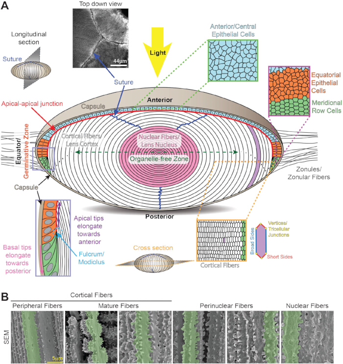
그림 1: 수정체 해부학과 구조. (A) 수정체는 전반구를 덮는 연한 파란색, 주황색, 연한 녹색 상피세포 단층과 흰색과 분홍색 수정체 섬유의 대량으로 구성되어 있습니다. 얇은 콜라겐 막인 수정체 캡슐(탄)이 조직을 감싸고 있습니다. 수정체는 안구 전방에 안구 전방에 안경 섬유(조눌)가 삽입되어 있습니다. 앞쪽/중앙 상피 세포(연한 파란색, 상단)는 직방면체형이며 단면은 자갈돌 모양입니다. 적도 상피 세포(주황색)는 발아대에 있는 무작위 세포(주황색)에서 조직된 오두막 세포(녹색)로 변형됩니다. 정경선 세포는 수정체 말초에서 새로운 피질 2차 섬유 세포층(흰색)으로 길어집니다. 새로 형성된 2차 섬유의 끝부분은 지렛대/모디오룰스(페리윙클 박스, 왼쪽 아래)에 고정되어 있습니다. 길게 뻗은 2차 섬유의 끝부분은 수정체 상피세포의 끝부분을 따라 앞극 쪽으로 이동하며, 특이한 첨단-끝 접합부(빨간 선)를 형성합니다. 길게 늘어진 섬유의 기저 끝은 후방 캡슐을 따라 후방 극 쪽으로 이동합니다. 2차 섬유가 완전히 길어지면 끝이 전상피 또는 후방 캡슐에서 떨어져 나가 수정체 봉합선을 형성합니다(짙은 파란색 선과 왼쪽 위 이미지). 렌즈 섬유 세포는 단면이 길쭉한 육각형(오른쪽 아래)이며, 2개의 긴 넓은 면, 4개의 짧은 면, 6개의 삼세포 접합부를 가지고 있습니다. 섬유세포 성숙 과정에서는 모든 세포 소기관이 제거되어 소기관 없는 영역(짙은 녹색 양면 화살표)을 형성하여 빛의 산란을 최소화합니다. 가장 오래된 수정체 섬유 세포는 조직 중심에 압축되어 수정체 핵(분홍색)을 형성합니다. 만화는 축척에 맞춰 그려지지 않습니다. (B) 렌즈 내 다양한 깊이에 있는 섬유 세포의 주사 전자현미경 이미지는 분화 및 성숙 과정에서 세포 형태 변화를 보여줍니다. 단일 셀은 강조를 위해 초록색으로 강조되어 있습니다. 피질 말초 섬유는 긴 변을 따라 구와 소켓 교차 모양의 교차점과 짧은 변을 따라 작은 돌출부를 가지고 있습니다. 섬유 세포 성숙 과정에는 작은 돌기와 큰 돌기가 장식된 큰 패들(패들)이 성숙한 세포의 짧은 면을 장식합니다. 핵주위 섬유는 전체 막을 따라 혀와 홈 모양의 교차 모양과 짧은 면을 따라 큰 돌출부를 가지고 있습니다. 핵섬유는 짧은 변을 따라 큰 돌출부와 구상막 형태를 가지고 있습니다. 스케일 바, 5 μm. 16,328에서 수정됨. 이 그림의 더 큰 버전을 보시려면 여기를 클릭해 주세요.
렌즈의 복잡성은 고도로 질서 정연한 3차원(3D) 세포 배열, 모든 세포 소기관을 파괴하고 제거하는 특수한 섬유 세포 성숙, 그리고 조직의 투명성과 높은 굴절률을 확립하고 유지하는 분자 및 세포 메커니즘에 있습니다. 대부분의 수정체 섬유 세포에서 세포 소기관이 부족하다는 점은, 유기체가 평생 동안 만들어진 모든 수정체 세포의 건강과 수명을 유지하는 데 어려움을 더합니다. 렌즈는 초기 배아 발달 중에 가장 중심 세포가 만들어졌고, 생애 내내 조직 말부에서 새로운 세포를 생성하여 연령 및 연령 관련 변화를 연구하는 데 매우 우수합니다. 이는 나이 들어 나이 든 세포와 노년에 만들어진 세포를 연구할 수 있게 해줍니다. 수정체의 미세 초점 기능은 서로 다른 거리의 물체에서 망막에 선명한 이미지를 형성할 수 있게 하며, 이는 수정체가 형태를 바꾸거나 수용할 수 있는 능력에 달려 있습니다. 수정체는 결합성이 없고 근육이 아닌 조직으로, 정상적인 기능을 위해 생체역학적 특성이 필요합니다.
시각 장애의 주요 원인은17세 노화입니다. 나이 관련 변화로 인해 두 가지 주요 수정체 병리, 즉 백내장과 노안이발생합니다. 백내장이나 수정체 불투명도는 여전히 전 세계에서 실명의 주요 원인으로 남아 있으며, 자외선, 흡연, 당뇨병, 산화 스트레스 등 유전적·환경적 요인에 의해 단백질 응집 및/또는 세포 결함을 유발하는 경우가 많습니다. 이는 이전 리뷰에서 설명된 것이죠 18,20,21,22,23,24,25. 노안은 나이에 따른 수용 감소로, 가까운 물체에 초점을 맞출 때 수정체의 형태 변화 능력이 감소하여 발생하며, 확장해서 독서용 안경의 필요성 증가로 인해 발생합니다 26,27,28. 세계보건기구(WHO)는 전 세계적으로 해결되지 않은 시각 장애를 가진 사람이 최소 10억 명에 달하며, 이 중 백내장 환자 6,520만 명, 노안(17) 환자 8억 2,600만 명이 있다고 추정합니다. 고령화 인구가 진행됨에 따라 렌즈의 생리학과 생물학을 더 잘 이해할 필요가 긴급합니다.
이 리뷰는 포유류 수정체의 기능과 항상성에 중요한 조직 구조, 세포 배열, 분자 메커니즘을 다루며, 다른 척추동물의 수정체에 관한 정보도 포함합니다. 목표는 수정체 생물학과 생리학에 대한 일반적인 개요와 추가적인 상세 리뷰 참고문헌을 제공하는 것입니다. 주제는 배아 수정체 발달, 출생 후 수정체 성장, 3D 세포 및 조직 배열, 조직의 생체역학적 특성, 미세 순환 등을 포함합니다. 주요 렌즈 단백질, 즉 결정체, 세포골격 네트워크, 세포 간 통신에 대해 아래에서 논의합니다. 이 리뷰는 이 독특한 조직을 연구하기 위한 현재의 도전 과제와 새로운 방법들로 마무리될 것입니다.
액세스가 제한되었습니다. 이 콘텐츠를 보려면 로그인하거나 체험판을 시작하세요.
리뷰 및 관점
배아 수정체 발달 및 출생 후 수정체 성장
척추동물에서는 수정체가 표면 외배엽 세포에서 발달하며, 미래 수정체는 시낭 6,29,30 위 표면 외배엽이 두꺼워지면서 시작됩니다(그림 2A). 광경컵 형성과 함께 두꺼워진 외배엽 또는 수정체 판은 수정체구멍 6, 29, 30 안으로 침투한다(그림 2B). 액틴 필라멘트(F-actin)는 후방 수정체 구덩이를 발달 중인 시선컵의 앞면에 부착하여 이 발달 중인 조직들의 움직임과 형태 변화를 조정합니다. 31,32,33,34이며, 수정체 구멍 함몰을 위해서는 수정체 상피 세포의 꼭지 수축이 필요합니다. 35,36,37,38,39,40 (그림 2C). 수정체 구멍의 입구는 구멍이 닫힐 때까지 계속 좁아지며, 수정체 줄기 세포는 세포멸사(apoptosis 41, 42, 43)를 겪습니다. 수정체 소포는 미래의 각막 외배엽에서 분리되어 수정체 소포를 형성하는데, 이는 상피세포 6,29,30의 속이 빈 구체입니다(그림 2D). 수정체 소포의 후상 상피 세포는 전방 상피 쪽으로 길게 뻗어 1차 섬유 세포로 분화하여 강을 채우고 배아 수정체를 형성합니다(그림 2E). 완전히 형성된 배아 수정체에는 후반 상피세포가 없지만, 전반구를 덮는 단핵세포 단층은 유지된다(그림 2F). 1차 섬유 세포는 수정체 핵의 가장 중앙 부분을 형성하며, 생애 6,29,30 동안 수정체 주변부 또는 피질에서 추가되는 새로운 세대의 2차 수정체 섬유에 둘러싸여 있습니다. 대부분의 척추동물 수정체는 함몰에 의해 형성되지만, 제브라피시에서는 판이 두꺼워졌다가 분해되어 수정체44를 형성합니다.
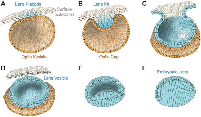
그림 2: 포유류 눈의 배아 수정체 발달. 표면 외배엽 세포는 흰색이고, 수정체 세포는 연한 파란색이며, 시각컵/소포세포는 모래색입니다. (A) 배아 수정체 판(연한 파란색)은 표면 외배엽에서 발달합니다. 표면의 외배엽과 수정체 판의 뒤쪽에는 시세포 소포가 있습니다. (B) 수정체 판의 내몰이 수정체 구멍을 형성하고, 시낭의 내몰이 시선컵을 형성합니다. 앞쪽 시선컵은 홍채와 섬모체로 발전하며, 후방 시선컵은 망막과 망막 색소 상피를 형성합니다. (C) 렌즈 플라코드 개구부가 닫힐 때까지 좁아집니다. (D) 수정체 소포는 표면 외배엽에서 분리되며, 수정체 상피 세포로 둘러싸인 속이 빈 구체입니다. (E) 수정체 소포 내 후방 수정체 상피세포가 길어지기 시작하며 1차 수정체 섬유로 분화합니다. (F) 1차 수정체 섬유가 수정체 소포의 공동을 채워 배아 수정체를 형성합니다. 만화는 축척에 맞춰 그려지지 않습니다. 411을 모델로 했습니다. 이 그림의 더 큰 버전을 보시려면 여기를 클릭해 주세요.
배아 발달 동안 수정체는 6,45,46 혈관으로 이루어진 바구니 같은 네트워크로 둘러싸여 있습니다. 이 혈관들은 망막의 유리동동맥에서 시작되어 수정체 구덩이 뒤쪽에서 네트워크를 형성하기 시작하고, 결국 배아 수정체에 영양을 공급하기 위해 배아의 수정체에 영양을 공급하는 질서 있는 망을 형성합니다 6,45. 이 혈관들은 출생 전이나 눈이 뜨기 전에 명확한 빛 경로를 확보하기 위해 후퇴하고 사라지는데, 종에 따라 6,45,46,47에 따라 다릅니다.
Pax6는 초기 눈 현상(48, 49, 50, 51, 52)에 필요하며, 수정체 발달53, 54, 55, 56에 필수적이고 충분합니다. Pax6의 인산화와 SUMOylation은 뇌, 중추신경계, 안구 발달 과정에서 이 전사 인자의 기능을 조절합니다 57,58,59,60. 배아 눈과 수정체 발달에 필요한 다른 중요한 신호 전달 경로는 Pax6 신호와 교차하는데, 여기에는 강화제[Meis, 골 형태발생 단백질(Bmp), 섬유아세포 성장인자(FGF)] 및 하위 단백질(Foxe3 및 레티노산(RA)]이 포함되며, 이는 이전 리뷰61에서 설명되었습니다. Meis1과 Meis2는 Pax6 유전자의 증강체 영역에 결합하며, 척추동물62의 수정체 유도에 필수적입니다. 유전학 연구에 따르면 Bmp7은 Pax6 발현63,64를 조절하여 수정체 판 발달에 필요하다. Foxe3는 Pax6 신호 전달의 하류에 위치하며, 수정체65, 66의 발생, 특히 수정체 소포가 형성될 때 수정체 줄기와 표면 외배엽을 깨끗이 분리하는 데 필요합니다. Foxe3의 상실은 선천성 안구 결함인 Peters Anomaly67로 이어집니다. FGF 수용체(FGFR) 신호 전달은 Bmp7과 협력하여 Pax6 발현을 조절하며, 이는 다시 발생 중인 수정체68의 Foxe3 수치에 영향을 미칩니다. 억제성 FGFR의 발현은 수정체 판 형성 지연, 수정체 줄기의 불완전한 분리, 그리고 작은 수정체소포 68을 초래합니다. RA 신호 전달의 교란은 눈 형성 결함을 유발하며, Pax6가 파괴되면 RA의 발현이 감소하여 두 신호 경로 간에 일부 상호작용이 있음을 시사한다70.
배아 수정체가 완전히 형성된 후, 적도 상피세포의 증식 및 분화를 통한 지속적인 수정체 성장은 주로 포유류 수정체에서 FGF를 통한 성장 인자 신호전달에 의해 촉진되는 것으로 추정되며, 이는 이전 리뷰71에서 설명되었습니다. FGF는 방수혈보다 유리체에서 더 풍부하게 존재하여, 렌즈 적도72,73 근처에서 성장 및 분화 신호가 증가하는 구배를 형성합니다. 수정체는 4개의 FGFR 중 3개인 FGFR1, FGFR2, FGFR3을 발현하며, 적도74,75 후쪽 렌즈 지점에서 발현이 증가합니다. FGFs의 과발현은 정상적으로 조용한 전부 상피세포가 비정상적으로 신장되고 섬유 유사 세포로 분화되도록 유도합니다 76,77,78. FGF 신호 전달 외에도, 억제제 분석은 Bmp 경로가 섬유세포 신장에 관여하는 것으로 나타났으며, 79,80, 그리고 비정상적인 형질전환 성장인자 베타(TGFβ) 수용체가 정상적인 섬유세포 성숙을 방해한다는 것을 확인했습니다81.
렌즈 투명도와 굴절률
렌즈 섬유 세포는 성숙 과정에서 모든 세포 소기관을 제거하여 빛 경로 내 광 산란 대상을 제거하고 소기관 자유 영역(OFZ)82를 생성하는 특수한 과정을 거칩니다(그림 1A). 섬유세포가 성숙 프로그래밍 83,84,85,86,87,88에 도달하면 모든 세포 소기관의 동기화된 손실이 빠르게 발생합니다. 비정상적인 소기관 분해는 백내장을 유발하며, 여러 녹아웃(KO) 제브라피시 및 마우스 모델에서 염색질 및 DNA 분해 유전자인 89,90,91,92,93, 사이클린 의존성 키나제94,95, 열충격 인자4 91,96,97,98, PLATT 포스폴리파제99 유전자를 확인했습니다 이는 OFZ 형성에 중요한 요소들입니다. 세포막 및 세포골격 단백질 100,101,102,103,104, 결정체105,106,107, 종양유전자108,109의 비정상 발현 또는 돌연변이도 소기관 제거와 수정체 투명성에 영향을 줄 수 있습니다. 그러나 섬유세포에서 소기관 분해를 유발하는 정확한 신호는 여전히 명확하지 않습니다. 최근 연구는 수정체 형성과 카타락토기생에서 RNA 결합 단백질의 중요성을 밝혀냈으며, 이는 이전 리뷰110에서 설명되었습니다. Tdrd7111, caprin2112, Rbm24113, Celf1114, 115, 116 등 RNA 결합 단백질의 교란은 비정상적인 수정체 발달, 소기관 분해 장애, 백내장을 초래합니다.
체내 대부분의 세포 유형과 비교할 때, 포유류 수정체 섬유 세포는 매우 높은 단백질 농도를 가지고 있습니다(200–450 mg/ml)117,118,119,120. 단백질은 전체 수정체 무게의 30%–35%를 차지하며, 따라서 수정체의 수분 함량은 상대적으로 낮아 약 65%–70%입니다. 렌즈 단백질의 질량 기준 90%가 결정체123이며, 포유류 렌즈에는 α, β, γ 세 가지 결정체 단백질 계열이 있습니다. 이는 이전 리뷰124,125에서 설명된 바와 같습니다. 높은 단백질 함량은 일반적으로 빛의 절반 파장보다 큰 응집체의 위험을 증가시켜 빛 산란을 유발하지만, 결정체 단백질은 섬유세포 세포질 내에서 단거리 질로 조직되어 높은 굴절률과 투명도를 촉진합니다117,127. 단백질 응집은 아래에서 논의하는 α결정체의 샤페론 유사 기능에 의해 약화됩니다. 렌즈 전체의 굴절률(GRIN)은 이전 리뷰에서 설명한128,129와 같이 빛의 초점을 촉진하며, 렌즈 중심부의 최대 굴절률은 핵섬유 전지의 압축 130,131,132와 상관관계가 있습니다. 어류 렌즈에서는 핵의 높은 굴절률이 단백질이 조직 중심으로 이동하는 결과임을 데이터가 시사합니다133. 결정질 단백질이 세포 전반과 섬유 껍질 사이에 균일하게 분포하는 것은 섬유 세포막을 따라 막 융합을 통한 거대분자 수송 경로에 의해 촉진된다고 가설화됩니다134,135. 그러나 쥐 렌즈 136,137,138에서는 막 융합이 명확히 관찰되지 않았으며, 여러 종의 수정체에서 임피던스 연구를 통해 막 융합 139,140,141,142번은 검출되지 못했습니다. 간극 접합 통신이 방해된 마우스 모델의 데이터는 세포 간 통신과 섬유 세포 신장도 고분자 수송 경로에 기여함을 시사한다143.
결정체는 분자량 ~20–30 kDa이며, 수정체에서 구조적·굴절 역할을 합니다. 알파-결정체는 αA와 αB 소단위체로 구성되며, 이들은 55%–60%의 상동성을 공유합니다 144,145,146. αB는 대부분의 세포에서 널리 발현되는 반면, αA-결정체는 주로 수정체에서 발현되며, 비장과 흉선을 포함한 다른 조직에서도 낮은 수준으로 검출된 바 있습니다 147,148. 최근 연구는 망막 색소 상피세포와 망막 신경절 세포가 스트레스나 질병 시 αA-결정체를 발현할 수 있음을 보여주었으며, 이는 신경 보호 효과를 제공하기 위한 것으로 추정됩니다 149,150,151. 알파-결정체는 일반적으로 30–35개의 혼합 αA와 αB 소단위체로 이루어진 대형 이형다중체로 결합한다123. 쥐, 소, 인간의 수정체 상피에서는 전방 세포에서 αB만 검출되는 반면, 적도 세포에서는 αA와 αB가 1:3 비율로 존재합니다; 이 비율은 2차 섬유세포 분화 및 신장 시 3:1로 전환됩니다 123,152,153. 알파-결정체는 작은 열 충격 단백질 계열에 속하며, 샤페론유사 기능을 가지고 있어 비정상적이거나 접힘되지 않은 단백질을 결합하고 격리하여 추가 단백질 응집을 방지합니다. 작은 α결정 펩타이드는 단백질 응집에 의한 질병, 특히 백내장을 예방하기 위해 연구되어 왔으며, 이는 이전 리뷰155에서 설명되었습니다. 그러나 마우스 모델에서 α결정체의 샤페론 유사 활성이 증가하거나 감소하는 것은 두 경우 모두 백내장 중증도를 증가시킨다는 것을 보여주며, α결정체의 적절한 샤페론 유사 활성이 수정체 투명도를 위해 매우 중요함을 시사한다156. 베타γ-및 -결정체는 자체 β/γ-결정체 단백질 계열에 속하며, 섬유 세포 특이적이다; 포유류에서는 β결정체의 아이소폼이 7가지(이머, 사머, 또는 고차원 올리고머로 존재하며), 이전 리뷰125에서 설명한 바와 같이 단량체인 γ결정체의 아이소폼이 7가지가 있습니다. 구조적 기능 외에도 β 및 γ-결정체는 칼슘 이온에 결합하여 렌즈 섬유 내 칼슘 이온 가용성과 저장량을 조절합니다 157,158,159,160,161.
렌즈 세포골격 및 막 단백질
수정체 상피세포와 섬유세포는 이전 리뷰에서 설명한 F-액틴, 케라틴 또는 비멘틴 중간 필라멘트, 특수 구슬 중간 필라멘트, 미세소관 네트워크를 가지고 있습니다. 162, 163, 164, 165, 166. 눈과 수정체 발생에서의 기능 외에도, 수정체 상피 및 섬유 세포 내 다양한 F-액틴 네트워크가 수정체 투명성에 중요합니다167,168,169,170,171,172. 최근 연구는 F-액틴 네트워크의 변화가 렌즈 생체역학에 영향을 미친다는 것을 보여주었다 173,174,175,176. 비멘틴 또는 케라틴으로 구성된 중간 필라멘트는 각각 렌즈 섬유 세포 분화 초기 단계나 배아 발달 중에만 존재하며, 각각 177,178,179,180,181; 반면, 모든 렌즈 섬유는 필렌신(또는 CP95/CP115, Bsfp1으로 인코딩됨)과 CP49(일명 파키닌, Bsfp2로 인코딩됨)의 이종이체로 이루어진 비즈 형태의 중간 필라멘트를 가지고 있습니다. 이 이종 이질체의 구슬 모양은 필라멘트 코어185,186을 장식하는 CP49와 필렌신으로 구성된 올리고머와 이 특수 중간 필라멘트187,188,189,190,191에 결합된 α결정체 결합 때문입니다. filensin 또는 CP49 중 하나가 손실되면 이 네트워크가 완전히 사라집니다 192,193,194,195명이 있습니다. 비멘틴 중간 필라멘트 또는 구슬 중간 필라멘트의 파절은 인간과 동물 모델에서 백내장을 유발합니다. 192,193,196,197,198,199,200,201,202,203,204이 구조체들은 수정체 투명성을 위해 이러한 세포골격 네트워크의 중요성을 보여줍니다. 최근 연구는 미세소관이 수정체 섬유 세포 신장205,206과 소기관 자유 영역177 형성에 중요함을 시사합니다.
액틴 세포골격과 상호작용하는 부착 접합부는 수정체형성 207과 섬유세포 분화에 중요합니다. E-카데린과 N-카데린은 모두 수정체 상피세포 208,209,210,211에서 발현되며, E-카데린은 섬유세포 분화212 시작 시 차단됩니다. N-카데린은 렌즈 섬유 세포에서 매우 많이 발현되며, 8,209 신장섬유 세포의 기저 육각족발 중간점에서 풍부하게 나타납니다. 1차 섬유세포 형성 후 N-카데린의 손실은 분화 섬유의 꼭두리 끝이 전부 상피세포의 꼭대기 표면을 따라 비정상적으로 이동하여 2차 섬유세포 신장 교란을 일으킨다213. 수정체 내 Eph-ephrin 양방향 신호 전달 교란은 부착부 접합부 4,7,214,215,216에 결함을 일으킵니다.
다른 주요 수정체 막 단백질로는 아쿠아포린; 코넥신; 이전 리뷰217에서 설명한 MP20; 갈렉틴 3; 갈렉틴 관련 섬유 간 단백질(GRIFIN); 이전 리뷰218에서 설명한 인테그린; 그리고 산성 단백질과 시스테인이 풍부한 단백질(SPARC)을 분비합니다. 아쿠아포린과 콘넥신은 다음 섹션에서 다룹니다. MP20(일명 MP17 또는 Lim2)은 세포 간 부착 기능 85,219를 가진 렌즈 특이적 막 단백질로, 섬유세포 내에 풍부하게 존재합니다220,221; MP20의 돌연변이는 백내장 222,223,224,225,226,227,228,229,230을 유발합니다. 최근 연구에서는 MP20과 또 다른 중요한 세포 간 부착 분자인 갈렉틴 3 간의 상호작용이 밝혀졌습니다 231,232,233. 갈렉틴의 렌즈 특이적 아이소폼인 그리핀은 세포 섬유막에 국소되어 있고, Pax6, 결정체, 카데린 234,235,236,237과 상호작용합니다. 인테그린은 세포외기질(ECM)과 세포골격 사이에 중요한 연결체로, 정상적인 수정체 발달에 필수적입니다. 수정체에서 발현된 인테그린 아이소폼은 라미닌, 콜라겐, 또는 피브로넥틴과 같은 캡슐 성분의 수용체 역할을 합니다. 8,238,239,240,241,242,243,244,245. 각 매트릭스 구성 요소는 여러 인테그린과 상호작용할 수 있어, 수정체 발달 및 성장 과정에서 인테그린의 시공간적 발현을 통해 다양한 신호 전달을 이끌어낸다218. 특히 백내장 수술 후 잔류 수정체 상피세포에서 상피에서 중간엽으로 전환되는 과정에서 TGFβ, 인테그린, ECM 구성 요소의 변화를 이해하는 데 관심이 많으며, 이는 이전 리뷰218에서 설명된 바와 같이 후관절낭 불투명화로 이어집니다. SPARC는 세포 간 부착 및 증식 분자로, 수정체 상피에 국한되어 있으며 분화 섬유에는 존재하지 않습니다246. SPARC의 상실은 백내장(246,247,248)을 유발하며, 최근 연구에서는 SPARC가 샤페론 유사 기능을 가질 수 있음이 밝혀졌다(249), 인테그린과 ECM 단백질(250,251,252), 미세순환 경로(253)에 영향을 미친다.
무혈류 조직의 미세순환 경로
혈액 공급이 부족하기 때문에, 완전히 형성된 수정체는 영양분을 전달하고 노폐물을 제거하는 자체 미세순환 경로를 생성하는데, 이는 이전 리뷰254,255,256,257에서 설명된 바와 같습니다. 이온 전류는 전극과 후극에서 렌즈로 들어와 적도258에서 빠져나갑니다. 내향 전류는 각 극에서 수정체 세포 사이의 세포외 공간으로 나트륨이 활성 수송에 의해 생성된 농도 구배를 따라 이동하는 데 의존하며, 외부 전류는 세포막의 틈 접합 채널이 렌즈 적도 근처에 집중되어 나트륨 이온이 수정체 적도259에서 배출되도록 촉진됩니다. 적도 상피 및 말초 섬유 세포는 세포 밖 환경 260,261,262에서 나온 2개의 칼륨 이온으로 세포 밖에서 나온 3개의 나트륨 이온을 적극적으로 교환하는 Na+/K+ ATPase 펌프의 높은 발현을 가지고 있습니다. 렌즈는 높은 칼륨 농도(135mM)와 낮은 나트륨 농도(16mM)를 유지하여 극지방에서 나트륨이 다시 렌즈로 들어올 수 있도록 조건을 조성합니다. Na+/K+ ATPase 외에도, 이온 공수송체 Na-K-2Cl 공수송체 1(NKCC1)은 이전 리뷰264에서 설명한 바와 같이 기계감응 채널에 의한 활성화를 통해 렌즈 이온 농도를 조절하는 데 도움을 줍니다.
렌즈의 갭 접합부는 세 개의 연결체, 즉 Connexin 43(Cx43 또는 α1), Cx46(또는 α3), Cx50(또는 α8)265,266으로 구성됩니다. Cx43은 주로 수정체 상피세포267에서 발현되며, Cx43 KO 렌즈에서는 섬유세포 형성 시작이 정상이지만, 상피세포와 섬유세포 간 경계면이 확장되어 세포외 공간과 세포 내 공포가 커져, Cx43이 삼투 균형268에 기능함을 시사한다. Cx46은 수정체 섬유 세포에 집중되어 있으며, Cx50은 상피세포와 섬유세포 모두에 존재한다269. Cx23은 마우스 수정체의 섬유 세포에서도 발현되지만, 인간 수정체는 270,271,272,273. Cx23에서 우성 돌연변이가 생쥐에서 섬유세포 결함을 유발하지만, Cx23 KO 렌즈에는 눈에 띄는 결함이 없다274. 여섯 개의 코넥신 소단위체가 코넥슨 또는 헤미채널을 형성하며, 한 세포의 코넥슨은 인접 세포의 코넥슨과 도킹하여 갭 접합을 형성하여 인접 세포 사이에 ~10 옹스트롬 기공을 만들어 소분자, 이온, 물275,276의 교환을 가능하게 합니다. Connexin hemichannel은이전 리뷰에서 설명한 대로 세포 내 세포질과 세포외 환경 사이의 물질 통과를 허용하며, hemichannel은 미세 순환279, 영양소 및 항산화제의 기계적 민감성 수송 280,281,282,283, 산화 스트레스 반응284,285에 중요합니다,286,287,288. 간극 접합부는 섬유세포막에 큰 플라크로 모여 있습니다; 수정체 내에서 플라크는 마이크로 크기일 수 있으며, 인접한 섬유 세포289의 긴 변을 따라 정렬되어 있습니다. 마우스 수정체에서 Cx46 손실은 수정체 미세순환의 유출 경로 차단으로 인해 조밀한 핵 백내장을 초래합니다290,291. 섬유세포막 위의 대형 Cx46 플라크의 조립 및/또는 안정성은 F-액틴과 구슬 중간 필라멘트 네트워크(289)를 필요로 합니다. Cx50 KO는 소안증, 경미한 핵백내장, 그리고 비정상적인 상피세포 증식으로 인한 작은 수정체를 유발합니다292,293. Cx50의 손실은 수정체 섬유 세포139에 남아 있는 Cx46 갭 접합 플라크의 게이팅에 영향을 미칩니다. 내인성 Cx50 좌위 아래에 발현되는 노크인 Cx46(4개의 Cx46)으로 Cx50을 대체하면 Cx50 손실로 인한 백내장을 구하고 섬유 간 간극 결합 결합을 증가시키지만, 이 투명한 녹인 렌즈는 여전히 작아142,294개로 남아 있습니다. 따라서 Cx46은 Cx50의 수정체 성장 함수를 대체할 수 없습니다. 노크인 전략은 γB-결정 돌연변이 단백질에 의해 발생하는 핵 백내장을 구제하는 데에도 사용되었으며, 이는 격차 항상성 개선과 백내장 예방을 위한 전략이 될 수 있음을 시사한다295.
나트륨 이온에 의해 생성된 이온 플럭스는 아쿠아포린 채널을 통해 렌즈 세포로 물이 흐르는 것과 함께 있습니다259. 아쿠아포린 채널은 상피세포에서 Aqp1로 구성되며, 섬유세포에서는 세포의 성숙도에 따라 Aqp0(일명 MIP)와 Aqp5가 수중 채널을 구성하며, 이전 리뷰에서 설명된 264,296,297명입니다. 간극 접합 채널을 통한 물의 이동은 수정체 전체에 정수압 구배298,299를 생성하며, 이 구배는 각각 수정체 정수압299,300,301,302를 증가시키거나 감소시키는 과도 수용체 전위 바닐로이드 1(TRPV1)과 TRPV4 기계민감 채널을 통해 수정체 삼투압을 유지합니다 . 수정체에 가해지는 양측 장력이 감소하면 정수압이 감소하여 TRPV1과 NKCC1이 활성화되어 수정체 내 이온 농도를 증가시키고, 수정체로 물이 흐르도록 유도하여 정상 정수압300,301을 회복시킵니다. 반대로, 대위 장력과 수정체 부피가 증가하면 TRPV4가 활성화되고, 이어서 Na+/K+ ATPase가 활성화되어 수정체 이온 농도를 낮추고 정수압 300,301,302를 감소시킵니다.
필수 영양소와 아미노산은 미세순환 경로303을 통해 수정체로 운반되며, 이 운반은 특히 OFZ 섬유에 중요합니다. 촉진성 포도당 수송체(GLUT)는 수정체 상피세포와 섬유세포에서 발견됩니다; GLUT1은 두 세포 유형 모두에서 발현되며, GLUT3는 상피세포 304,305,306,307에서 발현됩니다. GLUT1 KO 마우스는 백내장이 발생하는데, 이는 수정체 투명성308에 포도당 흡수가 필요함을 시사합니다. 글루타치온(GSH)은 수정체 투명성 309,310의 중요한 항산화제로, 수정체 내에서 생성되며 방수 311,312,313,314,315에서 수정체로 운반될 수 있습니다. 시스테인, 글루타메이트, 글리신은 GSH 합성에 필수 아미노산이며, 이 아미노산들은 수정체314,315의 피질과 핵 영역에 축적됩니다. 연령 관련 GSH 합성 감소나 GSH 사용 증가는 수정체 내 GSH 가용성 감소, 특히 핵 316,317,318,319,320,321에서 감소할 수 있습니다. 글루타티온 과산화효소(GPX)는 GSH를 보효소로 사용하여 하이드로퍼옥사이드를 중화시키며, 최근 연구에서는 독성 지질 과산화물로 인한 페로토토시스가 GPX4322,323의 교란으로 인해 백내장을 유발할 수 있음을 보여줍니다.
3D 렌즈 구조 및 생체역학
수정체 섬유가 육각형 세포 행으로 조직화된 패턴화는 적도 상피 세포가 자오선 행으로 패턴화되는 과정에서 비롯됩니다(그림 1A). 무작위로 배치된 적도 상피 세포들이 육각형 자경선 행 세포로 조직되는 메커니즘은 액틴 세포골격을 중심으로 전개됩니다. 수용체 티로신 키나아제인 EphA2의 신호전달은 육방 자수선 세포의 정점에 F-액틴이 축적되어세포 조직을 촉진하는 데 필요합니다. F-액틴에 결합하는 수축성 단백질인 마이오신 IIA도 육각형 세포형태 8,325,326 촉진에 중요합니다. 오랫동안 이 고도로 질서정연한 섬유 세포 패킹이 렌즈 투명성을 위해 필요하다고 이론화되어 왔으나, EphA2 7,324 손실, 마이오신 IIA325,326의 돌연변이, 또는 노년에 형성된 기형세포130으로 인한 오스패킹은 투명성을 위해 육각형 세포 패킹이 필요하지 않음을 시사한다.
앞극에서 후극까지 뻗은 섬유 세포는 동물 종과 수정체 크기에 따라 수 밀리미터 길이일 수있습니다. 일반적으로 섬유 셀은 단면적 327에서 4–7 μm에 불과하며, 이 길고 가느다란 세포들은 이전 리뷰328에서 설명한 바와 같이 인접한 섬유 세포 간의 3D 지퍼를 형성하는 동적 세포 간 교차 결합으로 결합됩니다. 수정체 섬유 세포 교차 화질은 세포가 분화하고 성숙함에 따라 변화하는 복잡한 패턴을 가지며, 1) 짧은 변에 작은 돌기가 있고 긴 변에 볼과 소켓 돌출이 있는 말초 새로 형성된 섬유, 2) 짧은 변에 작은 맞물리는 돌기가 있는 큰 패들 소포를 가진 성숙한 섬유세포, 3) 짧은 변을 따라 큰 맞물리는 돌출부와 전체 막에 걸친 혀와 홈 교차 모양을 가진 핵 주위 섬유, 4) 짧은 변에 큰 돌출부가 있고 전체 막을 따라 구상막 형태가 있는 핵 섬유137,328,329,330,331,332,333,333,334,335, 336,337,338,339 (그림 1B). 최근 연구는 F-액틴 네트워크에 의존하는 수정체 섬유 세포 간 상호화가 조직강성에 중요하다는 직접적인 증거를 제공했으며, 이는 오랜 가설을 뒷받침합니다. 부착체 접합부의 구성 요소인 ARVCF 상실은 비정상적인 섬유세포 교차, 세포외 공간 확장, 조기 발병 피질 백내장340을 초래합니다. 수정체 핵의 강성과 크기의 결함은 혀와 홈의 상호지화 변화와 구상막으로의 지연된 전이와 상관관계가 있다131.
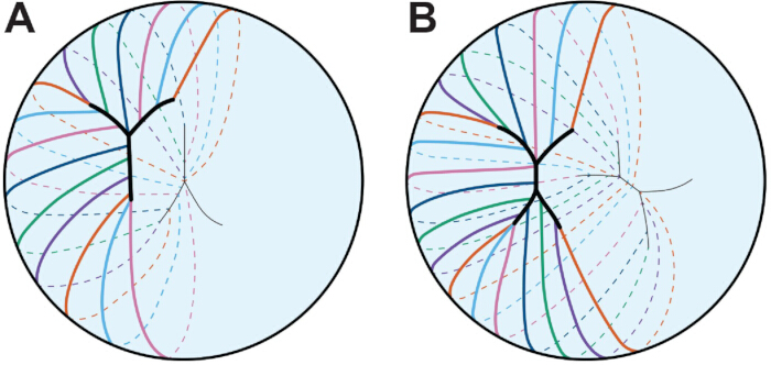
그림 3: 수정체 봉합사 패턴. 각 색깔 선은 단일 광섬유 셀을 나타냅니다. (A) 배아 인간 수정체 내 섬유 세포가 Y-봉합선을 형성합니다. Y자 봉합선은 앞극과 후극에 대칭 처리되어 있습니다. (B) 인간 수정체가 계속 발달하고 성장함에 따라 봉합선이 더 가지가 많아집니다. 단순함을 위해 4가지 갈래 봉합선이 묘사되어 있습니다. 봉합 패턴은 두 극 사이에 어긋나 있습니다. 만화는 축척에 맞춰 그려지지 않습니다. 45년을 모델로 했습니다. 이 그림의 더 큰 버전을 보시려면 여기를 클릭해 주세요.
수정체의 전체 형태는 종에 따라 다르며, 섬유세포 곡률과 봉합선 유형이 수정체모양 341,342를 결정하는 데 이론화됩니다. 봉합선은 앞극과 후극에서 길게 뻗은 섬유 세포 끝이 만나는 지점으로 형성되며, 봉합선은 두 극 11,12,13,343 사이에 대칭 구조되어 있습니다(그림 3). 쥐의 수정체에서는 봉합선이 Y자형이며, 영장류와 인간의 수정체에서는 봉합선이 Y자형으로 시작해11, 12, 45세에 따라 더 가지가 많아집니다. 흥미롭게도, 마우스 수정체의 Y 봉합선은 기계적 하중이 제거된 후 수정체의 탄력성 또는 탄력성을 제한하며, KO 수정체에서 Eph 에프린 신호 전달이 교란된 분기 봉합사는 회복력 증가와 상관관계가 있다344. 수정체 봉합의 정상 형성은 간극 접합345를 필요로 하며, 쥐 수정체의 연령 관련 전방 백내장은 불완전한 봉합 형성130의 결과입니다.
렌즈의 생체역학적 특성은 이전 리뷰에서 설명된 바와 같이 다양한 분자, 세포, 조직 요인에 의해 영향을 받을 수 있습니다. 노화346의 주요 원인은 수정체 강성 증가로 여겨집니다. 인간은 먼 물체를 볼 때 섬모근의 이완으로 캡슐에 붙어 있는 대척 섬유가 늘어나면서 수정체가 납작해지게 됩니다. 가까이서 초점을 맞출 때는 섬모근이 수축하여 조울이 이완되고, 렌즈가 둥글게 올라가가까운 물체의 빛을 집중시킵니다. 이 조절 과정은 수정체의 생체역학적 특성, 섬모근 수축, 그리고 대척 장력을 필요로 합니다. 나이가 들면서 섬모근 수축력과 대추 유연성 및 긴장력에 거의 변화가 없거나 전혀 없는 것으로 나타났으며, 3억 4,348,349,350명입니다. 여러 연구들은 수정체 크기와 핵 크기, 강성이 26,351,352,353,354년에 걸쳐 전체 수정체의 전반적인 강성에 기여한다고 제안했습니다. 그러나 쥐에서 수정체 강성에 따른 나이 연구에서는 수정체와 핵 크기 사이에 전체 수정체 강성130,132 사이에 거의 상관관계가 없으며, 이는 연령 관련 수정체 강성에 영향을 미치는 다른 요인들이 있음을 시사합니다. 최근 연구는 세포골격 네트워크의 변화가 수정체 강성에 영향을 미친다는 것을 보여주었습니다; F-액틴 안정화 단백질인 트로포미오신 3.5의 손실은 섬유세포 막에서 F-액틴 네트워크의 구성과 배열에 영향을 미치고 수정체 강성을 감소시킵니다173. 마찬가지로, 여러 근친교배 야생형 마우스 계통에서 자발적 돌연변이가 발생하면 CP49192,355 개와 더 부드러운 렌즈356,357개가 발생한다. 수정체 섬유 세포막은 비정상적으로 높은 콜레스테롤 함량을 가지고 있는데, 358,359,360,361,362,363으로, 막 탄성을 조절할 수 있습니다. 막 콜레스테롤 함량과 α결정체가 섬유세포막에 결합하는 변화가 나이에 따라 수정체 강성을 증가시키는 것으로 이론화됩니다 365,366,367,368,369,370,371,372.
렌즈 연구를 위한 도전과 새로운 방법
수정체의 3D 구조는 세포 간 배열을 이해하고 조직 구조를 재현하는 적합한 시험관 내 모델을 만드는 데 어려움을 초래합니다. 신호전달과 돌연변이 단백질 위치화에 관한 귀중한 통찰은 불멸화 또는 1차 배양 세포를 통해 밝혀졌으나, 분리된 수정체 상피세포는 고유한 입방체 형태를 가지지 못하며, 토종 수정체 상피세포와 동일한 유전자 발현 또는 세포골격 구조를 갖지 못합니다373. 이전 리뷰에서 설명한 시험관 내 렌토이드374와 렌즈렛 모델375는 각각 작은 수정체와 유사한 섬유 같은 세포와 세포 덩어리를 만듭니다. 이 모델들은 빠른 약물 스크리닝에 유용할 수 있는 투명한 렌즈 같은 구조를 만들지만, 어느 모델도 정상적인 섬유세포 조직이나 렌즈의 3D 구조를 재현하지는 않습니다. 수정체 조직 절편에서 단백질을 위치시키거나 면역염색을 하기 위한 많은 프로토콜이 개발되었으며, 이 방법은 단백질이 특정 세포 구획에서 발현되는지 여부를 이해하는 가장 효율적인 방법으로 남아 있습니다. 그러나 조직학 및 면역염색을 위한 조직 절절은 특히 노령 포유류 샘플에서 렌즈137의 불완전 고정 때문에 어려울 수 있습니다; 그리고 2D 단면에서 3D 구조를 연구하면 세포의 형태, 세포 간 상호작용, 단백질의 위치 파악을 이해하는 데 중요한 세부 사항들이 제거됩니다. 전자현미경(EM)이 섬유 세포 형태 연구의 금본위로 남아 있지만, 전자현미경 샘플 내 단백질 위치 파악은 여전히 도전적입니다(37,637,378). 최근 상피세포 단층의 평면 마운트 작업 4,7,324,373, 단일 섬유 세포 염색 8,5173,174,379, 전체 렌즈 영상 4,7,324,344,380,381 세포 간 유착, 수정체 생체역학, 수정체 섬유 내 변화하는 세포골격 네트워크에 관한 중요한 정보를 밝혀냈습니다.
수정체 상피세포와 섬유세포는 기능, 전사체, 단백질체가 다르며, 이전 연구들은 상피세포가 전반구를 단일층으로 덮고 있어 많은 샘플을 모으지 않고 수집할 수 있는 RNA나 단백질의 양이 제한되어 있어 서로 다른 세포 구획을 비교하는 연구는 어려웠습니다. RNA 추출 기술, 실시간 PCR, 자동 모세관 전기영동 서부의 발전으로 쥐 한 쌍의 눈에서 채취한 수정체 상피세포 샘플(130,132,373,382 또는 수정체 상피, 피질 섬유, 핵섬유383 등 미세하게 해리된 영역)에서 추출한 수정체 상피세포 샘플의 유전자 및 단백질 분석이 가능해졌습니다. 단세포 전사체학은 제브라피시 배아와 유충 및 병아리 배아384,385의 렌즈 세포를 심층적으로 프로파일링할 수 있게 합니다. 이러한 새로운 방법은 정상적인 노화 및 병리 과정에서 각 수정체 세포 구획을 유지하는 분자 메커니즘을 밝혀낼 수 있는 문을 열어줍니다.
이 분야가 발전함에 따라, 마우스 유전적 배경은 수정 병리학386,387과 생체역학355,356,357에서 중요한 역할을 할 수 있음을 기억하는 것이 중요하며, 따라서 실험을 위한 형제끼 대조군은 데이터 분석에 매우 중요합니다. 돌연변이 및 KO 마우스 모델은 연령 관련 백내장과 수정체 강성 증가를 연구하는 시스템을 제공합니다 130,356,357,388,389,390,391,392,393,394,395,396,397쥐의 수명이 비교적 짧기 때문에, 노화 연구는 젊은 동물의 데이터를 외삽하기보다는 노령 동물에 대해 수행되어야 합니다.
액세스가 제한되었습니다. 이 콘텐츠를 보려면 로그인하거나 체험판을 시작하세요.
결론
전사체학, 단백질체학, 영상 촬영, 생체역학 검사 기술의 발전으로 수정체 생물학, 생리학, 병리학에 대한 이해가 향상되었습니다. 수정체 상피세포와 섬유세포의 뚜렷한 집단 간 차이에 대한 우리의 확장된 지식은 노화 과정 변화와 단백질 및 세포 수명의 메커니즘을 이해하는 데 중요할 것입니다. 백내장 및/또는 노안의 예방 또는 지연, 그리고 백내장 수술 후 자연스러운 수정체 재생 가능성에 대한 약물 개입 개발에 대한 관심이 증가하고 있습니다. 백내장이나 노안을 예방하거나 지연시키기 위한 약물 전달은 수정체 캡슐을 통한 전달을 증가시키기 위해 특정 화학 특성을 가진 안약을 제조해야 하며, 수정체 내 다양한 깊이에 침투하고 축적되는 약물의 양을 신중히 연구해야 합니다 398,399,400,401,402<...
액세스가 제한되었습니다. 이 콘텐츠를 보려면 로그인하거나 체험판을 시작하세요.
공개 사항
저자들은 공개할 것이 없습니다.
감사의 글
이 연구는 국립안과 연구소의 R01 EY032056(CC에 대한 보조금)로 지원받았습니다. 저자들은 아이제아 J. 이니스와 그리핀 M. 플라워스에게 유익한 의견을 제공해 주신 것에 감사드립니다.
액세스가 제한되었습니다. 이 콘텐츠를 보려면 로그인하거나 체험판을 시작하세요.
참고문헌
- Freddo, T. F., Chaum, E. Anatomy of the Eye and Orbit: The Clinical Essentials, 1e. , Lippincott Williams & Wilkins, a Wolters Kluwer business. (2018).
- Danysh, B. P., Duncan, M. K. The lens capsule. Exp Eye Res. 88 (2), 151-164 (2009).
- Zampighi, G. A., Eskandari, S., Kreman, M. Epithelial organization of the mammalian lens. Exp Eye Res. 71 (4), 415-435 (2000).
- Cheng, C., Gong, X. Diverse roles of Eph/ephrin signaling in the mouse lens. PLoS One. 6 (11), e28147(2011).
- Parreno, J., Cheng, C., Nowak, R. B., Fowler, V. M. The effects of mechanical strain on mouse eye lens capsule and cellular microstructure. Mol Biol Cell. 29 (16), 1963-1974 (2018).
- Lovicu, F. J., Robinson, M. L. Development of the Ocular Lens. , Cambridge University Press. (2011).
- Cheng, C., Ansari, M. M., Cooper, J. A., Gong, X. EphA2 and Src regulate equatorial cell morphogenesis during lens development. Development. 140 (20), 4237-4245 (2013).
- Bassnett, S., Missey, H., Vucemilo, I. Molecular architecture of the lens fiber cell basal membrane complex. J Cell Sci. 112 (Pt 13), 2155-2165 (1999).
- Bassnett, S., Sikic, H. The lens growth process. Prog Retin Eye Res. 60, 181-200 (2017).
- Sikic, H., Shi, Y., Lubura, S., Bassnett, S. A full lifespan model of vertebrate lens growth. R Soc Open Sci. 4 (1), 160695(2017).
- Kuszak, J. R. The development of lens sutures. Progress in Retinal and Eye Research. 14 (2), 567-591 (1995).
- Kuszak, J. R., Mazurkiewicz, M., Zoltoski, R. Computer modeling of secondary fiber development and growth: I. Nonprimate lenses. Mol Vis. 12, 251-270 (2006).
- Kuszak, J. R., Zoltoski, R. K., Tiedemann, C. E. Development of lens sutures. Int J Dev Biol. 48 (8-9), 889-902 (2004).
- Kuszak, J. R. The ultrastructure of epithelial and fiber cells in the crystalline lens. Int Rev Cytol. 163, 305-350 (1995).
- Kuszak, J. R., Macsai, M. S., Rae, J. L. Stereo scanning electron microscopy of the crystalline lens. Scan Electron Microsc. (Pt 3), 1415-1426 (1983).
- Cheng, C. Tissue, cellular, and molecular level determinants for eye lens stiffness and elasticity. Front Ophthalmol (Lausanne). 4, 1456474(2024).
- World Health Organization. World report on vision. , (2019).
- Michael, R., Bron, A. J. The ageing lens and cataract: a model of normal and pathological ageing. Philos Trans R Soc Lond B Biol Sci. 366 (1568), 1278-1292 (2011).
- Asbell, P. A., et al. Age-related cataract. Lancet. 365 (9459), 599-609 (2005).
- Hejtmancik, J. F. Oxidative Stress in Genetic Cataract Formation. Antioxidants (Basel). 13 (11), (2024).
- Shiels, A., Hejtmancik, J. F. Biology of Inherited Cataracts and Opportunities for Treatment. Annu Rev Vis Sci. 5, 123-149 (2019).
- Truscott, R. J. W., Friedrich, M. G. Molecular Processes Implicated in Human Age-related Nuclear Cataract. Invest Ophthalmol Vis Sci. 60 (15), 5007-5021 (2019).
- Fan, X., Monnier, V. M. Protein posttranslational modification (PTM) by glycation: Role in lens aging and age-related cataractogenesis. Exp Eye Res. 210, 108705(2021).
- Budnar, P., Tangirala, R., Bakthisaran, R., Rao, C. M. Protein Aggregation and Cataract: Role of Age-related Modifications and Mutations in alpha Crystallins. Biochemistry (Mosc). 87 (3), 225-241 (2022).
- Shiels, A., Hejtmancik, J. F. Inherited cataracts: Genetic mechanisms and pathways new and old. Exp Eye Res. 209, 108662(2021).
- Heys, K. R., Cram, S. L., Truscott, R. J. Massive increase in the stiffness of the human lens nucleus with age: the basis for presbyopia? Mol Vis. 10, 956-963 (2004).
- Glasser, A., Campbell, M. C. Biometric, optical and physical changes in the isolated human crystalline lens with age in relation to presbyopia. Vision Res. 39 (11), 1991-2015 (1999).
- Weeber, H. A., et al. Dynamic mechanical properties of human lenses. Exp Eye Res. 80 (3), 425-434 (2005).
- Piatigorsky, J. Lens differentiation in vertebrates. A review of cellular and molecular features. Differentiation. 19 (3), 134-153 (1981).
- Gunhaga, L. The lens: a classical model of embryonic induction providing new insights into cell determination in early development. Philos Trans R Soc Lond B Biol Sci. 366 (1568), 1193-1203 (2011).
- Chauhan, B. K., et al. Cdc42 and IRSp53 dependent contractile filopodia tether presumptive lens and retina to coordinate epithelial invagination. Development. 136 (21), 3657-3667 (2009).
- McAvoy, J. W. Cytoplasmic processes interconnect lens placode and optic vesicle during eye morphogenesis. Exp Eye Res. 31 (5), 527-534 (1980).
- Mann, I. The development of the human eye. , 3d, Grune & Stratton. (1964).
- Eiraku, M., et al. Self organizing optic cup morphogenesis in three dimensional culture. Nature. 472 (7341), 51-56 (2011).
- Lang, R. A., Herman, K., Reynolds, A. B., Hildebrand, J. D., Plageman, T. F. Jr p120 catenin dependent junctional recruitment of Shroom3 is required for apical constriction during lens pit morphogenesis. Development. 141 (16), 3177-3187 (2014).
- Plageman, T. F. Jr, et al. A Trio RhoA Shroom3 pathway is required for apical constriction and epithelial invagination. Development. 138 (23), 5177-5188 (2011).
- Borges, R. M., Lamers, M. L., Forti, F. L., Santos, M. F., Yan, C. Y. Rho signaling pathway and apical constriction in the early lens placode. Genesis. 49 (5), 368-379 (2011).
- Quintin, S., Gally, C., Labouesse, M. Epithelial morphogenesis in embryos: asymmetries, motors and brakes. Trends Genet. 24 (5), 221-230 (2008).
- Muccioli, M., Qaisi, D., Herman, K., Plageman, T. F. Jr Lens placode planar cell polarity is dependent on Cdc42 mediated junctional contraction inhibition. Dev Biol. 412 (1), 32-43 (2016).
- Houssin, N. S., Martin, J. B., Coppola, V., Yoon, S. O., Plageman, T. F. Jr Formation and contraction of multicellular actomyosin cables facilitate lens placode invagination. Dev Biol. 462 (1), 36-49 (2020).
- Glucksmann, A. Cell deaths in normal vertebrate ontogeny. Biol Rev Camb Philos Soc. 26 (1), 59-86 (1951).
- Ozeki, H., Ogura, Y., Hirabayashi, Y., Shimada, S. Suppression of lens stalk cell apoptosis by hyaluronic acid leads to faulty separation of the lens vesicle. Exp Eye Res. 72 (1), 63-70 (2001).
- Yan, Q., Liu, J. P., Li, D. W. Apoptosis in lens development and pathology. Differentiation. 74 (5), 195-211 (2006).
- Easter, S. S. Jr, Nicola, G. N. The development of vision in the zebrafish (Danio rerio). Dev Biol. 180 (2), 646-663 (1996).
- Mann, I. The development of the human eye. , The University Press. (1928).
- Darche, M., et al. Three dimensional characterization of developing and adult ocular vasculature in mice using in toto clearing. Communications Biology. 5 (1), 1135(2022).
- Wang, Z., Liu, C. H., Huang, S., Chen, J. Assessment and Characterization of Hyaloid Vessels in Mice. J Vis Exp. (147), e59222(2019).
- Hill, R. E., et al. Mouse small eye results from mutations in a paired like homeobox containing gene. Nature. 354 (6354), 522-525 (1991).
- Jordan, T., et al. The human PAX6 gene is mutated in two patients with aniridia. Nat Genet. 1 (5), 328-332 (1992).
- Hogan, B. L., et al. Small eyes (Sey): a homozygous lethal mutation on chromosome 2 which affects the differentiation of both lens and nasal placodes in the mouse. J Embryol Exp Morphol. 97, 95-110 (1986).
- Hanson, I. M., et al. Mutations at the PAX6 locus are found in heterogeneous anterior segment malformations including Peters' anomaly. Nat Genet. 6 (2), 168-173 (1994).
- Grindley, J. C., Davidson, D. R., Hill, R. E. The role of Pax 6 in eye and nasal development. Development. 121 (5), 1433-1442 (1995).
- Fujiwara, M., Uchida, T., Osumi Yamashita, N., Eto, K. Uchida rat (rSey): a new mutant rat with craniofacial abnormalities resembling those of the mouse Sey mutant. Differentiation. 57 (1), 31-38 (1994).
- Collinson, J. M., Hill, R. E., West, J. D. Different roles for Pax6 in the optic vesicle and facial epithelium mediate early morphogenesis of the murine eye. Development. 127 (5), 945-956 (2000).
- Quinn, J. C., West, J. D., Hill, R. E. Multiple functions for Pax6 in mouse eye and nasal development. Genes Dev. 10 (4), 435-446 (1996).
- Ashery Padan, R., Marquardt, T., Zhou, X., Gruss, P. Pax6 activity in the lens primordium is required for lens formation and for correct placement of a single retina in the eye. Genes Dev. 14 (21), 2701-2711 (2000).
- Kim, E. A., et al. Phosphorylation and transactivation of Pax6 by homeodomain interacting protein kinase 2. J Biol Chem. 281 (11), 7489-7497 (2006).
- Mikkola, I., Bruun, J. A., Bjorkoy, G., Holm, T., Johansen, T. Phosphorylation of the transactivation domain of Pax6 by extracellular signal regulated kinase and p38 mitogen activated protein kinase. J Biol Chem. 274 (21), 15115-15126 (1999).
- Yan, Q., et al. Sumoylation activates the transcriptional activity of Pax 6, an important transcription factor for eye and brain development. Proc Natl Acad Sci U S A. 107 (49), 21034-21039 (2010).
- Yan, Q., et al. Protein phosphatase 1 modulates the function of Pax 6, a transcription factor controlling brain and eye development. J Biol Chem. 282 (19), 13954-13965 (2007).
- Cvekl, A., Zhang, X. Signaling and Gene Regulatory Networks in Mammalian Lens Development. Trends Genet. 33 (10), 677-702 (2017).
- Zhang, X., Friedman, A., Heaney, S., Purcell, P., Maas, R. L. Meis homeoproteins directly regulate Pax6 during vertebrate lens morphogenesis. Genes Dev. 16 (16), 2097-2107 (2002).
- Dudley, A. T., Lyons, K. M., Robertson, E. J. A requirement for bone morphogenetic protein 7 during development of the mammalian kidney and eye. Genes Dev. 9 (22), 2795-2807 (1995).
- Wawersik, S., et al. BMP7 acts in murine lens placode development. Dev Biol. 207 (1), 176-188 (1999).
- Blixt, A., et al. A forkhead gene, FoxE3, is essential for lens epithelial proliferation and closure of the lens vesicle. Genes Dev. 14 (2), 245-254 (2000).
- Brownell, I., Dirksen, M., Jamrich, M. Forkhead Foxe3 maps to the dysgenetic lens locus and is critical in lens development and differentiation. Genesis. 27 (2), 81-93 (2000).
- Dimanlig, P. V., Faber, S. C., Auerbach, W., Makarenkova, H. P., Lang, R. A. The upstream ectoderm enhancer in Pax6 has an important role in lens induction. Development. 128 (22), 4415-4424 (2001).
- Faber, S. C., et al. Fgf receptor signaling plays a role in lens induction. Development. 128 (22), 4425-4438 (2001).
- Bavik, C., Ward, S. J., Chambon, P. Developmental abnormalities in cultured mouse embryos deprived of retinoic by inhibition of yolk sac retinol binding protein synthesis. Proc Natl Acad Sci U S A. 93 (7), 3110-3114 (1996).
- Enwright, J. F. 3rd, Grainger, R. M. Altered retinoid signaling in the heads of small eye mouse embryos. Dev Biol. 221 (1), 10-22 (2000).
- Lovicu, F. J., McAvoy, J. W. Growth factor regulation of lens development. Dev Biol. 280 (1), 1-14 (2005).
- Schulz, M. W., Chamberlain, C. G., de Iongh, R. U., McAvoy, J. W. Acidic and basic FGF in ocular media and lens: implications for lens polarity and growth patterns. Development. 118 (1), 117-126 (1993).
- Lovicu, F. J., Chamberlain, C. G., McAvoy, J. W. Differential effects of aqueous and vitreous on fiber differentiation and extracellular matrix accumulation in lens epithelial explants. Invest Ophthalmol Vis Sci. 36 (7), 1459-1469 (1995).
- de Iongh, R. U., Lovicu, F. J., Chamberlain, C. G., McAvoy, J. W. Differential expression of fibroblast growth factor receptors during rat lens morphogenesis and growth. Invest Ophthalmol Vis Sci. 38 (9), 1688-1699 (1997).
- de Iongh, R. U., Lovicu, F. J., Hanneken, A., Baird, A., McAvoy, J. W. FGF receptor 1 (flg) expression is correlated with fibre differentiation during rat lens morphogenesis and growth. Dev Dyn. 206 (4), 412-426 (1996).
- Lovicu, F. J., Overbeek, P. A. Overlapping effects of different members of the FGF family on lens fiber differentiation in transgenic mice. Development. 125 (17), 3365-3377 (1998).
- Robinson, M. L., et al. Extracellular FGF 1 acts as a lens differentiation factor in transgenic mice. Development. 121 (2), 505-514 (1995).
- Robinson, M. L., et al. Disregulation of ocular morphogenesis by lens specific expression of FGF 3/int 2 in transgenic mice. Dev Biol. 198 (1), 13-31 (1998).
- Faber, S. C., Robinson, M. L., Makarenkova, H. P., Lang, R. A. Bmp signaling is required for development of primary lens fiber cells. Development. 129 (15), 3727-3737 (2002).
- Belecky Adams, T., et al. Pax 6, Prox 1, and Chx10 homeobox gene expression correlates with phenotypic fate of retinal precursor cells. Invest Ophthalmol Vis Sci. 38 (7), 1293-1303 (1997).
- de Iongh, R. U., et al. Requirement for TGFbeta receptor signaling during terminal lens fiber differentiation. Development. 128 (20), 3995-4010 (2001).
- Bassnett, S. Lens organelle degradation. Exp Eye Res. 74 (1), 1-6 (2002).
- Bassnett, S. The fate of the Golgi apparatus and the endoplasmic reticulum during lens fiber cell differentiation. Invest Ophthalmol Vis Sci. 36 (9), 1793-1803 (1995).
- Bassnett, S., Beebe, D. C. Coincident loss of mitochondria and nuclei during lens fiber cell differentiation. Dev Dyn. 194 (2), 85-93 (1992).
- Bassnett, S., Shi, Y., Vrensen, G. F. Biological glass: structural determinants of eye lens transparency. Philos Trans R Soc Lond B Biol Sci. 366 (1568), 1250-1264 (2011).
- Counis, M. F., et al. Analysis of nuclear degradation during lens cell differentiation. Cell Death Differ. 5 (4), 251-261 (1998).
- Vrensen, G. F., Graw, J., De Wolf, A. Nuclear breakdown during terminal differentiation of primary lens fibres in mice: a transmission electron microscopic study. Exp Eye Res. 52 (6), 647-659 (1991).
- Costello, M. J., et al. Autophagy and mitophagy participate in ocular lens organelle degradation. Exp Eye Res. 116, 141-150 (2013).
- De Maria, A., Bassnett, S. DNase IIbeta distribution and activity in the mouse lens. Invest Ophthalmol Vis Sci. 48 (12), 5638-5646 (2007).
- He, S., et al. Chromatin remodeling enzyme Brg1 is required for mouse lens fiber cell terminal differentiation and its denucleation. Epigenetics Chromatin. 3 (1), 21(2010).
- Cui, X., et al. HSF4 regulates DLAD expression and promotes lens de nucleation. Biochim Biophys Acta. 1832 (8), 1167-1172 (2013).
- Nakahara, M., et al. Degradation of nuclear DNA by DNase II like acid DNase in cortical fiber cells of mouse eye lens. FEBS J. 274 (12), 3055-3064 (2007).
- Nishimoto, S., et al. Nuclear cataract caused by a lack of DNA degradation in the mouse eye lens. Nature. 424 (6952), 1071-1074 (2003).
- Chaffee, B. R., et al. Nuclear removal during terminal lens fiber cell differentiation requires CDK1 activity: appropriating mitosis related nuclear disassembly. Development. 141 (17), 3388-3398 (2014).
- Lyu, L., et al. Unfolded protein response associated stabilization of p27(Cdkn1b) interferes with lens fiber cell denucleation, leading to cataract. FASEB J. 30 (3), 1087-1095 (2016).
- Gao, M., et al. HSF4 regulates lens fiber cell differentiation by activating p53 and its downstream regulators. Cell Death Dis. 8 (10), e3082(2017).
- Fujimoto, M., et al. HSF4 is required for normal cell growth and differentiation during mouse lens development. EMBO J. 23 (21), 4297-4306 (2004).
- Min, J. N., Zhang, Y., Moskophidis, D., Mivechi, N. F. Unique contribution of heat shock transcription factor 4 in ocular lens development and fiber cell differentiation. Genesis. 40 (4), 205-217 (2004).
- Morishita, H., et al. Organelle degradation in the lens by PLAAT phospholipases. Nature. 592 (7855), 634-638 (2021).
- Capetanaki, Y., Smith, S., Heath, J. P. Overexpression of the vimentin gene in transgenic mice inhibits normal lens cell differentiation. J Cell Biol. 109 (4 Pt 1), 1653-1664 (1989).
- Hamai, Y., Kuwabara, T. Early cytologic changes of Fraser cataract. An electron microscopic study. Invest Ophthalmol. 14 (7), 517-527 (1975).
- Muggleton Harris, A. L., Festing, M. F., Hall, M. A gene location for the inheritance of the cataract Fraser (CatFr) mouse congenital cataract. Genet Res. 49 (3), 235-238 (1987).
- Hamai, Y., Fukui, H. N., Kuwabara, T. Morphology of hereditary mouse cataract. Exp Eye Res. 18 (6), 537-546 (1974).
- Takehana, M. Hereditary cataract of the Nakano mouse. Exp Eye Res. 50 (6), 671-676 (1990).
- Uga, S., Kador, P. F., Kuwabara, T. Cytological study of Philly mouse cataract. Exp Eye Res. 30 (1), 79-92 (1980).
- Nakamura, M., Russell, P., Carper, D. A., Inana, G., Kinoshita, J. H. Alteration of a developmentally regulated, heat stable polypeptide in the lens of the Philly mouse. Implications for cataract formation. J Biol Chem. 263 (35), 19218-19221 (1988).
- Wang, K., et al. GammaD crystallin associated protein aggregation and lens fiber cell denucleation. Invest Ophthalmol Vis Sci. 48 (8), 3719-3728 (2007).
- Cavalheiro, G. R., et al. N myc regulates growth and fiber cell differentiation in lens development. Dev Biol. 429 (1), 105-117 (2017).
- Khillan, J. S., et al. Defects in lens fiber differentiation are linked to c mos overexpression in transgenic mice. Genes Dev. 1 (10), 1327-1335 (1987).
- Lachke, S. A. RNA binding proteins and post transcriptional regulation in lens biology and cataract: Mediating spatiotemporal expression of key factors that control the cell cycle, transcription, cytoskeleton and transparency. Exp Eye Res. 214, 108889(2022).
- Lachke, S. A., et al. Mutations in the RNA granule component TDRD7 cause cataract and glaucoma. Science. 331 (6024), 1571-1576 (2011).
- Dash, S., Dang, C. A., Beebe, D. C., Lachke, S. A. Deficiency of the RNA binding protein caprin2 causes lens defects and features of Peters anomaly. Dev Dyn. 244 (10), 1313-1327 (2015).
- Dash, S., et al. The master transcription factor SOX2, mutated in anophthalmia/microphthalmia, is post transcriptionally regulated by the conserved RNA binding protein RBM24 in vertebrate eye development. Hum Mol Genet. 29 (4), 591-604 (2020).
- Siddam, A. D., et al. The RNA binding protein Celf1 post transcriptionally regulates p27Kip1 and Dnase2b to control fiber cell nuclear degradation in lens development. PLoS Genet. 14 (3), e1007278(2018).
- Siddam, A. D., et al. High Throughput Transcriptomics of Celf1 Conditional Knockout Lens Identifies Downstream Networks Linked to Cataract Pathology. Cells. 12 (7), (2023).
- Aryal, S., et al. The cataract linked RNA binding protein Celf1 post transcriptionally controls the spatiotemporal expression of the key homeodomain transcription factors Pax6 and Prox1 in lens development. Hum Genet. 139 (12), 1541-1554 (2020).
- Takemoto, L., Sorensen, C. M. Protein protein interactions and lens transparency. Exp Eye Res. 87 (6), 496-501 (2008).
- Oyster, C. W. The human eye : structure and function. , Sinauer Associates, Inc.. (1999).
- Flaugh, S. L., Kosinski Collins, M. S., King, J. Contributions of hydrophobic domain interface interactions to the folding and stability of human gammaD crystallin. Protein Sci. 14 (3), 569-581 (2005).
- Papanikolopoulou, K., et al. Formation of amyloid fibrils in vitro by human gammaD crystallin and its isolated domains. Mol Vis. 14, 81-89 (2008).
- Graw, J. Genetics of crystallins: cataract and beyond. Exp Eye Res. 88 (2), 173-189 (2009).
- Kosinski Collins, M. S., King, J. In vitro unfolding, refolding, and polymerization of human gammaD crystallin, a protein involved in cataract formation. Protein Sci. 12 (3), 480-490 (2003).
- Horwitz, J., Bova, M. P., Ding, L. L., Haley, D. A., Stewart, P. L. Lens alpha crystallin: function and structure. Eye (Lond). 13 (Pt 3b), 403-408 (1999).
- Hejtmancik, J. F., et al. Progress in Molecular Biology and Translational Science. Hejtmancik, J. F., Nickerson, J. M. 134, Academic Press. 169-201 (2015).
- Bloemendal, H., et al. Ageing and vision: structure, stability and function of lens crystallins. Prog Biophys Mol Biol. 86 (3), 407-485 (2004).
- Benedek, G. B. Theory of transparency of the eye. Appl Opt. 10 (3), 459-473 (1971).
- Delaye, M., Tardieu, A. Short range order of crystallin proteins accounts for eye lens transparency. Nature. 302 (5907), 415-417 (1983).
- Bassnett, S., Costello, M. J. The cause and consequence of fiber cell compaction in the vertebrate lens. Exp Eye Res. 156, 50-57 (2017).
- Pierscionek, B. K., Regini, J. W. The gradient index lens of the eye: an opto biological synchrony. Prog Retin Eye Res. 31 (4), 332-349 (2012).
- Cheng, C., et al. Age-related changes in eye lens biomechanics, morphology, refractive index and transparency. Aging (Albany NY). 11 (24), 12497-12531 (2019).
- Cheng, C., et al. EphA2 Affects Development of the Eye Lens Nucleus and the Gradient of Refractive Index. Invest Ophthalmol Vis Sci. 63 (1), 2(2022).
- Flowers, G. M., et al. Eph ephrin signaling affects lens growth and shape, nucleus size, and gradient refractive index in adult mice. Frontiers in Ophthalmology. 5, (2025).
- Kozlowski, T. M., Kroger, R. H. H. Constant lens fiber cell thickness in fish suggests crystallin transport to denucleated cells. Vision Res. 162, 29-34 (2019).
- Shestopalov, V. I., Bassnett, S. Expression of autofluorescent proteins reveals a novel protein permeable pathway between cells in the lens core. J Cell Sci. 113 (Pt 11), 1913-1921 (2000).
- Shestopalov, V. I., Bassnett, S. Development of a macromolecular diffusion pathway in the lens. J Cell Sci. 116 (Pt 20), 4191-4199 (2003).
- Dunia, I., et al. Structural and immunocytochemical alterations in eye lens fiber cells from Cx46 and Cx50 knockout mice. Eur J Cell Biol. 85 (8), 729-752 (2006).
- Blankenship, T., Bradshaw, L., Shibata, B., Fitzgerald, P. Structural specializations emerging late in mouse lens fiber cell differentiation. Invest Ophthalmol Vis Sci. 48 (7), 3269-3276 (2007).
- Lo, W. K., Reese, T. S. Multiple structural types of gap junctions in mouse lens. J Cell Sci. 106 (Pt 1), 227-235 (1993).
- Baldo, G. J., et al. Gap junctional coupling in lenses from alpha(8) connexin knockout mice. J Gen Physiol. 118 (5), 447-456 (2001).
- Baldo, G. J., Mathias, R. T. Spatial variations in membrane properties in the intact rat lens. Biophys J. 63 (2), 518-529 (1992).
- Duncan, G., Patmore, L., Pynsent, P. B. Impedance of the amphibian lens. J Physiol. 312, 17-27 (1981).
- Martinez Wittinghan, F. J., et al. Lens gap junctional coupling is modulated by connexin identity and the locus of gene expression. Invest Ophthalmol Vis Sci. 45 (10), 3629-3637 (2004).
- Cheng, C., et al. Gap junction communication influences intercellular protein distribution in the lens. Exp Eye Res. 86 (6), 966-974 (2008).
- Bloemendal, H. The vertebrate eye lens. Science. 197 (4299), 127-138 (1977).
- van der Ouderaa, F. J., de Jong, W. W., Bloemendal, H. The amino acid sequence of the alphaA2 chain of bovine alpha crystallin. Eur J Biochem. 39 (1), 207-222 (1973).
- Bloemendal, H., de Jong, W. W. Lens proteins and their genes. Prog Nucleic Acid Res Mol Biol. 41, 259-281 (1991).
- Kato, K., et al. Immunoreactive alpha A crystallin in rat non lenticular tissues detected with a sensitive immunoassay method. Biochim Biophys Acta. 1080 (2), 173-180 (1991).
- Srinivasan, A. N., Nagineni, C. N., Bhat, S. P. alpha A crystallin is expressed in non ocular tissues. J Biol Chem. 267 (32), 23337-23341 (1992).
- Nath, M., Fort, P. E. alphaA Crystallin Mediated Neuroprotection in the Retinal Neurons Is Independent of Protein Kinase B. Front Neurosci. 16, 912757(2022).
- Skeie, J. M., Mahajan, V. B. Proteomic interactions in the mouse vitreous retina complex. PLoS One. 8 (11), e82140(2013).
- Ruebsam, A., et al. A specific phosphorylation regulates the protective role of alphaA crystallin in diabetes. JCI Insight. 3 (4), (2018).
- Vermorken, A. J., Hilderink, J. M., van de Ven, W. J., Bloemendal, H. Lens differentiation. Crystallin synthesis in isolated epithelia from calf lenses. J Cell Biol. 76 (1), 175-183 (1978).
- Boyle, D. L., Takemoto, L., Brady, J. P., Wawrousek, E. F. Morphological characterization of the Alpha A and Alpha B crystallin double knockout mouse lens. BMC Ophthalmol. 3, 3(2003).
- Horwitz, J. Alpha crystallin. Exp Eye Res. 76 (2), 145-153 (2003).
- Raju, M., Santhoshkumar, P., Krishna Sharma, K. Alpha crystallin derived peptides as therapeutic chaperones. Biochim Biophys Acta. 1860 (1 Pt B), 246-251 (2016).
- Cheng, C., et al. Altered chaperone like activity of alpha crystallins promotes cataractogenesis. J Biol Chem. 285 (52), 41187-41193 (2010).
- Aravind, P., et al. The betagamma crystallin superfamily contains a universal motif for binding calcium. Biochemistry. 48 (51), 12180-12190 (2009).
- Srivastava, S. S., Mishra, A., Krishnan, B., Sharma, Y. Ca2+ binding motif of betagamma crystallins. J Biol Chem. 289 (16), 10958-10966 (2014).
- Vanmarle, J., Jonges, R., Vrensen, G. F., Dewolf, A. Calcium and its localization in human lens fibres: an electron tomographic study. Exp Eye Res. 65 (1), 83-88 (1997).
- Jobby, M. K., Sharma, Y. Calcium binding to lens betaB2 and betaA3 crystallins suggests that all beta crystallins are calcium binding proteins. FEBS J. 274 (16), 4135-4147 (2007).
- Bergman, M. R., Deravi, L. F. Manipulating polydispersity of lens beta crystallins using divalent cations demonstrates evidence of calcium regulation. Proc Natl Acad Sci U S A. 119 (48), e2212051119(2022).
- Cheng, C., Nowak, R. B., Fowler, V. M. The lens actin filament cytoskeleton: Diverse structures for complex functions. Exp Eye Res. 156, 58-71 (2017).
- Quinlan, R. A., Clark, J. I. Insights into the biochemical and biophysical mechanisms mediating the longevity of the transparent optics of the eye lens. J Biol Chem. 298 (11), 102537(2022).
- Song, S., et al. Functions of the intermediate filament cytoskeleton in the eye lens. J Clin Invest. 119 (7), 1837-1848 (2009).
- FitzGerald, P. G. Lens intermediate filaments. Exp Eye Res. 88 (2), 165-172 (2009).
- Logan, C. M., Menko, A. S. Microtubules: Evolving roles and critical cellular interactions. Exp Biol Med (Maywood). 244 (15), 1240-1254 (2019).
- More, M. I., Kirsch, F. P., Rathjen, F. G. Targeted ablation of NrCAM or ankyrin B results in disorganized lens fibers leading to cataract formation. J Cell Biol. 154 (1), 187-196 (2001).
- Lin, Q., et al. Genetic variations and polymorphisms in the ezrin gene are associated with age-related cataract. Mol Vis. 19, 1572-1579 (2013).
- Hao, J., Kunishima, S., Guo, X., Hu, R., Gao, W. A large family with MYH9 disorder caused by E1841K mutation suffering from serious kidney and hearing impairment and cataracts. Ann Hematol. 91 (7), 1147-1148 (2012).
- De Rocco, D., et al. MYH9 related disease: five novel mutations expanding the spectrum of causative mutations and confirming genotype/phenotype correlations. Eur J Med Genet. 56 (1), 7-12 (2013).
- Economou, M., et al. MYH9 related disorders: report on a patient of Greek origin presenting with macroscopic hematuria and presenile cataract, caused by an R1165C mutation. J Pediatr Hematol Oncol. 34 (6), 412-415 (2012).
- Saposnik, B., et al. Mutation spectrum and genotype phenotype correlations in a large French cohort of MYH9 Related Disorders. Mol Genet Genomic Med. 2 (4), 297-312 (2014).
- Cheng, C., et al. Tropomyosin 3.5 protects the F-actin networks required for tissue biomechanical properties. J Cell Sci. 131 (23), (2018).
- Cheng, C., et al. Tropomodulin 1 Regulation oF-actin Is Required for the Formation of Large Paddle Protrusions Between Mature Lens Fiber Cells. Invest Ophthalmol Vis Sci. 57 (10), 4084-4099 (2016).
- Maddala, R., Walters, M., Brophy, P. J., Bennett, V., Rao, P. V. Ankyrin B directs membrane tethering of periaxin and is required for maintenance of lens fiber cell hexagonal shape and mechanics. Am J Physiol Cell Physiol. 310 (2), C115-C126 (2016).
- Won, G. J., Fudge, D. S., Choh, V. The effects of actomyosin disruptors on the mechanical integrity of the avian crystalline lens. Mol Vis. 21, 98-109 (2015).
- Kasper, M., Moll, R., Stosiek, P., Karsten, U. Patterns of cytokeratin and vimentin expression in the human eye. Histochemistry. 89 (4), 369-377 (1988).
- Kasper, M., Viebahn, C. Cytokeratin expression and early lens development. Anat Embryol (Berl). 186 (3), 285-290 (1992).
- Ellis, M., Alousi, S., Lawniczak, J., Maisel, H., Welsh, M. Studies on lens vimentin. Exp Eye Res. 38 (2), 195-202 (1984).
- Ramaekers, F. C., Dunia, I., Dodemont, H. J., Benedetti, E. L., Bloemendal, H. Lenticular intermediate sized filaments: biosynthesis and interaction with plasma membrane. Proc Natl Acad Sci U S A. 79 (10), 3208-3212 (1982).
- Ramaekers, F. C., et al. Identification of the cytoskeletal proteins in lens forming cells, a special epitheloid cell type. Exp Cell Res. 127 (2), 309-327 (1980).
- FitzGerald, P. G. Immunochemical characterization of a Mr 115 lens fiber cell specific extrinsic membrane protein. Curr Eye Res. 7 (12), 1243-1253 (1988).
- Ireland, M. E., et al. Up regulation of novel intermediate filament proteins in primary fiber cells: an indicator of all vertebrate lens fiber differentiation? Anat Rec. 258 (1), 25-33 (2000).
- Quinlan, R. A., Stewart, M. Molecular interactions in intermediate filaments. Bioessays. 13 (11), 597-600 (1991).
- Goulielmos, G., et al. Filensin and phakinin form a novel type of beaded intermediate filaments and coassemble de novo in cultured cells. J Cell Biol. 132 (4), 643-655 (1996).
- Georgatos, S. D., Gounari, F., Goulielmos, G., Aebi, U. To bead or not to bead? Lens specific intermediate filaments revisited. J Cell Sci. 110 (Pt 21), 2629-2634 (1997).
- Ireland, M., Maisel, H. A cytoskeletal protein unique to lens fiber cell differentiation. Exp Eye Res. 38 (6), 637-645 (1984).
- Bloemendal, H., et al. Interaction of crystallins with the cytoskeletal plasma membrane complex of the bovine lens. Ciba Found Symp. 106, 177-190 (1984).
- Carter, J. M., Hutcheson, A. M., Quinlan, R. A. In vitro studies on the assembly properties of the lens proteins CP49, CP115: coassembly with alpha crystallin but not with vimentin. Exp Eye Res. 60 (2), 181-192 (1995).
- Chaves, J. M., Gupta, R., Srivastava, K., Srivastava, O. Human alpha A crystallin missing N terminal domain poorly complexes with filensin and phakinin. Biochem Biophys Res Commun. 494 (1-2), 402-408 (2017).
- FitzGerald, P. G., Graham, D. Ultrastructural localization of alpha A crystallin to the bovine lens fiber cell cytoskeleton. Curr Eye Res. 10 (5), 417-436 (1991).
- Sandilands, A., et al. Bfsp2 mutation found in mouse 129 strains causes the loss of CP49' and induces vimentin dependent changes in the lens fibre cell cytoskeleton. Exp Eye Res. 78 (4), 875-889 (2004).
- Sandilands, A., et al. Knockout of the intermediate filament protein CP49 destabilises the lens fibre cell cytoskeleton and decreases lens optical quality, but does not induce cataract. Exp Eye Res. 76 (3), 385-391 (2003).
- Alizadeh, A., et al. Targeted deletion of the lens fiber cell specific intermediate filament protein filensin. Invest Ophthalmol Vis Sci. 44 (12), 5252-5258 (2003).
- Alizadeh, A., et al. Targeted genomic deletion of the lens specific intermediate filament protein CP49. Invest Ophthalmol Vis Sci. 43 (12), 3722-3727 (2002).
- Perng, M. D., Zhang, Q., Quinlan, R. A. Insights into the beaded filament of the eye lens. Exp Cell Res. 313 (10), 2180-2188 (2007).
- Muller, M., et al. Dominant cataract formation in association with a vimentin assembly disrupting mutation. Hum Mol Genet. 18 (6), 1052-1057 (2009).
- Bornheim, R., et al. A dominant vimentin mutant upregulates Hsp70 and the activity of the ubiquitin proteasome system, and causes posterior cataracts in transgenic mice. J Cell Sci. 121 (Pt 22), 3737-3746 (2008).
- Jakobs, P. M., et al. Autosomal dominant congenital cataract associated with a deletion mutation in the human beaded filament protein gene BFSP2. Am J Hum Genet. 66 (4), 1432-1436 (2000).
- Conley, Y. P., et al. A juvenile onset, progressive cataract locus on chromosome 3q21 q22 is associated with a missense mutation in the beaded filament structural protein 2. Am J Hum Genet. 66 (4), 1426-1431 (2000).
- Zhang, Q., et al. Clinical description and genome wide linkage study of Y sutural cataract and myopia in a Chinese family. Mol Vis. 10, 890-900 (2004).
- Cui, X., et al. The E233del mutation in BFSP2 causes a progressive autosomal dominant congenital cataract in a Chinese family. Mol Vis. 13, 2023-2029 (2007).
- Ma, X., et al. A new mutation in BFSP2 (G1091A) causes autosomal dominant congenital lamellar cataracts. Mol Vis. 14, 1906-1911 (2008).
- Ramachandran, R. D., Perumalsamy, V., Hejtmancik, J. F. Autosomal recessive juvenile onset cataract associated with mutation in BFSP1. Hum Genet. 121 (3-4), 475-482 (2007).
- Logan, C. M., Bowen, C. J., Menko, A. S. Functional role for stable microtubules in lens fiber cell elongation. Exp Cell Res. 362 (2), 477-488 (2018).
- Basu, S., Rajakaruna, S., Reyes, B., Van Bockstaele, E., Menko, A. S. Suppression of MAPK/JNK MTORC1 signaling leads to premature loss of organelles and nuclei by autophagy during terminal differentiation of lens fiber cells. Autophagy. 10 (7), 1193-1211 (2014).
- Pontoriero, G. F., et al. Co operative roles for E-cadherin and N-cadherin during lens vesicle separation and lens epithelial cell survival. Dev Biol. 326 (2), 403-417 (2009).
- Leonard, M., Zhang, L., Bleaken, B. M., Menko, A. S. Distinct roles for N-cadherin linked c Src and fyn kinases in lens development. Dev Dyn. 242 (5), 469-484 (2013).
- Leonard, M., et al. Modulation of N-cadherin junctions and their role as epicenters of differentiation specific actin regulation in the developing lens. Dev Biol. 349 (2), 363-377 (2011).
- Maddala, R., Nagendran, T., Lang, R. A., Morozov, A., Rao, P. V. Rap1 GTPase is required for mouse lens epithelial maintenance and morphogenesis. Dev Biol. 406 (1), 74-91 (2015).
- Wigle, J. T., Chowdhury, K., Gruss, P., Oliver, G. Prox1 function is crucial for mouse lens fibre elongation. Nat Genet. 21 (3), 318-322 (1999).
- Xu, L., Overbeek, P. A., Reneker, L. W. Systematic analysis of E , N and P cadherin expression in mouse eye development. Exp Eye Res. 74 (6), 753-760 (2002).
- Logan, C. M., et al. N-cadherin regulates signaling mechanisms required for lens fiber cell elongation and lens morphogenesis. Dev Biol. 428 (1), 118-134 (2017).
- Cooper, M. A., et al. Loss of ephrin A5 function disrupts lens fiber cell packing and leads to cataract. Proc Natl Acad Sci U S A. 105 (43), 16620-16625 (2008).
- Son, A. I., et al. Further analysis of the lens of ephrin A5 / mice: development of postnatal defects. Mol Vis. 19, 254-266 (2013).
- Biswas, S., Son, A. I., Yu, Q., Zhou, R., Lo, W. K. Breakdown of interlocking domains may contribute to formation of membranous globules and lens opacity in ephrin A5( / ) mice. Exp Eye Res. 145, 130-139 (2016).
- Maher, G. J., Black, G. C., Manson, F. D. Focus on molecules: lens intrinsic membrane protein (LIM2/MP20). Exp Eye Res. 103, 115-116 (2012).
- Walker, J., Menko, A. S. Integrins in lens development and disease. Exp Eye Res. 88 (2), 216-225 (2009).
- Shi, Y., et al. Further analysis of the lens phenotype in Lim2 deficient mice. Invest Ophthalmol Vis Sci. 52 (10), 7332-7339 (2011).
- Mulders, J. W., et al. MP17, a fiber specific intrinsic membrane protein from mammalian eye lens. Curr Eye Res. 7 (2), 207-219 (1988).
- Voorter, C. E., et al. Distribution of MP17 in isolated lens fibre membranes. Curr Eye Res. 8 (7), 697-706 (1989).
- Steele, E. C. Jr, et al. Identification of a mutation in the MP19 gene, Lim2, in the cataractous mouse mutant To3. Mol Vis. 3, 5(1997).
- Irum, B., et al. Mutation in LIM2 Is Responsible for Autosomal Recessive Congenital Cataracts. PLoS One. 11 (11), e0162620(2016).
- Wang, X., et al. Elongated axial length and myopia related fundus changes associated with the Arg130Cys mutation in the LIM2 gene in four Chinese families with congenital cataracts. Ann Transl Med. 9 (3), 235(2021).
- Berry, V., et al. A recurrent variant in LIM2 causes an isolated congenital sutural/lamellar cataract in a Japanese family. Ophthalmic Genet. 43 (5), 622-626 (2022).
- Pei, R., et al. A novel mutation of LIM2 causes autosomal dominant membranous cataract in a Chinese family. Int J Ophthalmol. 13 (10), 1512-1520 (2020).
- Ponnam, S. P., Ramesha, K., Tejwani, S., Matalia, J., Kannabiran, C. A missense mutation in LIM2 causes autosomal recessive congenital cataract. Mol Vis. 14, 1204-1208 (2008).
- Pras, E., et al. A missense mutation in the LIM2 gene is associated with autosomal recessive presenile cataract in an inbred Iraqi Jewish family. Am J Hum Genet. 70 (5), 1363-1367 (2002).
- Goyal, S., Singh, R., Singh, J. R., Vanita, V. Novel and known variants in GJA3 and LIM2 in congenital cataract families from North India. BMC Genomics. 25 (1), 31(2024).
- Puk, O., Ahmad, N., Wagner, S., de Angelis, M. H., Graw, J. Microphakia and congenital cataract formation in a novel Lim2(C51R) mutant mouse. Mol Vis. 17, 1164-1171 (2011).
- Dahm, R., Bramke, S., Dawczynski, J., Nagaraj, R. H., Kasper, M. Developmental aspects of galectin 3 expression in the lens. Histochem Cell Biol. 119 (3), 219-226 (2003).
- Gonen, T., Donaldson, P., Kistler, J. Galectin 3 is associated with the plasma membrane of lens fiber cells. Invest Ophthalmol Vis Sci. 41 (1), 199-203 (2000).
- Gonen, T., Grey, A. C., Jacobs, M. D., Donaldson, P. J., Kistler, J. MP20, the second most abundant lens membrane protein and member of the tetraspanin superfamily, joins the list of ligands of galectin 3. BMC Cell Biol. 2, 17(2001).
- Barton, K. A., Hsu, C. D., Petrash, J. M. Interactions between small heat shock protein alpha crystallin and galectin related interfiber protein (GRIFIN) in the ocular lens. Biochemistry. 48 (18), 3956-3966 (2009).
- Garcia Caballero, G., et al. Chicken lens development: complete signature of expression of galectins during embryogenesis and evidence for their complex formation with alpha , beta , delta , and tau crystallins, N CAM, and N-cadherin obtained by affinity chromatography. Cell Tissue Res. 379 (1), 13-35 (2020).
- Garcia Caballero, G., et al. Chicken GRIFIN: binding partners, developmental course of localization and activation of its lens specific gene expression by L Maf/Pax6. Cell Tissue Res. 375 (3), 665-683 (2019).
- Ogden, A. T., et al. GRIFIN, a novel lens specific protein related to the galectin family. J Biol Chem. 273 (44), 28889-28896 (1998).
- Menko, A. S., Philip, N. J. Beta 1 integrins in epithelial tissues: a unique distribution in the lens. Exp Cell Res. 218 (2), 516-521 (1995).
- Menko, S., Philp, N., Veneziale, B., Walker, J. Integrins and development: how might these receptors regulate differentiation of the lens. Ann N Y Acad Sci. 842, 36-41 (1998).
- Walker, J. L., Menko, A. S. alpha6 Integrin is regulated with lens cell differentiation by linkage to the cytoskeleton and isoform switching. Dev Biol. 210 (2), 497-511 (1999).
- Walker, J. L., Zhang, L., Zhou, J., Woolkalis, M. J., Menko, A. S. Role for alpha 6 integrin during lens development: Evidence for signaling through IGF 1R and ERK. Dev Dyn. 223 (2), 273-284 (2002).
- Wederell, E. D., et al. Laminin binding integrins in rat lens morphogenesis and their regulation during fibre differentiation. Exp Eye Res. 81 (3), 326-339 (2005).
- Barbour, W., et al. Expression patterns of beta1 related alpha integrin subunits in murine lens during embryonic development and wound healing. Curr Eye Res. 29 (1), 1-10 (2004).
- Wu, J. E., Santoro, S. A. Complex patterns of expression suggest extensive roles for the alpha 2 beta 1 integrin in murine development. Dev Dyn. 199 (4), 292-314 (1994).
- Duncan, M. K., Kozmik, Z., Cveklova, K., Piatigorsky, J., Cvekl, A. Overexpression of PAX6(5a) in lens fiber cells results in cataract and upregulation of (alpha)5(beta)1 integrin expression. J Cell Sci. 113 (Pt 18), 3173-3185 (2000).
- Bassuk, J. A., et al. Disruption of the Sparc locus in mice alters the differentiation of lenticular epithelial cells and leads to cataract formation. Exp Eye Res. 68 (3), 321-331 (1999).
- Gilmour, D. T., et al. Mice deficient for the secreted glycoprotein SPARC/osteonectin/BM40 develop normally but show severe age onset cataract formation and disruption of the lens. EMBO J. 17 (7), 1860-1870 (1998).
- Kantorow, M., Horwitz, J., Carper, D. Up regulation of osteonectin/SPARC in age-related cataractous human lens epithelia. Mol Vis. 4, 17(1998).
- Emerson, R. O., Sage, E. H., Ghosh, J. G., Clark, J. I. Chaperone like activity revealed in the matricellular protein SPARC. J Cell Biochem. 98 (4), 701-705 (2006).
- Weaver, M. S., Sage, E. H., Yan, Q. Absence of SPARC in lens epithelial cells results in altered adhesion and extracellular matrix production in vitro. J Cell Biochem. 97 (2), 423-432 (2006).
- Weaver, M. S., Workman, G., Sage, E. H. The copper binding domain of SPARC mediates cell survival in vitro via interaction with integrin beta1 and activation of integrin linked kinase. J Biol Chem. 283 (33), 22826-22837 (2008).
- Yan, Q., Perdue, N., Blake, D., Sage, E. H. Absence of SPARC in murine lens epithelium leads to increased deposition of laminin 1 in lens capsule. Invest Ophthalmol Vis Sci. 46 (12), 4652-4660 (2005).
- Greiling, T. M., Stone, B., Clark, J. I. Absence of SPARC leads to impaired lens circulation. Exp Eye Res. 89 (3), 416-425 (2009).
- Giannone, A. A., Li, L., Sellitto, C., White, T. W. Physiological Mechanisms Regulating Lens Transport. Front Physiol. 12, 818649(2021).
- Donaldson, P. J., Chen, Y., Petrova, R. S., Grey, A. C., Lim, J. C. Regulation of lens water content: Effects on the physiological optics of the lens. Prog Retin Eye Res. 95, 101152(2023).
- Donaldson, P. J., Grey, A. C., Maceo Heilman, B., Lim, J. C., Vaghefi, E. The physiological optics of the lens. Prog Retin Eye Res. 56, e1-e24 (2017).
- Mathias, R. T., Kistler, J., Donaldson, P. The lens circulation. J Membr Biol. 216 (1), 1-16 (2007).
- Robinson, K. R., Patterson, J. W. Localization of steady currents in the lens. Curr Eye Res. 2 (12), 843-847 (1982).
- Mathias, R. T., Rae, J. L., Baldo, G. J. Physiological properties of the normal lens. Physiol Rev. 77 (1), 21-50 (1997).
- Gao, J., Sun, X., Yatsula, V., Wymore, R. S., Mathias, R. T. Isoform specific function and distribution of Na/K pumps in the frog lens epithelium. J Membr Biol. 178 (2), 89-101 (2000).
- Alvarez, L. J., Candia, O. A., Grillone, L. R. Na+ K+ ATPase distribution in frog and bovine lenses. Curr Eye Res. 4 (2), 143-152 (1985).
- Delamere, N. A., Dean, W. L. Distribution of lens sodium potassium adenosine triphosphatase. Invest Ophthalmol Vis Sci. 34 (7), 2159-2163 (1993).
- Paterson, C. A. Distribution and movement of ions in the ocular lens. Doc Ophthalmol. 31 (1), 1-28 (1972).
- Donaldson, P. J., Petrova, R. S., Nair, N., Chen, Y., Schey, K. L. Regulation of water flow in the ocular lens: new roles for aquaporins. J Physiol. 602 (13), 3041-3056 (2024).
- Goodenough, D. A. The crystalline lens. A system networked by gap junctional intercellular communication. Semin Cell Biol. 3 (1), 49-58 (1992).
- Mathias, R. T., White, T. W., Gong, X. Lens gap junctions in growth, differentiation, and homeostasis. Physiol Rev. 90 (1), 179-206 (2010).
- Beyer, E. C., Kistler, J., Paul, D. L., Goodenough, D. A. Antisera directed against connexin43 peptides react with a 43 kD protein localized to gap junctions in myocardium and other tissues. J Cell Biol. 108 (2), 595-605 (1989).
- Gao, Y., Spray, D. C. Structural changes in lenses of mice lacking the gap junction protein connexin43. Invest Ophthalmol Vis Sci. 39 (7), 1198-1209 (1998).
- Rong, P., et al. Disruption of Gja8 (alpha8 connexin) in mice leads to microphthalmia associated with retardation of lens growth and lens fiber maturation. Development. 129 (1), 167-174 (2002).
- Puk, O., et al. Mutation in a novel connexin like gene (Gjf1) in the mouse affects early lens development and causes a variable small eye phenotype. Invest Ophthalmol Vis Sci. 49 (4), 1525-1532 (2008).
- Sonntag, S., et al. Mouse lens connexin23 (Gje1) does not form functional gap junction channels but causes enhanced ATP release from HeLa cells. Eur J Cell Biol. 88 (2), 65-77 (2009).
- Bassnett, S., Wilmarth, P. A., David, L. L. The membrane proteome of the mouse lens fiber cell. Mol Vis. 15, 2448-2463 (2009).
- Wang, Z., Han, J., David, L. L., Schey, K. L. Proteomics and phosphoproteomics analysis of human lens fiber cell membranes. Invest Ophthalmol Vis Sci. 54 (2), 1135-1143 (2013).
- Berthoud, V. M., et al. Connexin23 deletion does not affect lens transparency. Exp Eye Res. 146, 283-288 (2016).
- Fleishman, S. J., Unger, V. M., Yeager, M., Ben Tal, N. A Calpha model for the transmembrane alpha helices of gap junction intercellular channels. Mol Cell. 15 (6), 879-888 (2004).
- Unger, V. M., Kumar, N. M., Gilula, N. B., Yeager, M. Three dimensional structure of a recombinant gap junction membrane channel. Science. 283 (5405), 1176-1180 (1999).
- Hu, Z., Riquelme, M. A., Gu, S., Jiang, J. X. Regulation of Connexin Gap Junctions and Hemichannels by Calcium and Calcium Binding Protein Calmodulin. Int J Mol Sci. 21 (21), (2020).
- Quan, Y., Du, Y., Tong, Y., Gu, S., Jiang, J. X. Connexin Gap Junctions and Hemichannels in Modulating Lens Redox Homeostasis and Oxidative Stress in Cataractogenesis. Antioxidants (Basel). 10 (9), (2021).
- Ebihara, L., Korzyukov, Y., Kothari, S., Tong, J. J. Cx46 hemichannels contribute to the sodium leak conductance in lens fiber cells. Am J Physiol Cell Physiol. 306 (5), C506-C513 (2014).
- Liu, J., et al. Mechanosensitive collaboration between integrins and connexins allows nutrient and antioxidant transport into the lens. J Cell Biol. 219 (12), (2020).
- Tong, Y., et al. Mechano activated connexin hemichannels and glutathione transport protect lens fiber cells against oxidative insults. Redox Biol. 73, 103216(2024).
- Wang, G., Quan, Y., Ma, B., Gu, S., Jiang, J. X. Mechano activated connexin hemichannels mediate intercellular glutathione transport and support lens redox homeostasis. Redox Biol. 85, 103767(2025).
- Shi, W., Riquelme, M. A., Gu, S., Jiang, J. X. Connexin hemichannels mediate glutathione transport and protect lens fiber cells from oxidative stress. J Cell Sci. 131 (6), (2018).
- Ramachandran, S., Xie, L. H., John, S. A., Subramaniam, S., Lal, R. A novel role for connexin hemichannel in oxidative stress and smoking induced cell injury. PLoS One. 2 (8), e712(2007).
- Quan, Y., Du, Y., Wu, C., Gu, S., Jiang, J. X. Connexin hemichannels regulate redox potential via metabolite exchange and protect lens against cellular oxidative damage. Redox Biol. 46, 102102(2021).
- Quan, Y., Du, Y., Wu, C., Gu, S., Jiang, J. X. Data on Connexin 43 hemichannels regulation of cellular redox in lens. Data Brief. 39, 107572(2021).
- Ren, Q., et al. Cataract causing mutation of human connexin 46 impairs gap junction, but increases hemichannel function and cell death. PLoS One. 8 (9), e74732(2013).
- Siller Jackson, A. J., et al. Adaptation of connexin 43 hemichannel prostaglandin release to mechanical loading. J Biol Chem. 283 (39), 26374-26382 (2008).
- Cheng, C., et al. Lens ion homeostasis relies on the assembly and/or stability of large connexin 46 gap junction plaques on the broad sides of differentiating fiber cells. Am J Physiol Cell Physiol. 308 (10), C835-C847 (2015).
- Gong, X., et al. Disruption of alpha3 connexin gene leads to proteolysis and cataractogenesis in mice. Cell. 91 (6), 833-843 (1997).
- Gong, X., Baldo, G. J., Kumar, N. M., Gilula, N. B., Mathias, R. T. Gap junctional coupling in lenses lacking alpha3 connexin. Proc Natl Acad Sci U S A. 95 (26), 15303-15308 (1998).
- White, T. W., Goodenough, D. A., Paul, D. L. Targeted ablation of connexin50 in mice results in microphthalmia and zonular pulverulent cataracts. J Cell Biol. 143 (3), 815-825 (1998).
- Sellitto, C., Li, L., White, T. W. Connexin50 is essential for normal postnatal lens cell proliferation. Invest Ophthalmol Vis Sci. 45 (9), 3196-3202 (2004).
- White, T. W. Unique and redundant connexin contributions to lens development. Science. 295 (5553), 319-320 (2002).
- Li, L., et al. Connexin mediated cataract prevention in mice. PLoS One. 5 (9), (2010).
- Schey, K. L., et al. Lens Aquaporins in Health and Disease: Location is Everything! Front Physiol. 13, 882550(2022).
- Schey, K. L., Petrova, R. S., Gletten, R. B., Donaldson, P. J. The Role of Aquaporins in Ocular Lens Homeostasis. Int J Mol Sci. 18 (12), (2017).
- Gao, J., et al. Lens intracellular hydrostatic pressure is generated by the circulation of sodium and modulated by gap junction coupling. J Gen Physiol. 137 (6), 507-520 (2011).
- Gao, J., Sun, X., White, T. W., Delamere, N. A., Mathias, R. T. Feedback Regulation of Intracellular Hydrostatic Pressure in Surface Cells of the Lens. Biophys J. 109 (9), 1830-1839 (2015).
- Shahidullah, M., et al. TRPV1 activation stimulates NKCC1 and increases hydrostatic pressure in the mouse lens. Am J Physiol Cell Physiol. 318 (5), C969-C980 (2020).
- Petrova, R. S., et al. Modulation of Membrane Trafficking of AQP5 in the Lens in Response to Changes in Zonular Tension Is Mediated by the Mechanosensitive Channel TRPV1. Int J Mol Sci. 24 (10), (2023).
- Chen, Y., Petrova, R. S., Qiu, C., Donaldson, P. J. Intracellular hydrostatic pressure regulation in the bovine lens: a role in the regulation of lens optics? Am J Physiol Regul Integr Comp Physiol. 322 (3), R263-R279 (2022).
- Donaldson, P. J., Musil, L. S., Mathias, R. T. Point: A critical appraisal of the lens circulation model an experimental paradigm for understanding the maintenance of lens transparency? Invest Ophthalmol Vis Sci. 51 (5), 2303-2306 (2010).
- Merriman Smith, B. R., Krushinsky, A., Kistler, J., Donaldson, P. J. Expression patterns for glucose transporters GLUT1 and GLUT3 in the normal rat lens and in models of diabetic cataract. Invest Ophthalmol Vis Sci. 44 (8), 3458-3466 (2003).
- Merriman Smith, R., Donaldson, P., Kistler, J. Differential expression of facilitative glucose transporters GLUT1 and GLUT3 in the lens. Invest Ophthalmol Vis Sci. 40 (13), 3224-3230 (1999).
- Zahraei, A., et al. Mapping Glucose Uptake, Transport and Metabolism in the Bovine Lens Cortex. Front Physiol. 13, 901407(2022).
- Lim, J. C., Perwick, R. D., Li, B., Donaldson, P. J. Comparison of the expression and spatial localization of glucose transporters in the rat, bovine and human lens. Exp Eye Res. 161, 193-204 (2017).
- Swarup, A., et al. Deletion of GLUT1 in mouse lens epithelium leads to cataract formation. Exp Eye Res. 172, 45-53 (2018).
- Reddy, V. N. Metabolism of glutathione in the lens. Exp Eye Res. 11 (3), 310-328 (1971).
- Truscott, R. J. Age-related nuclear cataract oxidation is the key. Exp Eye Res. 80 (5), 709-725 (2005).
- Kannan, R., Yi, J. R., Zlokovic, B. V., Kaplowitz, N. Molecular characterization of a reduced glutathione transporter in the lens. Invest Ophthalmol Vis Sci. 36 (9), 1785-1792 (1995).
- Mackic, J. B., et al. Transport of circulating reduced glutathione at the basolateral side of the anterior lens epithelium: physiologic importance and manipulations. Exp Eye Res. 62 (1), 29-37 (1996).
- Li, B., Li, L., Donaldson, P. J., Lim, J. C. Dynamic regulation of GSH synthesis and uptake pathways in the rat lens epithelium. Exp Eye Res. 90 (2), 300-307 (2010).
- Lim, J., Li, L., Jacobs, M. D., Kistler, J., Donaldson, P. J. Mapping of glutathione and its precursor amino acids reveals a role for GLYT2 in glycine uptake in the lens core. Invest Ophthalmol Vis Sci. 48 (11), 5142-5151 (2007).
- Li, L., Lim, J., Jacobs, M. D., Kistler, J., Donaldson, P. J. Regional differences in cystine accumulation point to a sutural delivery pathway to the lens core. Invest Ophthalmol Vis Sci. 48 (3), 1253-1260 (2007).
- Rathbun, W. B. Activity of glutathione synthesis enzymes in the rhesus monkey lens related to age: a model for the human lens. Curr Eye Res. 5 (2), 161-166 (1986).
- Sethna, S. S., Holleschau, A. M., Rathbun, W. B. Activity of glutathione synthesis enzymes in human lens related to age. Curr Eye Res. 2 (11), 735-742 (1982).
- Giblin, F. J., et al. Nuclear light scattering, disulfide formation and membrane damage in lenses of older guinea pigs treated with hyperbaric oxygen. Exp Eye Res. 60 (3), 219-235 (1995).
- Padgaonkar, V. A., et al. Hyperbaric oxygen in vivo accelerates the loss of cytoskeletal proteins and MIP26 in guinea pig lens nucleus. Exp Eye Res. 68 (4), 493-504 (1999).
- Padgaonkar, V., Giblin, F. J., Reddy, V. N. Disulfide cross linking of urea insoluble proteins in rabbit lenses treated with hyperbaric oxygen. Exp Eye Res. 49 (5), 887-899 (1989).
- Lou, M. F., Dickerson, J. E. Jr Protein thiol mixed disulfides in human lens. Exp Eye Res. 55 (6), 889-896 (1992).
- Wei, Z., et al. Aging lens epithelium is susceptible to ferroptosis. Free Radic Biol Med. 167, 94-108 (2021).
- Wei, Z., et al. Deficiency in glutathione peroxidase 4 (GPX4) results in abnormal lens development and newborn cataract. Proc Natl Acad Sci U S A. 121 (48), e2407842121(2024).
- Cheng, C., Fowler, V. M., Gong, X. EphA2 and ephrin A5 are not a receptor ligand pair in the ocular lens. Exp Eye Res. 162, 9-17 (2017).
- Islam, S. T., Cheheltani, S., Cheng, C., Fowler, V. M. Disease related non muscle myosin IIA D1424N rod domain mutation, but not R702C motor domain mutation, disrupts mouse ocular lens fiber cell alignment and hexagonal packing. Cytoskeleton (Hoboken). 81 (12), 789-805 (2024).
- Islam, S. T., Cheng, C., Parreno, J., Fowler, V. M. Nonmuscle Myosin IIA Regulates the Precise Alignment of Hexagonal Eye Lens Epithelial Cells During Fiber Cell Formation and Differentiation. Invest Ophthalmol Vis Sci. 64 (4), 20(2023).
- Forrester, J. V., Dick, A. D., McMenamin, P. G., Roberts, F., Pearlman, E. The Eye (Fourth Edition). Forrester, J. V. , W.B. Saunders. 1-102.e102 (2016).
- D’Amore, P. A. Encyclopedia of the Eye (Second Edition). , Elsevier. 514-520 (2025).
- Vrensen, G. F. Aging of the human eye lens a morphological point of view. Comp Biochem Physiol A Physiol. 111 (4), 519-532 (1995).
- Vrensen, G. F., Duindam, H. J. Maturation of fiber membranes in the human eye lens. Ultrastructural and Raman microspectroscopic observations. Ophthalmic Res. 27 Suppl 1, 78-85 (1995).
- Willekens, B., Vrensen, G. The three dimensional organization of lens fibers in the rabbit. A scanning electron microscopic reinvestigation. Albrecht Von Graefes Arch Klin Exp Ophthalmol. 216 (4), 275-289 (1981).
- Willekens, B., Vrensen, G. The three dimensional organization of lens fibers in the rhesus monkey. Graefes Arch Clin Exp Ophthalmol. 219 (3), 112-120 (1982).
- Willekens, B., Vrensen, G. Lens fiber organization in four avian species: a scanning electron microscopic study. Tissue Cell. 17 (3), 359-377 (1985).
- Lo, W. K., et al. Aquaporin 0 targets interlocking domains to control the integrity and transparency of the eye lens. Invest Ophthalmol Vis Sci. 55 (3), 1202-1212 (2014).
- Lo, W. K., Harding, C. V. Square arrays and their role in ridge formation in human lens fibers. J Ultrastruct Res. 86 (3), 228-245 (1984).
- Kistler, J., et al. Membrane interlocking domains in the lens. Invest Ophthalmol Vis Sci. 27 (10), 1527-1534 (1986).
- Dickson, D. H., Crock, G. W. Interlocking patterns on primate lens fibers. Invest Ophthalmol. 11 (10), 809-815 (1972).
- Kuszak, J., Alcala, J., Maisel, H. The surface morphology of embryonic and adult chick lens fiber cells. Am J Anat. 159 (4), 395-410 (1980).
- Kuwabara, T. The maturation of the lens cell: a morphologic study. Exp Eye Res. 20 (5), 427-443 (1975).
- Martin, J. B., et al. Arvcf Dependent Adherens Junction Stability is Required to Prevent Age-related Cortical Cataracts. Front Cell Dev Biol. 10, 840129(2022).
- Chen, Y., Stump, R. J., Lovicu, F. J., Shimono, A., McAvoy, J. W. Wnt signaling is required for organization of the lens fiber cell cytoskeleton and development of lens three dimensional architecture. Dev Biol. 324 (1), 161-176 (2008).
- Sugiyama, Y., et al. Concave to convex curve conversion of fiber cells correlates with Y shaped suture formation at the poles of the rodent lens. Exp Eye Res. 248, 110066(2024).
- Kuszak, J. R., Sivak, J. G., Weerheim, J. A. Lens optical quality is a direct function of lens sutural architecture. Invest Ophthalmol Vis Sci. 32 (7), 2119-2129 (1991).
- Cheng, C. EphA2 and Ephrin A5 Guide Eye Lens Suture Alignment and Influence Whole Lens Resilience. Invest Ophthalmol Vis Sci. 62 (15), 3(2021).
- Xia, C. H., et al. Absence of alpha3 (Cx46) and alpha8 (Cx50) connexins leads to cataracts by affecting lens inner fiber cells. Exp Eye Res. 83 (3), 688-696 (2006).
- BEERS, A. P. A., van der HEIJDE, G. L. Age-related Changes in the Accommodation Mechanism. Optometry and Vision Science. 73 (4), 235-242 (1996).
- van Alphen, G. W., Graebel, W. P. Elasticity of tissues involved in accommodation. Vision Res. 31 (7-8), 1417-1438 (1991).
- Fisher, R. F. The ciliary body in accommodation. Trans Ophthalmol Soc U K (1962). 105 (Pt 2), 208-219 (1986).
- Graebel, W. P., van Alphen, G. W. H. M. The Elasticity of Sclera and Choroid of the Human Eye, and Its Implications on Scleral Rigidity and Accommodation. Journal of Biomechanical Engineering. 99 (4), 203-208 (1977).
- Tamm, S., Tamm, E., Rohen, J. W. Age-related changes of the human ciliary muscle. A quantitative morphometric study. Mech Ageing Dev. 62 (2), 209-221 (1992).
- Scarcelli, G., Kim, P., Yun, S. H. In vivo measurement of age-related stiffening in the crystalline lens by Brillouin optical microscopy. Biophys J. 101 (6), 1539-1545 (2011).
- Zhou, H., Yan, H., Yan, W., Wang, X., Li, Q. In vivo ultraosound elastographic evaluation of the age-related change of human lens nuclear stiffness. BMC Ophthalmol. 20 (1), 135(2020).
- Hermans, E., Dubbelman, M., van der Heijde, R., Heethaar, R. The shape of the human lens nucleus with accommodation. J Vis. 7 (10), 16.1-10 (2007).
- Fisher, R. F., Pettet, B. E. Presbyopia and the water content of the human crystalline lens. J Physiol. 234 (2), 443-447 (1973).
- Alizadeh, A., et al. Characterization of a mutation in the lens specific CP49 in the 129 strain of mouse. Invest Ophthalmol Vis Sci. 45 (3), 884-891 (2004).
- Fudge, D. S., et al. Intermediate filaments regulate tissue size and stiffness in the murine lens. Invest Ophthalmol Vis Sci. 52 (6), 3860-3867 (2011).
- Gokhin, D. S., et al. Tmod1 and CP49 synergize to control the fiber cell geometry, transparency, and mechanical stiffness of the mouse lens. PLoS One. 7 (11), e48734(2012).
- Duindam, J. J., Vrensen, G. F., Otto, C., Greve, J. Aging affects the conformation of cholesterol in the human eye lens. Ophthalmic Res. 28 Suppl 1, 86-91 (1996).
- Li, L. K., So, L., Spector, A. Age dependent changes in the distribution and concentration of human lens cholesterol and phospholipids. Biochim Biophys Acta. 917 (1), 112-120 (1987).
- Mainali, L., Raguz, M., O'Brien, W. J., Subczynski, W. K. Changes in the Properties and Organization of Human Lens Lipid Membranes Occurring with Age. Curr Eye Res. 42 (5), 721-731 (2017).
- Truscott, R. J. Age-related nuclear cataract: a lens transport problem. Ophthalmic Res. 32 (5), 185-194 (2000).
- Subczynski, W. K., Raguz, M., Widomska, J., Mainali, L., Konovalov, A. Functions of cholesterol and the cholesterol bilayer domain specific to the fiber cell plasma membrane of the eye lens. J Membr Biol. 245 (1), 51-68 (2012).
- Widomska, J., Subczynski, W. K. Why Is Very High Cholesterol Content Beneficial for the Eye Lens but Negative for Other Organs? Nutrients. 11 (5), (2019).
- Khadka, N. K., Mortimer, M. F., Marosvari, M., Timsina, R., Mainali, L. Membrane elasticity modulated by cholesterol in model of porcine eye lens lipid membrane. Exp Eye Res. 220, 109131(2022).
- Bracchi, P. G., Carta, F., Fasella, P., Maraini, G. Selective binding of aged alpha crystallin to lens fibre ghosts. Exp Eye Res. 12 (1), 151-154 (1971).
- Maraini, G., Fasella, P. Reversible binding of soluble lens proteins to lens fibre ghosts. Exp Eye Res. 10 (1), 133-139 (1970).
- Broekhuyse, R. M., Kuhlmann, E. D. Lens membranes. IV. Preparative isolation and characterization of membranes and various membrane proteins from calf lens. Exp Eye Res. 26 (3), 305-320 (1978).
- Friedrich, M. G., Truscott, R. J. Large scale binding of alpha crystallin to cell membranes of aged normal human lenses: a phenomenon that can be induced by mild thermal stress. Invest Ophthalmol Vis Sci. 51 (10), 5145-5152 (2010).
- Khadka, N. K., Timsina, R., Mainali, L. An AFM Approach Applied in a Study of alpha Crystallin Membrane Association: New Insights into Lens Hardening and Presbyopia Development. Membranes (Basel). 12 (5), (2022).
- Timsina, R., Khadka, N. K., Maldonado, D., Mainali, L. Interaction of alpha crystallin with four major phospholipids of eye lens membranes. Exp Eye Res. 202, 108337(2021).
- Timsina, R., Mainali, L. Association of Alpha Crystallin with Fiber Cell Plasma Membrane of the Eye Lens Accompanied by Light Scattering and Cataract Formation. Membranes (Basel). 11 (6), (2021).
- Timsina, R., Trossi Torres, G., O'Dell, M., Khadka, N. K., Mainali, L. Cholesterol and cholesterol bilayer domains inhibit binding of alpha crystallin to the membranes made of the major phospholipids of eye lens fiber cell plasma membranes. Exp Eye Res. 206, 108544(2021).
- Parreno, J., et al. Methodologies to unlock the molecular expression and cellular structure of ocular lens epithelial cells. Front Cell Dev Biol. 10, 983178(2022).
- Cvekl, A., Camerino, M. J. Generation of Lens Progenitor Cells and Lentoid Bodies from Pluripotent Stem Cells: Novel Tools for Human Lens Development and Ocular Disease Etiology. Cells. 11 (21), (2022).
- Murphy, P., et al. Light focusing human micro lenses generated from pluripotent stem cells model lens development and drug induced cataract in vitro. Development. 145 (1), (2018).
- Goldberg, M. W. High Resolution Scanning Electron Microscopy and Immuno Gold Labeling of the Nuclear Lamina and Nuclear Pore Complex. Methods Mol Biol. 1411, 441-459 (2016).
- Goldberg, M. W., Fiserova, J. Immunogold Labeling for Scanning Electron Microscopy. Methods Mol Biol. 1474, 309-325 (2016).
- Hermann, R., Walther, P., Muller, M. Immunogold labeling in scanning electron microscopy. Histochem Cell Biol. 106 (1), 31-39 (1996).
- Vu, M. P., Cheng, C. Preparation and Immunofluorescence Staining of Bundles and Single Fiber Cells from the Cortex and Nucleus of the Eye Lens. J Vis Exp. (196), e65638(2023).
- Emin, G., et al. Whole Mount Imaging to Visualize and Quantify Peripheral Lens Structure, Cell Morphology, and Organization. J Vis Exp. (203), e66017(2024).
- Zhou, Y., Shiels, A. Epha2 and Efna5 participate in lens cell pattern formation. Differentiation. 102, 1-9 (2018).
- Huynh, P. N., Cheng, C. Spatial temporal comparison of Eph/Ephrin gene expression in ocular lenses from aging and knockout mice. Front Ophthalmol (Lausanne). 4, 1410860(2024).
- Disatham, J., et al. Lens differentiation is characterized by stage specific changes in chromatin accessibility correlating with differentiation state specific gene expression. Dev Biol. 453 (1), 86-104 (2019).
- Farnsworth, D. R., Posner, M., Miller, A. C. Single cell transcriptomics of the developing zebrafish lens and identification of putative controllers of lens development. Exp Eye Res. 206, 108535(2021).
- Tangeman, J. A., et al. Integrated single cell multiomics uncovers foundational regulatory mechanisms of lens development and pathology. Development. 151 (1), (2024).
- Xia, C. H., et al. Periaxin gene variants are linked to age-related cataracts in Cx46 deficient lenses. Commun Biol. 8 (1), 1356(2025).
- Gong, X., Agopian, K., Kumar, N. M., Gilula, N. B. Genetic factors influence cataract formation in alpha 3 connexin knockout mice. Dev Genet. 24 (1-2), 27-32 (1999).
- Baradia, H., Nikahd, N., Glasser, A. Mouse lens stiffness measurements. Exp Eye Res. 91 (2), 300-307 (2010).
- Sindhu Kumari, S., et al. Role of Aquaporin 0 in lens biomechanics. Biochem Biophys Res Commun. 462 (4), 339-345 (2015).
- Cheng, C., Gokhin, D. S., Nowak, R. B., Fowler, V. M. Sequential Application of Glass Coverslips to Assess the Compressive Stiffness of the Mouse Lens: Strain and Morphometric Analyses. J Vis Exp. (111), e53986(2016).
- Wolf, N. S., Li, Y., Pendergrass, W., Schmeider, C., Turturro, A. Normal mouse and rat strains as models for age-related cataract and the effect of caloric restriction on its development. Exp Eye Res. 70 (5), 683-692 (2000).
- Martin Montalvo, A., et al. Metformin improves healthspan and lifespan in mice. Nat Commun. 4, 2192(2013).
- Pearson, K. J., et al. Resveratrol delays age-related deterioration and mimics transcriptional aspects of dietary restriction without extending life span. Cell Metab. 8 (2), 157-168 (2008).
- Pendergrass, W., Penn, P., Possin, D., Wolf, N. Accumulation of DNA, nuclear and mitochondrial debris, and ROS at sites of age-related cortical cataract in mice. Invest Ophthalmol Vis Sci. 46 (12), 4661-4670 (2005).
- Pendergrass, W. R., Penn, P. E., Li, J., Wolf, N. S. Age-related telomere shortening occurs in lens epithelium from old rats and is slowed by caloric restriction. Exp Eye Res. 73 (2), 221-228 (2001).
- Schriner, S. E., et al. Extension of murine life span by overexpression of catalase targeted to mitochondria. Science. 308 (5730), 1909-1911 (2005).
- Wolf, N., et al. Age-related cataract progression in five mouse models for anti oxidant protection or hormonal influence. Exp Eye Res. 81 (3), 276-285 (2005).
- Panja, S., et al. Aggrelyte 2 promotes protein solubility and decreases lens stiffness through lysine acetylation and disulfide reduction: Implications for treating presbyopia. Aging Cell. 22 (4), e13797(2023).
- Panja, S., Nam, M. H., Gaikwad, H., Rankenberg, J., Nagaraj, R. H. Topical ocular application of aggrelyte 2A reduces lens stiffness in mice. Frontiers in Ophthalmology. 3, (2023).
- Kador, P. F., Guo, C., Kawada, H., Randazzo, J., Blessing, K. Topical nutraceutical Optixcare EH ameliorates experimental ocular oxidative stress in rats. J Ocul Pharmacol Ther. 30 (7), 593-602 (2014).
- Kador, P. F., Webb, T. R., Bras, D., Ketring, K., Wyman, M. Topical KINOSTAT ameliorates the clinical development and progression of cataracts in dogs with diabetes mellitus. Vet Ophthalmol. 13 (6), 363-368 (2010).
- Kador, P. F., Wyman, M., Oates, P. J. Aldose reductase, ocular diabetic complications and the development of topical Kinostat((R)). Prog Retin Eye Res. 54, 1-29 (2016).
- Maden, M. Salamanders as Key Models for Development and Regeneration Research. Methods Mol Biol. 2562, 1-23 (2023).
- Grogg, M. W., Call, M. K., Tsonis, P. A. Signaling during lens regeneration. Semin Cell Dev Biol. 17 (6), 753-758 (2006).
- Tsonis, P. A., Makarev, E. On dorsal/ventral specific genes in the iris during lens regeneration. Cell Mol Life Sci. 65 (1), 41-44 (2008).
- Kha, C. X., Son, P. H., Lauper, J., Tseng, K. A. A model for investigating developmental eye repair in Xenopus laevis. Exp Eye Res. 169, 38-47 (2018).
- Gwon, A. E., Gruber, L. J., Mundwiler, K. E. A histologic study of lens regeneration in aphakic rabbits. Invest Ophthalmol Vis Sci. 31 (3), 540-547 (1990).
- Gwon, A., Gruber, L. J., Mantras, C. Restoring lens capsule integrity enhances lens regeneration in New Zealand albino rabbits and cats. J Cataract Refract Surg. 19 (6), 735-746 (1993).
- Medvedovic, M., Tomlinson, C. R., Call, M. K., Grogg, M., Tsonis, P. A. Gene expression and discovery during lens regeneration in mouse: regulation of epithelial to mesenchymal transition and lens differentiation. Mol Vis. 12, 422-440 (2006).
- Call, M. K., Grogg, M. W., Del Rio Tsonis, K., Tsonis, P. A. Lens regeneration in mice: implications in cataracts. Exp Eye Res. 78 (2), 297-299 (2004).
- Kuszak, J. R., Brown, H. G. Embryology and anatomy of the crystallin lens. , W.B. Saunders. 82-96 (1994).
액세스가 제한되었습니다. 이 콘텐츠를 보려면 로그인하거나 체험판을 시작하세요.