본 리뷰에서는 중증 천식의 메커니즘과 바이오마커를 검토하여 생물학적 제제 치료 선택을 안내하는 이들의 역할을 명확히 합니다. 또한 기 established된 바이오마커와 새롭게 등장하는 바이오마커를 현재의 생물학적 치료법 및 type 2-high 및 type 2-low 질환 전반에 걸쳐 진화하는 정밀 전략과 통합하여 분석합니다.
리뷰 논문
중증 천식의 생물학적 제제 치료: 바이오마커 기반 정밀 치료 및 면역병리학적 기전
* These authors contributed equally
이 논문에서
요약
초록
중증 천식은 임상 증상과 기저 염증 기전 모두에서 뚜렷한 이질성을 보이는 조절하기 어려운 기도 질환입니다. 본 리뷰에서는 중증 천식의 기전, 바이오마커 및 생물학적 제제 치료를 검토하며, 특히 T2-high (type 2-high) 및 T2-low (type 2-low) 질환에 대한 바이오마커 기반 치료 선택과 최신 정밀 전략에 초점을 맞춥니다. 생물학적 제제 치료의 개발은 특히 T2-high 염증 환자의 치료 패러다임을 변화시켰습니다. 면역글로불린 E (IgE), 인터루킨-5 (IL-5), 인터루킨-4 수용체 알파 (IL-4Rα) 및 흉선 기질 림프구 생성 촉진 인자 (TSLP) 관련 경로를 표적으로 함으로써, 이러한 약제들은 악화를 감소시키고 폐 기능과 증상 조절을 개선하며 삶의 질을 높일 수 있습니다. 혈중 호산구, 호기 산화질소 분율 (FeNO), 총 IgE, 객담 호산구를 포함한 바이오마커들이 환자 층화를 지원하기 위해 임상적 의사 결정 과정에 통합되었습니다. periostin, 상피 알라민, 유전자 발현 패턴, microRNA 및 멀티오믹스 시그니처와 같은 새로운 마커들이 더 정확한 표현형 분석 및 반응 예측을 위해 연구되고 있습니다. 이러한 발전에도 불구하고, 현재의 바이오마커들이 항상 충분한 예측 정확도를 제공하는 것은 아니며, T2-low 천식에 대한 표적 치료 옵션은 여전히 제한적이고, 생물학적 제제의 비용이 높으며, 장기적인 결과 데이터가 여전히 불충분합니다. 전반적으로 바이오마커 결과와 임상적 표현형, 동반 질환 및 치료 이력을 통합하는 것이 개별화된 생물학적 제제 선택의 핵심으로 남아 있습니다.
서론
천식은 기도 염증, 가변적인 호기성 기류 제한, 기도 반응성 증가를 특징으로 하는 만성 염증성 기도 질환이며, 전 세계적으로 여전히 주요한 건강 부담을 초래하고 있습니다1. 전 세계 인구의 약 4.4%가 영향을 받는 것으로 추정되며, 많은 국가에서 유병률이 증가하고 있는 것으로 보고되었습니다2,3. 대부분의 환자는 표준 흡입 요법, 특히 장기 작용 β₂-효능제(LABA)와 결합된 흡입 corticosteroid (ICS) 기반 요법으로 효과적으로 관리될 수 있습니다. 그러나 일부 하위 그룹은 치료가 어려운 천식을 앓고 있는데, 이는 처방된 중등도 또는 고용량 ICS-LABA 요법이나 유지 경구 corticosteroid (OCS) 치료에도 불구하고 조절되지 않거나, 적절한 조절 상태를 유지하기 위해 고용량 치료가 필요한 천식으로 정의됩니다. Global Initiative for Asthma (GINA)에 따르면, 중증 천식(SA)은 최적화된 고용량 ICS-LABA 요법의 양호한 복약 순응도와 기여 요인의 적절한 관리에도 불구하고 조절되지 않거나, 양호한 천식 조절 상태를 유지하기 위해 지속적인 고용량 치료가 필요한 하위 집단입니다4. 따라서 잘못된 흡입기 사용법, 낮은 순응도, 흡연 또는 치료되지 않은 동반 질환과 같이 수정 가능한 요인들이 해결된 후 조절 상태가 개선된다면, 이를 중증으로 분류해서는 안 됩니다. SA 환자들은 지속적인 호흡기 증상, 반복적인 악화 및 간헐적인 장기 OCS 의존성을 빈번하게 경험합니다5. 이들은 천식 환자 인구에서 상대적으로 적은 비율을 차지하지만, 천식 관련 이환율, 사망률 및 의료 비용의 상당 부분을 차지합니다6.
SA는 임상 및 면역학적 수준 모두에서 매우 이질적입니다. 제2형 고염증(T2-high) 염증은 가장 흔한 엔도타입(endotype)이며, 중증 질환을 앓는 성인 환자의 대다수에서 보고됩니다7,8. 천식의 분자적 기초에 대한 이해가 깊어짐에 따라 주요 염증 경로를 선택적으로 차단하는 생물학적 제제의 도입이 촉진되었습니다. 여기서 검토하는 생물학적 제제들은 미국, 유럽 연합 또는 두 지역 모두에서 천식 치료제로 승인되었습니다. 그러나 승인된 적응증, 대상 연령층, 투여 용법 및 가용성은 관할 구역마다 다릅니다. Omalizumab은 면역글로불린 E (IgE)를 표적으로 합니다9. Mepolizumab과 reslizumab은 인터루킨-5 (IL-5)를 중화하는 반면, benralizumab은 인터루킨-5 수용체 알파 (IL-5Rα)를 표적으로 합니다10,11. Dupilumab은 인터루킨-4 수용체 알파 (IL-4Rα)를 차단하며, tezepelumab은 흉선 기질 림프구 생성 촉진 인자 (TSLP)를 억제합니다12. Omalizumab의 경우, 전처치 전 총 IgE와 체중의 조합이 지역적으로 승인된 투여량 표의 범위 내에 있어야 하며, 그렇지 않으면 권장 용량을 할당할 수 없습니다13. Depemokimab은 6개월마다 투여하는 초장기 작용 anti-IL-5 항체입니다14. 선택된 환자들에게 생물학적 치료는 악화를 감소시키고, 폐 기능을 개선하며, 질병 조절 능력을 향상시킵니다15,16,17,18,19,20.
그럼에도 불구하고, 생물학적 제제의 효능은 개인마다 상당히 다르기 때문에 더 적절한 치료법 매칭의 필요성이 강조되고 있습니다. 중증 천식(SA)은 여러 가지 염증 패턴을 포함하므로, 단일한 치료 전략이 모든 환자에게 효과적일 가능성은 낮습니다. 이러한 맥락에서 질환의 하위 유형을 식별하고 치료 결정을 내리는 데 있어 바이오마커의 중요성이 점점 커지고 있습니다. 혈중 호산구, 호기 산화질소 분율(FeNO), 총 IgE 및 가래 호산구는 현재 진료에서 가장 흔히 사용되는 마커이며, 특히 T2 염증 상태를 평가하고 생물학적 제제 치료에 대한 반응성을 예측하는 데 활용됩니다21. 이와 병행하여 전사체학, 유전체학, 단백질체학 및 인공지능(AI) 기반 분석 모델과 같은 최신 접근 방식들이 더욱 정밀한 환자 분류를 위한 새로운 기회를 제공하고 있습니다. 본 리뷰에서는 SA의 면역 병태생리를 요약하고, 현재 사용 가능한 생물학적 제제의 기전을 설명하며, 정밀 치료에서 바이오마커의 현재 및 미래의 역할에 대해 논의합니다.
본 리뷰는 질병 기전과 개별 생물학적 제제에 대한 개요를 제공하는 것을 넘어, 염증 표현형 분석, 반응 예측, 생물학적 제제 선택 및 종단적 모니터링에서의 임상적 역할에 따라 확립된 바이오마커와 신규 바이오마커를 통합하여 제시합니다. 또한, 생물학적 제제 클래스 간의 후보 프로파일을 비교하고, 치료 선택을 어렵게 만들 수 있는 중첩되거나 시간적으로 가변적인 염증 패턴을 고찰합니다. 마지막으로, 복합 바이오마커 전략, 멀티오믹스 기술 및 AI 지원 표현형 분석이 특히 type 2-low (T2-low) 질환 및 불일치하는 바이오마커 프로파일을 가진 환자들을 위한 미래의 정밀 치료를 어떻게 개선할 수 있는지 살펴봅니다.
리뷰 및 관점
Literature Search and Selection
A focused narrative literature search was conducted using PubMed/MEDLINE, Embase, and Web of Science from database inception through May 2026. Search terms included “SA,” “biologic therapy,” “biomarker,” “blood eosinophils,” “FeNO,” “IgE,” “T2-high,” “T2-low,” and the names of individual biologics. English-language guidelines, systematic reviews, pivotal clinical trials, and clinically relevant original studies addressing disease mechanisms, biomarkers, biologic therapies, and precision treatment were prioritized. Relevant publications were selected after title, abstract, and full-text assessment, supplemented by manual screening of the reference lists of key articles. As this was a narrative review, no formal risk-of-bias assessment or quantitative synthesis was performed.
Immunopathological Mechanisms of SA
Major Inflammatory Endotypes:
SA can be broadly divided into two major inflammatory endotypes: T2-high and T2-low disease. T2-high asthma is dominated by immune activity driven by T helper 2 (Th2) cells and type 2 innate lymphoid cells (ILC2s), together with increased production of cytokines such as interleukin-4 (IL-4), IL-5, and interleukin-13 (IL-13)22. These mediators promote eosinophilic airway inflammation, stimulate IgE synthesis, and increase mucus production, thereby shaping the classical T2 inflammatory profile23. IL-4 and IL-13 signaling can also induce nitric oxide production in airway epithelial cells, resulting in elevated FeNO. Therefore, FeNO should be interpreted as a noninvasive marker of type 2 airway inflammation rather than as a direct measure of eosinophilic inflammation. Because of these biological features, T2-high asthma has become the main endotype for currently available biologic therapies23.
In contrast, T2-low asthma is a heterogeneous umbrella that includes neutrophilic and paucigranulocytic inflammatory patterns and may involve T helper 1 (Th1)- and T helper 17 (Th17)-related pathways and mediators such as interleukin-17 (IL-17), interferon-gamma (IFN-γ), interleukin-1 beta (IL-1β), and interleukin-8 (IL-8). Stratification remains difficult because validated, stable, and readily accessible biomarkers are lacking; induced-sputum phenotyping requires specialized expertise; and inflammatory patterns may be altered by corticosteroid exposure, respiratory infection, smoking, obesity, and environmental factors. Current management therefore emphasizes confirmation of the diagnosis, correction of modifiable factors, and phenotype-based treatment of comorbidities. Upstream alarmin blockade, including TSLP- and interleukin-33 (IL-33)/ST2-directed approaches, may provide broader mechanistic coverage, whereas IL-17/interleukin-23 (IL-23) signaling, C-X-C motif chemokine receptor 2 (CXCR2)-mediated neutrophil recruitment, and NOD-, LRR- and pyrin domain-containing protein 3 (NLRP3)/IL-1β inflammasome activation remain investigational targets with limited or inconsistent clinical evidence24. Importantly, T2-high and T2-low asthma should not be regarded as fixed or mutually exclusive categories because mixed inflammatory profiles can occur in clinical practice, and biomarkers such as blood eosinophils, FeNO, and sputum granulocyte counts may vary over time according to disease activity, corticosteroid exposure, respiratory infection, and environmental triggers21,24.
Key Immune Cells:
The inflammatory response in SA involves a wide range of structural and immune cells that contribute to both disease initiation and chronic persistence. Airway epithelial cells are among the earliest responders to environmental triggers, including allergens, viral infections, and air pollutants. Once activated, these cells release epithelial-derived cytokines such as TSLP, IL-33, and interleukin-25 (IL-25)25, which function as upstream alarm signals in airway inflammation. These mediators activate downstream effector populations, particularly Th2 cells and ILC2s, which in turn produce IL-4, IL-5, and IL-13. IL-5 is essential for eosinophil maturation, recruitment, and survival, whereas IL-4 and IL-13 support IgE class switching in B cells and amplify type 2 airway inflammation26. In addition, mast cells and basophils contribute to disease worsening by releasing histamine, leukotrienes, and other inflammatory mediators. Prostaglandin D2 (PGD2), a predominantly mast cell-derived lipid mediator, further promotes bronchoconstriction and type 2 inflammatory-cell activation, thereby intensifying airway inflammation27.
Key Cytokines and Signaling Pathways:
SA is driven by a complex cytokine milieu and interconnected intracellular signaling pathways. Among the most important mediators are IL-4, IL-5, IL-13, and IFN-γ, all of which participate in shaping distinct inflammatory responses28. IL-4 promotes both Th2-cell differentiation and IgE production by B cells29. IL-5 is primarily responsible for the expansion and persistence of eosinophils30. IFN-γ is more closely associated with non-T2 inflammation, where it enhances macrophage M1 polarization, strengthens proinflammatory activity, and favors Th1-skewed immune responses while counteracting Th2 differentiation31. IL-13 contributes to several characteristic features of asthma, including mucus hypersecretion, airway hyperresponsiveness, and structural airway remodeling32.
In addition to these downstream cytokines, epithelial-derived alarmins such as TSLP, IL-33, and IL-25 act at an earlier stage of the inflammatory cascade and can initiate or amplify T2 responses through activation of ILC2s and Th2 cells33. The biological effects of these mediators are transmitted through several major signaling routes, including the Janus kinase/signal transducer and activator of transcription (JAK/STAT), nuclear factor-kappa B (NF-κB), and mitogen-activated protein kinase (MAPK) pathways. Growing insight into these mechanisms has supported the development of biologic agents directed against key inflammatory targets. The major immunopathological features of SA are illustrated in Figure 1.
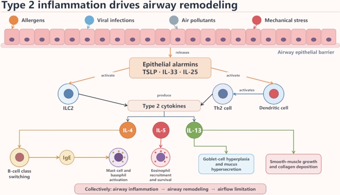
Figure 1. Cellular and cytokine networks driving type 2 airway inflammation and remodeling in severe asthma. Environmental triggers, including allergens, viral infections, air pollutants, and mechanical stress, stimulate the airway epithelial barrier and promote the release of epithelial alarmins, including thymic stromal lymphopoietin (TSLP), interleukin-33 (IL-33), and IL-25. These alarmins activate type 2 innate lymphoid cells (ILC2s) and, directly or through dendritic-cell activation, T helper 2 (Th2) cells, promoting the production of the type 2 cytokines IL-4, IL-5, and IL-13. IL-4 promotes B-cell class switching and immunoglobulin E (IgE)-mediated mast-cell and basophil activation; IL-5 promotes eosinophil recruitment and survival; and IL-13 contributes to goblet-cell hyperplasia, mucus hypersecretion, smooth-muscle growth, and collagen deposition. Together, these processes promote airway inflammation, airway remodeling, and airflow limitation. Abbreviations: IgE, immunoglobulin E; IL, interleukin; ILC2, type 2 innate lymphoid cell; Th2, T helper 2 cell; TSLP, thymic stromal lymphopoietin. Please click here to view a larger version of this figure.
Biomarkers for Precision Treatment
In recent years, biomarker-guided treatment has become a central component of precision management in SA. Several biomarkers are already incorporated into routine clinical practice, while a growing number of candidate markers are under investigation for more refined disease classification and therapeutic decision-making.
Established Biomarkers:
Among the biomarkers currently used in SA, blood eosinophil count, FeNO, IgE, and sputum eosinophils are the most commonly applied21. Blood eosinophil count is widely used in clinical settings because it is simple to obtain and easy to interpret21. Higher peripheral eosinophil levels generally reflect ongoing T2 inflammation and have been associated with exacerbation risk, disease severity, and eosinophilic airway activity34,35. Accumulating clinical evidence further suggests that patients with elevated blood eosinophils are more likely to benefit from biologics directed against IL-5 or IL-5Rα36. In practice, blood eosinophil counts of ≥150 cells/µL and ≥300 cells/µL are commonly used as eligibility or response-enrichment thresholds, with higher counts generally associated with a greater likelihood of benefit from eosinophil-targeted therapy4,36. However, these thresholds are not interchangeable and vary across clinical trials, guidelines, payer criteria, and individual biologic labels. Nevertheless, this marker is not entirely stable, as recent infection, systemic corticosteroid exposure, and diurnal variation may all affect the measured value37.
FeNO is another commonly used marker and provides a noninvasive measure of airway T2 inflammation38. Its elevation is largely related to IL-4- and IL-13-mediated induction of nitric oxide production in airway epithelial cells39. In clinical practice, increased FeNO reflects active type 2 airway inflammation, particularly IL-4/IL-13 pathway activity, rather than directly measuring eosinophilic inflammation, and it has therefore been used to support prediction of exacerbation risk and responsiveness to therapies targeting IL-4Rα40,41. FeNO values of approximately ≥20–25 parts per billion (ppb) are commonly used to support the presence of type 2 inflammation or to enrich for treatment response; however, the applicable cutoff varies across guidelines, clinical studies, and individual biologic labels. Nevertheless, FeNO levels can also be modified by smoking status, upper airway comorbidities such as rhinitis or sinusitis, and adherence to ICS42. For this reason, FeNO is generally more informative when interpreted together with other biomarkers rather than in isolation.
IgE is closely involved in allergic airway inflammation. By interacting with high-affinity receptors on mast cells and basophils, it promotes the release of inflammatory mediators and contributes to downstream airway inflammatory responses43. However, total IgE alone should not be regarded as a treatment-selection biomarker. When considering anti-IgE therapy, total IgE should be interpreted together with a clinically relevant allergic history and evidence of sensitization demonstrated by allergen-specific IgE testing and/or skin-prick testing. For omalizumab, total IgE and body weight are used primarily to determine treatment eligibility and dosing according to the applicable local product label, rather than to predict the magnitude of treatment response13. Sputum eosinophil count, in contrast, offers a more direct assessment of eosinophilic inflammation within the airways44. Although blood eosinophil counts and sputum eosinophil percentages are often correlated at the population level, they are not interchangeable at the individual-patient level. Discordant patterns may occur, including elevated blood eosinophil counts with low sputum eosinophil percentages or, conversely, low blood eosinophil counts with elevated sputum eosinophil percentages. An increased proportion of sputum eosinophils has been linked to a greater likelihood of asthma exacerbation and may also assist in treatment adjustment, including ICS titration and evaluation of biologic response45. Despite this advantage, sputum induction is technically demanding, not all patients can produce an adequate sample, and sample processing requires trained personnel, standardized procedures, and specialized laboratory facilities. These technical and logistical requirements limit the feasibility of sputum eosinophil assessment in routine clinical practice.
Emerging Biomarkers:
Advances in molecular biology and omics technologies have expanded the range of candidate biomarkers for SA and may improve the precision of inflammatory phenotyping. These candidates can be considered along a continuum of clinical maturity. Periostin and selected T2 gene-expression signatures have accumulated some translational evidence but have not been widely incorporated into routine practice, whereas direct measurements of epithelial alarmins, microRNAs (miRNAs), and multi-omics-derived signatures remain primarily investigational. Periostin is one of the most frequently studied emerging markers. This extracellular matrix protein is produced by airway epithelial cells and fibroblasts, and its expression is influenced by IL-4 and IL-13 signaling46. Elevated serum periostin has been associated with eosinophilic inflammation and airway remodeling, suggesting potential value in identifying patients with active T2 disease and in estimating response to therapies related to the IL-13 axis47. Even so, its broader clinical use remains limited by factors such as age-related variation and interference from bone metabolism, highlighting the need for further standardization.
Other upstream mediators, particularly epithelial-derived cytokines such as TSLP, IL-33, and IL-25, have also attracted considerable interest48. Although these alarmins are biologically relevant therapeutic targets, their direct measurement as clinical biomarkers remains investigational because standardized assays and validated interpretive thresholds are lacking. Because these molecules participate in the early phase of airway inflammation by activating ILC2s and Th2 cells, they may serve not only as indicators of disease activity but also as biologically meaningful therapeutic targets.
Gene-expression profiling has also provided new insights into asthma heterogeneity. Transcriptomic analyses of airway epithelial samples and peripheral blood have identified increased expression of genes such as POSTN, CLCA1, and SERPINB2 in association with eosinophilic inflammation and T2 immune activation49. These gene-expression signatures have shown translational potential for molecular phenotyping but are not yet sufficiently standardized for routine clinical use. In addition, composite T2 gene-expression signatures may allow more precise discrimination of inflammatory endotypes and may improve estimation of treatment response, especially for therapies directed at the IL-4/IL-13 pathway50,51,52.
miRNAs have emerged as another promising class of biomarkers because of their regulatory role in immune and inflammatory pathways, although their application currently remains primarily investigational. Molecules such as miR-21, miR-155, miR-146a, and miR-223 have been implicated in T2 inflammation and immune-cell function in SA53. For example, miR-21 appears to favor Th2 polarization and strengthen T2-associated inflammation, whereas miR-155 participates more broadly in immune activation and inflammatory signal regulation54,55. However, variability across specimen types and the lack of standardized analytical methods still hinder the clinical translation of miRNA-based biomarkers.
The expansion of high-throughput sequencing has further accelerated multi-omics research in asthma. Genomic studies have identified susceptibility-related variants in genes such as IL33, IL1RL1, TSLP, and ORMDL3, all of which are linked to the regulation of airway inflammation56,57. At the same time, transcriptomic and proteomic investigations have revealed broader molecular networks associated with inflammatory activity and disease heterogeneity52. Ultimately, integrating genomic, transcriptomic, proteomic, and metabolomic information may enable the development of composite biomarker panels with greater robustness than any single marker alone. Such integrated models could support a more comprehensive characterization of SA and facilitate more accurate patient stratification in precision treatment. At present, however, multi-omics-derived biomarker panels remain research tools and require prospective clinical validation, analytical standardization, and assessment of real-world feasibility before routine implementation.
Clinical Roles and Interpretation of Biomarkers:
From a clinical perspective, biomarkers in SA serve three complementary but distinct purposes. First, phenotyping biomarkers identify underlying inflammatory patterns. Blood and sputum eosinophils, FeNO, and total IgE, interpreted together with evidence of allergic sensitization, can help characterize eosinophilic, type 2, and allergic phenotypes. Second, predictive biomarkers estimate the likelihood of response to a particular biologic. For example, higher blood eosinophil counts are associated with greater expected benefit from anti-IL-5 or anti-IL-5Rα therapy, whereas elevated FeNO may predict a greater response to IL-4Rα blockade. Third, monitoring biomarkers are assessed longitudinally during follow-up. Serial blood eosinophil counts and FeNO measurements may provide adjunctive information regarding inflammatory activity but should be interpreted alongside exacerbation frequency, symptom control, lung function, and OCS use. These functions may overlap, and biomarker values can be affected by corticosteroid exposure, respiratory infection, smoking, comorbidities, and treatment adherence. Therefore, no individual biomarker should be interpreted in isolation21,36,40,41. Table 1 summarizes the specimen or detection methods, clinical relevance, and current clinical implementation status of established and emerging biomarkers in SA, thereby providing a practical framework for biomarker-informed clinical assessment and treatment selection.
| Biomarker | Specimen / detection method | Clinical relevance in severe asthma | Current clinical implementation status | |
| Blood eosinophils | Peripheral blood count | Marker of T2-high/eosinophilic inflammation; useful for exacerbation-risk estimation, patient stratification, and selection of eosinophil-targeted biologics. | Routine clinical practice. | |
| FeNO | Exhaled nitric oxide measurement using a FeNO analyzer | Noninvasive marker of IL-4/IL-13-driven airway inflammation; supports identification of T2-high disease and may assist in treatment monitoring and response assessment. | Routine clinical practice, subject to local availability. | |
| Total IgE | Serum immunoassay | Used in the evaluation of allergic phenotype when interpreted together with evidence of sensitization; helps inform eligibility assessment and biologic choice. | Routine clinical practice when interpreted together with allergic sensitization. | |
| Sputum eosinophils | Induced sputum cytology | Provides a direct measure of eosinophilic airway inflammation; useful for phenotype confirmation and longitudinal assessment of inflammatory control. | Limited clinical implementation; mainly available in specialized centers. | |
| Periostin | Serum enzyme-linked immunosorbent assay (ELISA); currently limited in routine practice | Candidate marker of IL-13-associated T2 activity and airway remodeling; mainly applied in research settings. | Investigational; not routinely used. | |
| Epithelial-derived alarmins (TSLP/IL-33/IL-25) | Research-based assays using airway samples, serum, or sputum | Reflect upstream epithelial immune activation and may support inflammatory endotyping; currently used mainly for research. | Research or exploratory use. | |
| T2 gene expression signatures (e.g., POSTN, CLCA1, SERPINB2) | Transcriptomic profiling, including RNA sequencing (RNA-seq) or microarray | May support molecular classification of T2-high disease and provide adjunctive information for patient stratification in research settings. | Research or exploratory use. | |
| MicroRNAs (e.g., miR-21, miR-155) | Quantitative polymerase chain reaction (qPCR) or sequencing; mainly research use | Associated with immune regulation and may have future value in diagnosis, phenotyping, and prediction of treatment response. | Research or exploratory use. | |
Table 1: Established and emerging biomarkers for severe asthma: detection methods, clinical relevance, and current clinical implementation status. This table summarizes the specimen or detection method, clinical relevance, and current clinical implementation status of established and emerging biomarkers for severe asthma. These biomarkers may support inflammatory phenotyping, biologic selection, exacerbation-risk assessment, and treatment monitoring. Most emerging biomarkers remain investigational and should be interpreted alongside clinical features and treatment history. Abbreviations: ELISA, enzyme-linked immunosorbent assay; FeNO, fractional exhaled nitric oxide; IgE, immunoglobulin E; IL, interleukin; miRNA, microRNA; qPCR, quantitative polymerase chain reaction; RNA-seq, RNA sequencing; T2, type 2; TSLP, thymic stromal lymphopoietin.
Approved Biologic Therapies and Their Mechanisms of Action
Compared with conventional anti-inflammatory therapies, biologics act more selectively by interrupting specific cytokines or immune pathways that drive airway inflammation. Current biologic strategies in SA mainly focus on IgE, the IL-5/IL-5 receptor axis, the IL-4/IL-13 pathway, and epithelial-derived upstream mediators. By targeting different components of the inflammatory cascade, these agents provide more individualized treatment options for patients with distinct inflammatory profiles.
Anti-IgE Therapy:
Omalizumab was the first biologic approved for asthma and is primarily used in patients with moderate-to-severe allergic asthma58. IgE is central to allergic airway inflammation because it binds to the high-affinity IgE receptor (FcεRI) expressed on mast cells and basophils, thereby promoting mediator release and downstream bronchoconstrictive and inflammatory responses. Omalizumab is a recombinant humanized monoclonal antibody that binds circulating free IgE and limits its interaction with FcεRI, leading to reduced activation of effector cells and attenuation of allergic inflammation59. Clinically, omalizumab is most suitable for patients with total IgE levels within the applicable treatment and dosing range, together with evidence of allergic sensitization, and it has been shown to lower exacerbation frequency while improving lung function and health-related quality of life60.
Anti-IL-5/IL-5R Pathway Therapy:
The IL-5 pathway is a central regulator of eosinophilic inflammation, as IL-5 supports eosinophil maturation, mobilization, recruitment, and persistence. This makes the IL-5 axis an important therapeutic target in eosinophilic SA. Mepolizumab, reslizumab, and benralizumab all reduce eosinophil-driven inflammation, although they do so through different mechanisms61,62,63. Mepolizumab and reslizumab directly bind IL-5 and prevent activation of the IL-5 receptor, thereby limiting eosinophil production and activity61. Benralizumab, by contrast, binds IL-5Rα and promotes near-complete eosinophil depletion through antibody-dependent cell-mediated cytotoxicity (ADCC)62,63. These therapies reduce exacerbations and can improve symptom control and lung function; mepolizumab and benralizumab also have established OCS-sparing evidence in selected patients64. Reslizumab requires weight-based intravenous infusion every 4 weeks, which may limit convenience and use in some patients65. By contrast, the extended half-life of depemokimab permits administration every 6 months14.
Anti-IL-4Rα Pathway Therapy:
IL-4 and IL-13 are key mediators of T2 inflammation and contribute to IgE synthesis, airway inflammation, mucus production, and structural remodeling. Dupilumab targets IL-4Rα, which is shared by the IL-4 and IL-13 signaling pathways. As a result, inhibition of IL-4Rα suppresses signaling from both cytokines66. Clinical evidence indicates that dupilumab can improve asthma control rapidly and reduce exacerbation burden while enhancing lung function67. It may be particularly useful in patients with persistent T2 inflammation or T2-related comorbidities, including chronic rhinosinusitis with nasal polyps and atopic dermatitis68,69. Dupilumab is administered subcutaneously every 2 weeks; this relatively frequent schedule may reduce convenience or adherence for some patients66.
Anti-Epithelial Cytokines:
Upstream epithelial mediators have emerged as important therapeutic targets in asthma. Among these, TSLP is recognized as a major initiator of airway inflammation48,70. In response to allergens, viral infection, or environmental insults, airway epithelial cells release TSLP, which in turn activates dendritic cells, ILC2s, and Th2 cells and promotes downstream production of IL-4, IL-5, and IL-13. Tezepelumab blocks TSLP signaling by preventing its interaction with the receptor complex, thereby reducing exacerbations71,72,73. Clinical benefit can occur in patients with low conventional T2 biomarkers, but the magnitude of benefit is generally greater in patients with higher baseline blood eosinophil counts and/or FeNO74,75,76. Additional upstream targets, including the IL-33/ST2 axis and IL-25, remain under investigation48,70.
Pivotal Phase III Evidence and Clinical Applications:
A number of pivotal phase III randomized trials have established the clinical role of biologic therapy in SA. In the INNOVATE trial, omalizumab reduced the adjusted rate of clinically significant exacerbations by 26% and the rate of severe exacerbations by 50% compared with placebo77. In MENSA, subcutaneous mepolizumab reduced clinically significant exacerbations by 53% and improved prebronchodilator forced expiratory volume in 1 second (FEV1) by 98 mL versus placebo78. In SIROCCO, benralizumab administered every 8 weeks reduced the annual exacerbation rate by 51% and improved prebronchodilator FEV1 by 159 mL versus placebo in patients with blood eosinophil counts ≥300 cells/µL79. In LIBERTY ASTHMA QUEST, dupilumab reduced the annualized rate of severe exacerbations by 47.7% and increased FEV₁ by 0.14 L versus placebo at week 1280. Likewise, in NAVIGATOR, tezepelumab reduced the annualized exacerbation rate by 56% and improved prebronchodilator FEV1 by 0.13 L versus placebo at week 5281. In SWIFT-1 and SWIFT-2, twice-yearly depemokimab reduced the annualized exacerbation rate by 54% versus placebo14. Additional pivotal and extension studies—including DREAM82, SIRIUS83, COMET84, phase III reslizumab studies85, CALIMA86, ZONDA87, BORA88, VENTURE89, and DESTINATION90—provide complementary evidence on biomarker-defined eligibility, OCS-sparing effects, and longer-term safety and efficacy. Their study-specific criteria are summarized in Table 2.
| Drug | Therapeutic target / pathway | Typical candidate profile in severe asthma | Common biomarker indicators | Representative phase III and extension studies | Key clinical features / remarks | Route and usual frequency | |||
| Omalizumab | IgE | Allergic severe asthma with clinically relevant sensitization and total IgE/body weight within the applicable local dosing table | Positive skin-prick test and/or allergen-specific IgE; pretreatment total IgE and body weight | INNOVATE77: total IgE 30–700 IU/mL and body weight within the dosing table | No recommended dose may be available outside local IgE/body-weight limits | SC every 2 or 4 weeks | |||
| Mepolizumab | IL-5 | Eosinophilic severe asthma | Blood eosinophils ≥150 cells/μL at screening or ≥300 cells/μL in the previous year | DREAM82; MENSA78; SIRIUS83; MUSCA17; COMET84 | Reduces eosinophilic inflammation through IL-5 neutralization and is well established in eosinophil-driven disease | SC every 4 weeks | |||
| Reslizumab | IL-5 | Eosinophilic severe asthma in adults | Blood eosinophils ≥400 cells/μL in the pivotal exacerbation trials | Phase III reslizumab studies65˒85 | Requires IV infusion every 4 weeks, which may limit convenience and use in some patients | IV every 4 weeks | |||
| Benralizumab | IL-5Rα | Eosinophilic severe asthma | Blood eosinophils ≥300 cells/μL in the primary SIROCCO/CALIMA population | SIROCCO79; CALIMA86; ZONDA87; BORA88 | Produces profound eosinophil depletion via antibody-dependent cell-mediated cytotoxicity (ADCC) | SC every 4 weeks for 3 doses, then every 8 weeks | |||
| Depemokimab | IL-5 | Severe asthma with an eosinophilic/type 2 phenotype | Blood eosinophils ≥150 cells/μL at screening or ≥300 cells/μL in the previous year | SWIFT-1 and SWIFT-214 | Ultra-long-acting anti-IL-5 biologic with twice-yearly dosing | SC every 6 months | |||
| Dupilumab | IL-4Rα, thereby inhibiting IL-4/IL-13 signaling | T2-high or OCS-dependent severe asthma, particularly with nasal polyps or atopic dermatitis | No minimum biomarker threshold in QUEST/VENTURE; greater benefit with higher eosinophils or FeNO | LIBERTY ASTHMA QUEST80; VENTURE89 | Every-2-week SC dosing may limit convenience or adherence in some patients | SC every 2 weeks | |||
| Tezepelumab | TSLP (upstream epithelial alarmin) | Severe asthma across multiple inflammatory phenotypes | No minimum blood eosinophil or FeNO threshold; higher levels predict greater benefit | NAVIGATOR81; DESTINATION90 | Can benefit some patients with low T2 biomarkers, but efficacy is generally greater in T2-high disease | SC every 4 weeks | |||
Table 2: Approved biologics for severe asthma: therapeutic targets, candidate profiles, associated biomarkers, and pivotal phase III clinical trials. This table summarizes the major biologics approved for asthma in the United States and/or the European Union, including their therapeutic targets, candidate patient groups, associated biomarkers, representative phase III and extension studies, key clinical features, and usual routes and frequencies of administration. Biomarker findings should be integrated with the clinical phenotype when selecting treatment.
From a comparative clinical perspective, omalizumab has the longest clinical experience and is best suited to patients with a clearly defined allergic phenotype and documented allergen sensitization. Anti-IL-5 and anti-IL-5Rα agents provide the most direct eosinophil-targeted approach and may be particularly attractive for patients with recurrent eosinophilic exacerbations or OCS dependence. Dupilumab provides simultaneous inhibition of IL-4 and IL-13 signaling and may be favored when elevated FeNO, chronic rhinosinusitis with nasal polyps, or atopic dermatitis is present. Tezepelumab acts further upstream and may cover a broader range of biomarker-defined phenotypes. Considerable eligibility overlap may occur because allergic sensitization, blood eosinophilia, and elevated FeNO frequently coexist. Because direct head-to-head evidence remains limited, treatment selection should prioritize the dominant treatable traits, relevant comorbidities, dosing considerations, safety, patient preference, cost, and access77,78,79,80,81.
Biomarker-Guided Treatment Strategies
The use of biomarkers has become a key component of precision treatment in SA. By identifying underlying inflammatory patterns, biomarkers can support biologic selection, improve treatment matching, and help avoid unnecessary exposure to ineffective therapies. At the same time, biomarker results should not be interpreted in isolation. Their clinical value is greatest when considered together with disease phenotype, previous exacerbation frequency, comorbid conditions, and treatment-related factors such as ICS or OCS use. This integrated approach may facilitate recognition of treatable traits and allow more flexible, individualized disease management.
Biomarker-Based Patient Stratification:
Among the currently recognized inflammatory phenotypes, T2-high SA is the most extensively characterized. In this review, allergic asthma refers to disease with clinically relevant sensitization demonstrated by skin-prick testing and/or allergen-specific IgE. For descriptive purposes, a T2-high profile is considered present when one or more of the following are identified: blood eosinophils ≥150 cells/µL, FeNO ≥20 ppb, sputum eosinophils ≥2%, or clinically allergen-driven asthma. Eosinophilic asthma is reported using the study-specific blood eosinophil threshold, most commonly ≥150, ≥300, or ≥400 cells/µL; these thresholds are not interchangeable with local regulatory or payer eligibility criteria4,21. Patients with T2-low asthma lack these typical biomarker features, although values can fluctuate and may be suppressed by corticosteroid exposure. Because no universally standardized biomarker framework has been established for T2-low disease, management still depends on repeated biomarker assessment, modifiable clinical factors, and treatable traits24.
Selection Among Biologics:
For patients with allergic asthma who have total serum IgE levels within the applicable treatment and dosing range, together with evidence of allergic sensitization, omalizumab remains a well-established treatment option9. Its long-term efficacy and safety have also been supported in pediatric asthma91. In eosinophilic disease, IL-5-pathway biologics—including mepolizumab78, reslizumab65, depemokimab14, and benralizumab92—are appropriate options. Benralizumab may be particularly useful in patients with marked eosinophilia and recurrent exacerbations because it induces eosinophil depletion through ADCC92,93. Dupilumab may be favored when FeNO is elevated or when T2-related comorbidities, such as chronic rhinosinusitis with nasal polyps or atopic dermatitis, are present94. Tezepelumab offers a broader upstream strategy and may benefit some patients with lower conventional T2 biomarker expression74,75,76.
Practical Clinical Decision Framework:
The following framework is intended as a practical guide rather than a rigid treatment hierarchy, and local regulatory, reimbursement, and product-label criteria should always be considered. After confirming SA and optimizing adherence, inhaler technique, and comorbidity management: (1) anti-IgE therapy may be prioritized for a clinically relevant allergic phenotype with documented sensitization and total IgE/body weight within the applicable dosing table9,13; (2) anti-IL-5 or anti-IL-5Rα therapy may be prioritized when eosinophilic inflammation is dominant, particularly with blood eosinophils ≥150 or ≥300 cells/µL, recurrent exacerbations, or OCS dependence65,78,92; (3) anti-IL-4Rα therapy may be favored with elevated FeNO and/or blood eosinophils, especially when chronic rhinosinusitis with nasal polyps or atopic dermatitis is present94; and (4) anti-TSLP therapy may be considered when conventional T2 biomarkers are low or discordant, although responses are generally greater with higher baseline eosinophils and/or FeNO76. When more than one biologic is suitable, comorbidities, previous response, dosing route and frequency, safety, patient preference, cost, and access should guide the final choice.
Combined Biomarkers and Individualized Therapy:
Increasing evidence suggests that treatment selection in SA may be improved by combining multiple biomarkers rather than relying on a single indicator95. A multidimensional assessment incorporating blood eosinophils, FeNO, allergic sensitization status, exacerbation history, and maintenance OCS exposure may provide a more comprehensive picture of inflammatory activity and improve treatment stratification95. Higher baseline blood eosinophil counts are generally associated with greater benefit from anti-IL-5 or anti-IL-5Rα therapy93,96, whereas elevated FeNO predicts a greater response to IL-4Rα blockade, partly independently of blood eosinophil levels97. In patients with recurrent exacerbations but low or atypical conventional T2 biomarkers, tezepelumab may provide clinical benefit; however, this should not be interpreted as uniform efficacy across all patients with T2-low asthma76. Emerging candidates such as periostin, genetic variants, and multi-omics-derived features require further prospective validation before routine clinical implementation98.
결론
생물학적 제제가 중증 천식(SA)의 결과를 개선시켰음에도 불구하고, 완전히 효과적인 정밀 치료를 가로막는 여러 장벽이 여전히 존재합니다. 현재의 바이오마커, 특히 혈중 호산구, FeNO 및 총 IgE는 예측 가치가 제한적이고 임상적 및 치료 관련 영향에 따라 수치가 변하기 때문에 여전히 불완전합니다98. T2-low 천식은 효과적인 표적 치료제가 부족하여 주요한 미충족 수요로 남아 있습니다99,100,101. 그럼에도 불구하고, 일반적인 T2 바이오마커가 상승하지 않은 경우에도 tezepelumab을 고려할 수 있으나, 일반적으로 기저 호산구 및/또는 FeNO 수치가 높을 때 그 이점이 더 큽니다76,102. 또한 교정 QT 연장, 비결핵 항산균 감염 및 항생제 내성을 고려한다면, 비호산구성 질환을 포함하여 지속적으로 조절되지 않는 천식을 앓는 일부 성인에게 저용량 azithromycin 추가 투여가 인정되는 옵션입니다102. 생물학적 이질성과 신뢰할 수 있는 층화 마커의 부재는 T2-low 관리를 더욱 복잡하게 만듭니다99,100,101. 생물학적 제제의 비용과 긴 치료 기간으로 인해 접근성은 보험 급여 및 경제적 부담 능력에 따라 달라집니다103,104. 또한 반응, 초반응(super-response), 관해 및 무반응에 대한 정의가 일관되지 않아 치료의 지속, 변경 및 중단 결정이 어렵습니다. 일부 환자들은 겉보기에 적절한 생물학적 제제를 선택했음에도 불구하고 적절하게 반응하지 않으며, 이러한 불완전한 반응의 기전은 여전히 충분히 이해되지 않은 상태입니다105,106.
중증 천식(SA)의 향후 발전은 새로운 표적, 치료 전략 및 기술의 진보에 달려 있을 가능성이 높습니다. IL-33/ST2, IL-25 및 차세대 TSLP 표적 기전과 같은 상위 경로에 대한 활발한 연구가 진행 중이며, astegolimab은 천식 악화 감소에 초기 가능성을 보여주었습니다107. 생물학적 제제의 전환 및 신중하게 선택된 치료 강도 완화가 점점 더 많은 관심을 받고 있는 반면, 병용 생물학적 치료는 여전히 연구 단계에 있습니다102,108. 이러한 접근 방식은 더 많은 생물학적 제제가 실제 임상에 도입됨에 따라 더욱 중요해질 수 있습니다. 또한 신흥 기술들이 바이오마커 혁신을 가속화할 것으로 기대됩니다. 멀티오믹스 접근법은 천식의 이질성을 더욱 포괄적으로 특성화하고 새로운 질환 아형 및 치료 표적의 식별을 지원할 수 있습니다109. 다음의 중요한 단계는 이러한 발견을 견고하고 해석 가능하며 임상적으로 검증된 복합 바이오마커 패널로 전환하는 것이 될 것입니다. 이와 병행하여, 머신러닝 모델은 천식 엔도타입 분류와 결과 예측을 개선함으로써 임상 진료를 보완할 수 있을 것입니다110.
종합적으로, 바이오마커는 이미 중증 천식(SA)에서 표현형 평가와 생물학적 제제 선택을 지원하고 있으나, 단일 마커만으로는 임상 현장에서 나타나는 중첩되고 이질적이며 역동적인 염증 패턴을 충분히 포착할 수 없습니다. 따라서 향후의 정밀 의료는 혈액 및 기도 바이오마커를 임상 표현형, 동반 질환, 악화 이력, 치료 부담 및 종단적 반응과 통합하는 검증된 다차원적 프레임워크를 우선시해야 합니다. 바이오마커 수치는 질환 활성도와 코르티코스테로이드 노출에 따라 변동될 수 있으므로 반복적인 평가가 필수적입니다. 향후 전향적 연구를 통해 T2-high 및 T2-low 질환의 정의를 정교화하고, 복합 예측 모델을 검증하며, 새로운 바이오마커와 디지털 도구가 생물학적 제제의 선택, 교체 및 치료 단계 축소(de-escalation)를 어떻게 안내할 수 있을지 결정해야 합니다.
연구 대상 군 및 표현형 기준: INNOVATE 연구에는 중증 알레르기성 천식을 앓고 있으며, 적어도 하나 이상의 다년성 공기 알레르겐에 감작되었고, 치료 전 총 IgE 수치가 30–700 IU/mL인 환자들이 등록되었습니다. DREAM 연구에서는 다음 중 하나 이상의 기준을 충족하여 호산구성 염증의 증거가 확인된 중증 호산구성 천식 환자를 대상으로 하였습니다: 객담 호산구 ≥3%, FeNO ≥50 ppb, 혈중 호산구 ≥300 cells/µL, 또는 코르티코스테로이드 감량 후의 즉각적인 상태 악화. MENSA 및 MUSCA 연구는 스크리닝 시 혈중 호산구 ≥150 cells/µL 또는 지난 1년 동안 ≥300 cells/µL인 환자를 대상으로 하였으며, SIRIUS 연구는 유지 OCS 치료가 필요한 환자에게 동일한 호산구 기준을 적용하였습니다. COMET 연구에는 mepolizumab을 ≥3년 동안 지속적으로 투여받은 환자들이 등록되었습니다. reslizumab의 중추적 연구들은 혈중 호산구 ≥400 cells/µL를 기준으로 하였습니다. SIROCCO 및 CALIMA 연구의 일차 호산구 대상 군은 혈중 호산구 ≥300 cells/µL였으며, ZONDA 연구는 혈중 호산구 ≥150 cells/µL인 OCS 의존성 환자를 등록하였고, BORA 연구는 SIROCCO 또는 CALIMA 연구를 완료한 환자들을 등록하였습니다. QUEST 및 VENTURE 연구는 최소 T2 바이오마커 요구 사항이 없었으나, 혈중 호산구 >1,500 cells/µL인 환자는 제외되었습니다. 특히 VENTURE 연구는 OCS 의존성 천식 환자들을 대상으로 하였습니다. NAVIGATOR 연구는 지난 1년 동안 ≥2회의 악화가 있었던 중증 비조절 천식 환자를 대상으로 하였으나 최소 바이오마커 임계값은 설정하지 않았으며, DESTINATION 연구는 NAVIGATOR 또는 SOURCE 연구에서 이어진 환자들을 등록하였습니다. SWIFT-1 및 SWIFT-2 연구는 스크리닝 시 혈중 호산구 수치 ≥150 cells/µL 또는 지난 1년 동안 ≥300 cells/µL이며, 중등도 또는 고용량 흡입 코르티코스테로이드 치료에도 불구하고 악화 이력이 있는 12세 이상의 환자들을 등록하였습니다. 이러한 기준은 각 연구별로 설정된 것이며, 보편적인 표현형 정의나 현재의 규제적 적격성 기준으로 해석되어서는 안 됩니다. 약어: ADCC, 항체 의존성 세포 매개 세포독성; FeNO, 호기 산화질소 분율; IgE, 면역글로불린 E; IL, 인터루킨; IL-4Rα, 인터루킨-4 수용체 알파; IL-5Rα, 인터루킨-5 수용체 알파; IU/mL, 밀리리터당 국제 단위; IV, 정맥 주사; OCS, 경구 코르티코스테로이드; ppb, 10억 분의 일; SC, 피하 주사; T2, 제2형; TSLP, 흉선 기질 림프구 생성물.
공개 사항
저자들은 이해상충이 없음을 밝힙니다. 본 원고의 작성 및 수정 과정에서 ChatGPT(OpenAI)는 문법, 명확성 및 스타일 개선을 포함한 영어 언어 교정 지원 목적으로만 사용되었습니다. AI의 지원을 받은 모든 텍스트는 저자들에 의해 비판적으로 검토 및 수정되었으며, 저자들은 원고의 정확성, 무결성 및 최종 내용에 대해 모든 책임을 집니다.
감사의 글
저자들은 이 원고를 준비하는 동안 가치 있는 제안과 의견을 제공해주신 모든 동료와 협력자들에게 감사드립니다. 본 연구의 수행, 저술 및 출판을 위해 지원받은 연구비는 없습니다.
참고문헌
- Papi A, Brightling C, Pedersen SE, Reddel HK. Asthma. Lancet. 2018;391(10122):783-800.
- Wang Z, et al. Global, regional, and national burden of asthma and its attributable risk factors from 1990 to 2019: a systematic analysis for the Global Burden of Disease Study 2019. Respir Res. 2023;24(1):169.
- Global Asthma Network Phase I Study Group. Worldwide trends in the burden of asthma symptoms in school-aged children: Global Asthma Network Phase I cross-sectional study. Lancet. 2021;398(10311):1569-1580.
- Global Initiative for Asthma. Global strategy for asthma management and prevention. 2026. Available at: https://ginasthma.org/2026-gina-strategy-report/. Accessed August 16, 2026.
- Reibman J, Tan L, Ambrose C, Tkacz J. Clinical and economic burden of severe asthma among US patients treated with biologic therapies. Ann Allergy Asthma Immunol. 2021;127(3):318-325.e2.
- Larenas-Linnemann D, et al. International Severe Asthma Registry (ISAR): 2017–2024 status and progress update. Tuberc Respir Dis (Seoul). 2025;88(2):193-215.
- Wenzel SE. Asthma phenotypes: the evolution from clinical to molecular approaches. Nat Med. 2012;18(5):716-725.
- Schleich F, et al. Biomarker profile and disease burden associated with intermittent and long-term oral corticosteroid use in patients with severe asthma prior to biologic initiation in real-life (STAR). World Allergy Organ J. 2025;18(7):101066.
- Cheng SL. Immunologic pathophysiology and airway remodeling mechanism in severe asthma: focused on IgE-mediated pathways. Diagnostics (Basel). 2021;11(1):83.
- García G, et al. Severe asthma: Adding new evidence - latin american thoracic society. ERJ Open Res. 2021;7(1):00318-2020.
- Brusselle GG, Koppelman GH. Biologic therapies for severe asthma. N Engl J Med. 2022;386(2):157-171.
- Pelaia C, et al. Biologics in severe asthma. Minerva Med. 2022;113(1):51-62.
- Genentech, Inc. Xolair (omalizumab) prescribing information. 2024. Available at: https://www.gene.com/download/pdf/xolair_prescribing.pdf. Accessed August 17, 2026.
- Jackson DJ, et al. Twice-yearly depemokimab in severe asthma with an eosinophilic phenotype. N Engl J Med. 2024;391(24):2337-2349.
- Cabrejos S, et al. FENOMA study: Achieving full control in patients with severe allergic asthma. J Asthma Allergy. 2020;13:159-166.
- Pelaia C, et al. Omalizumab lowers asthma exacerbations, oral corticosteroid intake and blood eosinophils: Results of a 5-year single-centre observational study. Pulm Pharmacol Ther. 2019;54:25-30.
- Chupp GL, et al. Efficacy of mepolizumab add-on therapy on health-related quality of life and markers of asthma control in severe eosinophilic asthma (MUSCA): A randomised, double-blind, placebo-controlled, parallel-group, multicentre, phase 3b trial. Lancet Respir Med. 2017;5(5):390-400.
- Varricchi G, et al. Reslizumab and eosinophilic asthma: One step closer to precision medicine? Front Immunol. 2017;8:242.
- Pelaia C, et al. Benralizumab in the treatment of severe asthma: Design, development and potential place in therapy. Drug Des Devel Ther. 2018;12:619-628.
- Corren J, et al. Tezepelumab in adults with uncontrolled asthma. N Engl J Med. 2017;377(10):936-946.
- Lee Y, Quoc QL, Park HS. Biomarkers for severe asthma: Lessons from longitudinal cohort studies. Allergy Asthma Immunol Res. 2021;13(3):375-389.
- Lambrecht BN, Hammad H, Fahy JV. The cytokines of asthma. Immunity. 2019;50(4):975-991.
- Fahy JV. Type 2 inflammation in asthma—present in most, absent in many. Nat Rev Immunol. 2015;15(1):57-65.
- Quoc QL, Choi Y, Hur GY, Park HS. New targets for type 2-low asthma. Korean J Intern Med. 2024;39(2):215-227.
- Hammad H, Lambrecht BN. The basic immunology of asthma. Cell. 2021;184(6):1469-1485.
- Holgate ST. Innate and adaptive immune responses in asthma. Nat Med. 2012;18(5):673-683.
- Fajt ML, et al. Prostaglandin D2 pathway upregulation: relation to asthma severity, control, and TH2 inflammation. J Allergy Clin Immunol. 2013;131(6):1504-1512.e12.
- Lambrecht BN, Hammad H. The immunology of asthma. Nat Immunol. 2015;16(1):45-56.
- Paul WE, Zhu J. How are T helper 2-type immune responses initiated and amplified? Nat Rev Immunol. 2010;10(4):225-235.
- Rothenberg ME, Hogan SP. The eosinophil. Annu Rev Immunol. 2006;24:147-174.
- Raundhal M, et al. High IFN-γ and low SLPI mark severe asthma in mice and humans. J Clin Invest. 2015;125(8):3037-3050.
- Wills-Karp M, et al. Interleukin-13: central mediator of allergic asthma. Science. 1998;282(5397):2258-2261.
- Roan F, Obata-Ninomiya K, Ziegler SF. Epithelial cell-derived cytokines: more than just signaling the alarm. J Clin Invest. 2019;129(4):1441-1451.
- Koefoed HJL, Gehring U, Vonk JM, Koppelman GH. Blood eosinophils associate with reduced lung function growth in adolescent asthmatics. Clin Exp Allergy. 2021;51(4):556-563.
- Price DB, et al. Blood eosinophil count and prospective annual asthma disease burden: a UK cohort study. Lancet Respir Med. 2015;3(11):849-858.
- Dhariwal J, et al. Real-world effectiveness of anti-IL-5/IL-5R therapy in severe atopic eosinophilic asthma with fungal sensitization. J Allergy Clin Immunol Pract. 2021;9(6):2315-2320.e2311.
- Spector SL, Tan RA. Is a single blood eosinophil count a reliable marker for eosinophilic asthma? J Asthma. 2012;49(8):807-810.
- Ulrik CS, Lange P, Hilberg O. Fractional exhaled nitric oxide as a determinant for the clinical course of asthma: A systematic review. Eur Clin Respir J. 2021;8(1):1891725.
- Maniscalco M, et al. Fractional exhaled nitric oxide in monitoring biological treatment for severe asthma in adults: Clinical implications and future perspectives. Respir Med. 2026;252:108647.
- Dweik RA, et al. An official ATS clinical practice guideline: interpretation of exhaled nitric oxide levels (FeNO) for clinical applications. Am J Respir Crit Care Med. 2011;184(5):602-615.
- Pianigiani T, et al. Exploring the interaction between fractional exhaled nitric oxide and biologic treatment in severe asthma: a systematic review. Antioxidants (Basel). 2023;12(2):400.
- Taylor DR, Pijnenburg MW, Smith AD, De Jongste JC. Exhaled nitric oxide measurements: clinical application and interpretation. Thorax. 2006;61(9):817-827.
- Galli SJ, Tsai M. IgE and mast cells in allergic disease. Nat Med. 2012;18(5):693-704.
- Hastie AT, et al. Biomarker surrogates do not accurately predict sputum eosinophil and neutrophil percentages in asthmatic subjects. J Allergy Clin Immunol. 2013;132(1):72-80.e12.
- Green RH, et al. Asthma exacerbations and sputum eosinophil counts: a randomised controlled trial. Lancet. 2002;360(9347):1715-1721.
- Licari A, et al. Measuring inflammation in paediatric severe asthma: Biomarkers in clinical practice. Breathe (Sheff). 2020;16(1):190301.
- Jia G, et al. Periostin is a systemic biomarker of eosinophilic airway inflammation in asthmatic patients. J Allergy Clin Immunol. 2012;130(3):647-654.e10.
- Pelaia C, et al. Monoclonal antibodies targeting alarmins: A new perspective for biological therapies of severe asthma. Biomedicines. 2021;9(9):1108.
- Woodruff PG, et al. T-helper type 2-driven inflammation defines major subphenotypes of asthma. Am J Respir Crit Care Med. 2009;180(5):388-395.
- Diver S, et al. FeNO differentiates epithelial gene expression clusters: Exploratory analysis from the MESOS randomized controlled trial. J Allergy Clin Immunol. 2022;150(4):830-840.
- Badi YE, et al. IL1RAP expression and the enrichment of IL-33 activation signatures in severe neutrophilic asthma. Allergy. 2023;78(1):156-167.
- Agache I, et al. Multidimensional endotyping using nasal proteomics predicts molecular phenotypes in the asthmatic airways. J Allergy Clin Immunol. 2023;151(1):128-137.
- Shaik NA, et al. Identification of miRNA-mRNA-TFs regulatory network and crucial pathways involved in asthma through advanced systems biology approaches. PLoS One. 2022;17(10):e0271262.
- Mao Z, et al. miR-21-5p modulates airway inflammation and epithelial-mesenchymal transition processes in a mouse model of combined allergic rhinitis and asthma syndrome. Int Arch Allergy Immunol. 2024;185(8):775-785.
- Pua HH, Ansel KM. MicroRNA regulation of allergic inflammation and asthma. Curr Opin Immunol. 2015;36:101-108.
- Moffatt MF, et al. A large-scale, consortium-based genomewide association study of asthma. N Engl J Med. 2010;363(13):1211-1221.
- Koh KD, et al. Genomic characterization and therapeutic utilization of IL-13-responsive sequences in asthma. Cell Genom. 2023;3(1):100229.
- Pelaia C, et al. Omalizumab, the first available antibody for biological treatment of severe asthma: more than a decade of real-life effectiveness. Ther Adv Respir Dis. 2018;12:1753466618810192.
- Chung KF, et al. Characteristics, phenotypes, mechanisms and management of severe asthma. Chin Med J (Engl). 2022;135(10):1141-1155.
- Huang WC, et al. The long-term effectiveness of omalizumab in adult patients with severe allergic asthma: Continuous treatment versus boosting treatment. J Clin Med. 2021;10(4):707.
- Principe S, et al. Treating severe asthma: targeting the IL-5 pathway. Clin Exp Allergy. 2021;51(8):992-1005.
- Menzella F, Lusuardi M, Galeone C, Zucchi L. Anti-IL-5 therapies for severe eosinophilic asthma: literature review and practical insights. J Asthma Allergy. 2020;13:301-313.
- Numata T, et al. Long-term efficacy and clinical remission after benralizumab treatment in patients with severe eosinophilic asthma: A retrospective study. J Asthma Allergy. 2022;15:1731-1741.
- Maglio A, et al. Real-life effectiveness of mepolizumab on forced expiratory flow between 25% and 75% of forced vital capacity in patients with severe eosinophilic asthma. Biomedicines. 2021;9(11):1550.
- Castro M, et al. Reslizumab for inadequately controlled asthma with elevated blood eosinophil counts: Results from two multicentre, parallel, double-blind, randomised, placebo-controlled, phase 3 trials. Lancet Respir Med. 2015;3(5):355-366.
- Ricciardolo FLM, Bertolini F, Carriero V. The role of dupilumab in severe asthma. Biomedicines. 2021;9(9):1096.
- Renner A, et al. Dupilumab rapidly improves asthma control in predominantly anti-IL-5/IL-5R pretreated Austrian real-life severe asthmatics. Immun Inflamm Dis. 2021;9(3):624-627.
- Canonica GW, et al. Defining type 2 asthma and patients eligible for dupilumab in Italy: A biomarker-based analysis. Clin Mol Allergy. 2021;19(1):5.
- Campisi R, et al. Real-world experience with dupilumab in severe asthma: One-year data from an Italian named patient program. J Asthma Allergy. 2021;14:575-583.
- Akenroye A, Boyce JA, Kita H. Targeting alarmins in asthma: from bench to clinic. J Allergy Clin Immunol. 2025;155(4):1133-1148.
- Dorey-Stein ZL, Shenoy KV. Tezepelumab as an emerging therapeutic option for the treatment of severe asthma: Evidence to date. Drug Des Devel Ther. 2021;15:331-338.
- Matera MG, Ora J, Rogliani P, Cazzola M. An overview of the preclinical discovery and development of tezepelumab for the treatment of asthma. Expert Opin Drug Discov. 2023;18(9):951-963.
- Roy P, et al. The impact of tezepelumab in uncontrolled severe asthma: A systematic review of randomized controlled trials. Cureus. 2022;14(12):e32156.
- Menzella F, et al. Tezepelumab: Patient selection and place in therapy in severe asthma. J Int Med Res. 2024;52(4):3000605241246740.
- Venegas Garrido C, Nair P, Dávila I, Pérez De Llano L. Role of thymic stromal lymphopoietin in the pathophysiology of asthma and clinical and biological effects of blockade with tezepelumab. J Investig Allergol Clin Immunol. 2024;34(5):293-302.
- Corren J, et al. Efficacy of tezepelumab in severe, uncontrolled asthma: pooled analysis of the PATHWAY and NAVIGATOR clinical trials. Am J Respir Crit Care Med. 2023;208(1):13-24.
- Humbert M, et al. Benefits of omalizumab as add-on therapy in patients with severe persistent asthma who are inadequately controlled despite best available therapy (GINA 2002 step 4 treatment): INNOVATE. Allergy. 2005;60(3):309-316.
- Ortega HG, et al. Mepolizumab treatment in patients with severe eosinophilic asthma. N Engl J Med. 2014;371(13):1198-1207.
- Bleecker ER, et al. Efficacy and safety of benralizumab for patients with severe asthma uncontrolled with high-dosage inhaled corticosteroids and long-acting β2-agonists (SIROCCO): A randomised, multicentre, placebo-controlled phase 3 trial. Lancet. 2016;388(10056):2115-2127.
- Castro M, et al. Dupilumab efficacy and safety in moderate-to-severe uncontrolled asthma. N Engl J Med. 2018;378(26):2486-2496.
- Menzies-Gow A, et al. Tezepelumab in adults and adolescents with severe, uncontrolled asthma. N Engl J Med. 2021;384(19):1800-1809.
- Pavord ID, et al. Mepolizumab for severe eosinophilic asthma (DREAM): A multicentre, double-blind, placebo-controlled trial. Lancet. 2012;380(9842):651-659.
- Bel EH, et al. Oral glucocorticoid-sparing effect of mepolizumab in eosinophilic asthma. N Engl J Med. 2014;371(13):1189-1197.
- Moore WC, et al. Stopping versus continuing long-term mepolizumab treatment in severe eosinophilic asthma (COMET study). Eur Respir J. 2022;59(1):2100396.
- Corren J, et al. Phase 3 study of reslizumab in patients with poorly controlled asthma: Effects across a broad range of eosinophil counts. Chest. 2016;150(4):799-810.
- FitzGerald JM, et al. Benralizumab, an anti-interleukin-5 receptor α monoclonal antibody, as add-on treatment for patients with severe, uncontrolled, eosinophilic asthma (CALIMA). Lancet. 2016;388(10056):2128-2141.
- Nair P, et al. Oral glucocorticoid-sparing effect of benralizumab in severe asthma. N Engl J Med. 2017;376(25):2448-2458.
- Busse WW, et al. Long-term safety and efficacy of benralizumab in patients with severe, uncontrolled asthma: 1-year results from the BORA phase 3 extension trial. Lancet Respir Med. 2019;7(1):46-59.
- Rabe KF, et al. Efficacy and safety of dupilumab in glucocorticoid-dependent severe asthma. N Engl J Med. 2018;378(26):2475-2485.
- Menzies-Gow A, et al. Long-term safety and efficacy of tezepelumab in people with severe, uncontrolled asthma (DESTINATION): A randomised, placebo-controlled extension study. Lancet Respir Med. 2023;11(5):425-438.
- Nakamura N, et al. Real-life long-term safety and effectiveness of omalizumab in japanese pediatric patients with severe allergic asthma: A post-marketing surveillance. Allergol Int. 2021;70(3):319-326.
- Jackson DJ, et al. Safety of eosinophil-depleting therapy for severe, eosinophilic asthma: Focus on benralizumab. Drug Saf. 2020;43(5):409-425.
- Fitzgerald JM, et al. Predictors of enhanced response with benralizumab for patients with severe asthma: Pooled analysis of the SIROCCO and CALIMA studies. Lancet Respir Med. 2018;6(1):51-64.
- Cottin S, Doyen V, Pilette C. Upper airway disease diagnosis as a predictive biomarker of therapeutic response to biologics in severe asthma. Front Med (Lausanne). 2023;10:1129300.
- Fouka E, et al. Recent insights in the role of biomarkers in severe asthma management. Front Med (Lausanne). 2022;9:992565.
- Yancey SW, Bradford ES, Keene ON. Disease burden and efficacy of mepolizumab in patients with severe asthma and blood eosinophil counts of ≥150–300 cells/µL. Respir Med. 2019;151:139-141.
- Pavord ID, et al. Baseline FeNO independently predicts the dupilumab response in patients with moderate-to-severe asthma. J Allergy Clin Immunol Pract. 2023;11(4):1213-1220.e2.
- Ray A, et al. Are we meeting the promise of endotypes and precision medicine in asthma? Physiol Rev. 2020;100(3):983-1017.
- Kyriakopoulos C, Gogali A, Bartziokas K, Kostikas K. Identification and treatment of T2-low asthma in the era of biologics. ERJ Open Res. 2021;7(2):00309-2020.
- Niessen NM, Fricker M, McDonald VM, Gibson PG. T2-low: What do we know? Past, present, and future of biologic therapies in noneosinophilic asthma. Ann Allergy Asthma Immunol. 2022;129(2):150-159.
- Chung KF. Type-2-low severe asthma endotypes for new treatments: the new asthma frontier. Curr Opin Allergy Clin Immunol. 2023;23(3):199-204.
- Global Initiative for Asthma. Difficult-to-treat and severe asthma in adolescent and adult patients: diagnosis and management. Version 7.0. 2026. Available at: https://ginasthma.org/2026-gina-severe-asthma-guide/. Accessed August 17, 2026.
- McQueen RB, et al. Cost-effectiveness of biological asthma treatments: a systematic review and recommendations for future economic evaluations. Pharmacoeconomics. 2018;36(8):957-971.
- Anderson WC III, Szefler SJ. Cost-effectiveness and comparative effectiveness of biologic therapy for asthma: to biologic or not to biologic? Ann Allergy Asthma Immunol. 2019;122(4):367-372.
- Khaleva E, et al. Definitions of non-response and response to biological therapy for severe asthma: a systematic review. ERJ Open Res. 2023;9(3):00444-2022.
- Papaioannou AI, et al. Defining response to therapy with biologics in severe asthma: from global evaluation to super-response and remission. Expert Rev Respir Med. 2023;17(6):481-493.
- Kelsen SG, et al. Astegolimab (anti-ST2) efficacy and safety in adults with severe asthma: a randomized clinical trial. J Allergy Clin Immunol. 2021;148(3):790-798.
- Keow S, et al. Patient outcomes and safety of combination biologic therapy with dupilumab: a systematic review. Ann Allergy Asthma Immunol. 2025;135(1):23-30.
- Kermani NZ, et al. Endotypes of severe neutrophilic and eosinophilic asthma from multi-omics integration of U-BIOPRED sputum samples. Clin Transl Med. 2024;14(7):e1771.
- Ray A, Das J, Wenzel SE. Determining asthma endotypes and outcomes: complementing existing clinical practice with modern machine learning. Cell Rep Med. 2022;3(12):100857.