This retrospective study shows that pregnancy outcomes in tubal factor infertility depend on the severity of tubal damage. Mild damage was associated with higher tubal recanalization and pregnancy rates, whereas severe damage increased infertility and ectopic pregnancy risk.
연구 논문
Prognostic Analysis of Combined Laparoscopic-Hysteroscopic Surgery for Tubal Factor Infertility: A Retrospective Study in Chinese Women
이 논문에서
요약
초록
To examine the clinical outcomes of tubal factor infertility (TFI) following combined laparoscopic-hysteroscopic surgery and evaluate the time to natural pregnancy, this retrospective study analyzed clinical data from TFI patients with isolated tubal obstruction treated at a Women and Children’s Health facility in China. Tubal condition was evaluated using the Gerard Mage scoring system based on the extent of obstruction, integrity of the fimbrial mucosa, and tubal wall quality, with damage classified into four stages (I–IV). Among 174 patients with bilateral fallopian tube obstruction, the overall tubal recanalization rate was 89.7%. Recanalization rates were highest in stages I and II (95.8% and 94.1%, respectively) and lowest in stage IV (80%). Intrauterine pregnancy rates decreased with increasing tubal damage severity, and no pregnancies occurred in stage IV patients (P < 0.05). Preoperative hysterosalpingography showed a tubal obstruction rate of 59.2%, with 95% agreement between diagnostic and surgical findings. The sensitivity and specificity were 94.4% and 86%, respectively. Factors affecting tubal recanalization included early age at first sexual intercourse, pelvic inflammatory disease, history of uterine procedures, tubal ectopic pregnancy treatment, pelvic or abdominal surgeries, and multiple sexual partners (P < 0.05). These findings demonstrate that increasing tubal damage severity is associated with reduced recanalization and pregnancy rates, along with higher infertility and ectopic pregnancy risk.
서론
Infertility is defined as the inability to conceive after one year of unprotected intercourse1. Female factors account for 40–55% and male factors for 30–40% of infertility cases2. Tubal factor infertility (TFI) accounts for more than 40% of female infertility cases3 and is often due to adhesions, hydrosalpinx, or obstruction4. Common causes and risk factors associated with TFI include pelvic inflammatory disease (PID), Chlamydia trachomatis and Ureaplasma urealyticum infections, endometriosis, pelvic or abdominal surgeries, tubal ectopic pregnancy, uterine procedures such as abortion or intrauterine device placement, and postoperative adhesions. Additional reproductive and behavioral factors, including early sexual activity and multiple sexual partners, may further increase the risk of tubal damage and infertility by promoting recurrent pelvic infections and tubal structural impairment. Accurate diagnosis and treatment selection are crucial for TFI patients.
Common diagnostic methods for tubal patency include hysterosalpingography (HSG), hysterosalpingo-contrast sonography (HyCoSy), magnetic resonance imaging hysterosalpingography (MRI-HSG), and laparoscopy. HSG is a widely used and minimally invasive method for evaluating tubal patency and demonstrates good diagnostic agreement with laparoscopic findings. Compared with HyCoSy, HSG provides a standardized radiographic assessment of tubal obstruction, whereas laparoscopy allows direct visualization of pelvic pathology and simultaneous surgical intervention. HyCoSy may be limited by operator skill requirements and bowel interference. TFI may be managed through surgical reconstruction procedures or assisted reproductive technologies (ART), such as in vitro fertilization (IVF), which bypass tubal dysfunction to achieve pregnancy. Laparoscopic surgery is associated with improved visualization, reduced bleeding, and faster healing5. However, not all patients achieve a natural pregnancy after surgery, and the evaluation of tubal pathology, outcome analysis, and postoperative management remains complex. Previous reports have shown that postoperative outcomes are related to the extent of tubal damage before surgery6. Patients with TFI have variable prognoses depending on the extent of tubal damage. This study, based on the Mage scoring system, was conducted at a referral hospital in China to assess pregnancy outcomes and the time to natural pregnancy.
The objective of this study was to evaluate the relationship between tubal damage severity, postoperative tubal recanalization, pregnancy outcomes, and time to natural pregnancy following combined laparoscopic-hysteroscopic surgery in Chinese women with tubal factor infertility. Increasing severity of tubal factor infertility was associated with progressively lower tubal recanalization and intrauterine pregnancy rates, along with higher infertility and ectopic pregnancy rates.
Although previous studies have investigated surgical treatment outcomes in tubal factor infertility (TFI), limited studies have provided stage-specific prognostic analysis based on tubal damage severity following combined laparoscopic-hysteroscopic treatment. This study differs from previous work by evaluating not only tubal recanalization rates, but also intrauterine pregnancy outcomes, ectopic pregnancy risk, infertility rates, and time to natural pregnancy according to the Gerard Mage tubal scoring system7 in Chinese women with isolated TFI. These findings may improve postoperative fertility counseling and individualized reproductive management.
프로토콜
This study was a retrospective analysis of anonymized clinical records of patients treated for tubal factor infertility at a Chinese Maternal and Child Health Hospital affiliated with a medical university between November 2015 and 2017. No direct patient contact or intervention occurred during the study, and patient identifiers were not available during data analysis. These patients, diagnosed with TFI based on preoperative HSG indicating tubal obstruction and with no other infertility causes, received combined hysteroscopic and laparoscopic treatment.
Case selection
Inclusion criteria were (1) couples desiring pregnancy who had regular unprotected intercourse for one year or more without conception; (2) normal menstrual cycles, regular ovulation, and normal levels of six key female endocrine hormones including follicle-stimulating hormone (FSH), luteinizing hormone (LH), estradiol, progesterone, testosterone, and prolactin; (3) HSG showing tubal obstruction; (4) complete clinical data available; and (5) receipt of combined laparoscopic-hysteroscopic therapy with postoperative follow-up to evaluate natural reproductive outcomes without ART assistance, including intrauterine pregnancy, ectopic pregnancy, or persistent infertility.
Exclusion criteria were (1) concomitant conditions found during laparoscopy (e.g., uterine fibroids, pelvic tuberculosis, ovarian cysts, endometriosis) or uterine pathologies detected during hysteroscopy (e.g., intrauterine adhesions, fibroids, endometrial polyps); (2) infertility due to endocrine or immune disorders (e.g., ovulatory dysfunction, polycystic ovary syndrome [PCOS], hyperprolactinemia, amenorrhea, thyroid disorders); (3) congenital malformations of the genital systems; (4) male infertility factors (e.g., impaired spermatogenesis, erectile dysfunction, azoospermia); (5) contraindications to laparoscopic or hysteroscopic surgery or anesthesia (e.g., severe organ dysfunction or inability to tolerate surgery).
Data collection
For patients with bilateral tubal obstruction at the same stage, general admission information was collected, including obstetric history, age at first sexual intercourse, number of sexual partners, history of pelvic infections, and previous pelvic or abdominal surgeries. Preoperative HSG results were collected from patients who met the inclusion criteria.
Analysis of hysteroscopic and laparoscopic surgical records
Tubal condition was assessed using the Gerard Mage tubal scoring method based on combined hysteroscopic and laparoscopic surgery records7. The assessment involved three factors: the extent of tubal obstruction, the completeness of the fimbrial mucosal folds, and the texture of the tubal walls. The options for surgical treatment were determined by the site of the tubal obstruction.
The extent of tubal obstruction was assessed based on the location and severity of proximal or distal blockage. Fimbrial mucosal integrity was evaluated according to the preservation and morphology of the fimbrial folds, which are important for ovum capture and transport. Tubal wall quality was assessed based on tubal thickness, fibrosis, elasticity, and the presence of adhesions or structural distortion. Based on these parameters, tubal damage severity was classified into four stages (I–IV), with higher stages indicating more severe structural and functional tubal impairment. Detailed stage definitions based on the Gerard Mage classification used in this study are provided in Supplementary Table S1.
Lower stages (I–II) represented relatively preserved tubal architecture with minor structural damage, whereas higher stages (III–IV) represented increasing fibrosis, structural distortion, loss of fimbrial integrity, and reduced reproductive potential.
Tubal damage severity was evaluated intraoperatively using the standardized Gerard Mage criteria to ensure consistency in staging across all patients.
All procedures were performed under general anesthesia by experienced gynecologic surgeons using a standardized combined laparoscopic-hysteroscopic approach. Hysteroscopy was initially performed to evaluate the uterine cavity and tubal ostia, followed by laparoscopic assessment of pelvic anatomy, tubal morphology, adhesions, and fimbrial condition. Tubal patency was assessed using dye infusion and catheterization techniques. Surgical management was standardized according to the location and severity of tubal obstruction. Proximal obstruction was treated using hysteroscopic tubal catheterization and fluid infusion, whereas distal obstruction was managed by laparoscopic adhesiolysis, fimbrial reconstruction, tubal dilation, or tubal anastomosis when indicated. Tubal damage severity was evaluated intraoperatively using the Gerard Mage scoring system to ensure consistent staging and operative decision-making across patients.
In cases of proximal obstruction, laparoscopic-hysteroscopic tubal catheterization was performed by fluid infusion. In cases of distal obstruction, laparoscopy-guided tubal reconstruction was performed, including tubal incision and anastomosis, fimbrial reconstruction, tubal dilation, and adhesiolysis or resection, depending on the lesion.
Postoperative follow-up and outcomes
Bilateral tubal recanalization was assessed during a 1 year follow-up after the operation. Follow-up also evaluated natural pregnancy outcomes, including intrauterine pregnancy, ectopic pregnancy, persistent infertility, and time to intrauterine pregnancy.
Statistical analyses
Categorical data were compared using the chi-square (χ2) test, and continuous data were compared using one-way analysis of variance (ANOVA). A P value below 0.05 was considered statistically significant. The diagnostic accuracy of HSG, including sensitivity, specificity, and false-positive and false-negative rates, was evaluated. The time to intrauterine pregnancy after surgery was also assessed.
결과
Analysis of general data
A total of 326 patients with tubal factor infertility (TFI) met the inclusion criteria, contributing 640 fallopian tubes for analysis. Preoperative hysterosalpingography (HSG) identified tubal obstruction in 379 of 640 evaluated tubes (59.2%), while intraoperative assessment confirmed obstruction in 361 tubes (56.4%). Among the included patients, both unilateral and bilateral tubal obstruction cases were observed. For stage-based prognostic analysis, 174 patients with bilateral tubal obstruction and identical tubal damage stages were selected. The distribution of tubal damage severity in these patients was as follows: Stage I, 24 patients; Stage II, 68 patients; Stage III, 52 patients; and Stage IV, 30 patients. Among the 174 selected patients, complete postoperative follow-up data for one year were available in 148 patients. Twenty-six patients were not included in pregnancy outcome analyses because complete outcome information was unavailable in historical records.
Postoperative follow-up outcomes
Comparison of postoperative tubal recanalization rates by stage (I–IV). A total of 348 obstructed tubes were treated, with 312 successfully restored, yielding a recanalization rate of 89.7% (312/348). Recanalization rates by stage were as follows: Stage I, 95.8% (46/48); Stage II, 94.1% (128/136); Stage III, 86.5% (90/104); and Stage IV, 80% (48/60). Differences in recanalization rates were statistically significant (χ2 = 12.016, P = 0.007). Pairwise comparisons revealed that Stage I and II rates were significantly higher than those of Stage III and IV (P < 0.05).
Comparison of postoperative intrauterine pregnancy rates by stage (I–IV)
The 1 year postoperative intrauterine pregnancy rates were as follows: Stage I, 81.8% (18/22); Stage II, 54.8% (34/62); Stage III, 22.7% (10/44); and Stage IV, 0% (0/20) (Figure 1). Differences between stages were statistically significant (χ2 = 39.734, P < 0.001). Pairwise comparisons also showed significant differences (P < 0.05), with pregnancy rates decreasing as the stage increased. No intrauterine pregnancies occurred in Stage IV.
Comparison of postoperative intrauterine pregnancy rates by stage (I–IV)
The 1-year postoperative ectopic pregnancy rates were as follows: Stage I, 0% (0/22); Stage II, 1.6% (1/62); Stage III, 11.4% (5/44); and Stage IV, 25.0% (5/20) (P < 0.05) (Figure 1). Pairwise comparisons showed significant increases in ectopic pregnancy rates as the stage advanced, with no ectopic pregnancies in Stage I and significantly higher rates in Stage III and IV compared with Stage I and II.
Postoperative infertility by stage (I–IV) comparison
The 1-year postoperative infertility rates were as follows: Stage I, 18.2% (4/22); Stage II, 43.6% (27/62); Stage III, 65.9% (29/44); and Stage IV, 75.0% (15/20) (Figure 1). The chi-square test showed that the differences were significant (χ2 = 19.372, P < 0.001), and infertility increased with disease stage. Pairwise comparisons showed significant differences across stages, with a greater likelihood of infertility in patients with more severe tubal damage.
Comparison of tubal scores by different pregnancy outcomes
Out of 148 TFI patients, 62 (41.9%) experienced intrauterine pregnancies, 11 (7.4%) experienced ectopic pregnancies, and 75 (50.7%) were infertile. The preoperative tubal scores were significantly different across outcomes (F = 118.800, P < 0.001). Patients with intrauterine pregnancies had the lowest tubal scores, followed by those with ectopic pregnancies, while infertile patients had the highest scores.
Comparison of time to intrauterine pregnancy by stage (I–IV)
Among the 62 patients who achieved intrauterine pregnancy, the time to pregnancy varied by stage. In Stage I, pregnancies peaked at month 6; in Stage II, pregnancies peaked at month 8. Between months 4 and 9, 84% of Stage I and 88.2% of Stage II patients became pregnant. Stage III had a lower rate, with 90.9% pregnant between months 6 and 9. No intrauterine pregnancies occurred in Stage IV patients (Figure 2).
Comparison of preoperative HSG and intraoperative tubal blockage assessment
The preoperative HSG showed a 59.2% tubal blockage rate (379/640 tubes), while the intraoperative assessment showed a 56.4% rate (361/640 tubes), with a diagnostic agreement of 95.2%. The sensitivity of HSG was 94.4%, specificity 86%, false-positive rate 13.6%, and false-negative rate 5.5%.
Factors influencing tubal patency results by stage of tubal damage (I–IV)
Several characteristics significantly differed across tubal damage stages (I–IV), including early first sexual intercourse (<19 years old), history of pelvic infections (Ureaplasma urealyticum [UU], Chlamydia trachomatis [CT], pelvic inflammatory disease [PID], cervicitis, or tuberculosis), history of uterine procedures (e.g., abortion or intrauterine device [IUD] placement), history of tubal ectopic pregnancy treatment, history of abdominal or pelvic surgery, and number of sexual partners (P < 0.05) (Table 1).
Data availability
This study was based on clinical records generated during routine patient care. The source data are not publicly available due to institutional policies on record retention and patient confidentiality. Supporting aggregate data are provided within the manuscript.
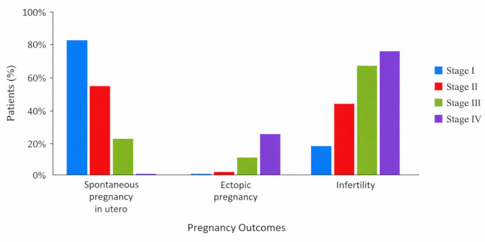
Figure 1: Comparison of pregnancy outcomes (intrauterine pregnancy, ectopic pregnancy, and infertility) according to tubal damage stage (I–IV) after surgery. The analysis included 148 patients with complete 1-year follow-up data: Stage I, n = 22; Stage II, n = 62; Stage III, n = 44; and Stage IV, n = 20. Differences across stages were assessed using chi-square tests, with P < 0.05 considered statistically significant. Please click here to view a larger version of this figure.
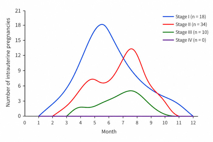
Figure 2: Time to intrauterine pregnancy according to tubal damage stage (I–IV) after surgery. The analysis included the 62 patients who achieved intrauterine pregnancy: Stage I, n = 18; Stage II, n = 34; Stage III, n = 10; and Stage IV, n = 0. The x-axis shows months after surgery, and the y-axis shows the number of intrauterine pregnancies. Please click here to view a larger version of this figure.
| Clinical Feature | Total Cases (N) | Stage I | Stage II | Stage III | Stage IV | χ² | P value |
| Age at First Sexual Intercourse (years) | |||||||
| <19 | 31 | 1 | 7 | 11 | 12 | 25.923 | P < 0.001 |
| ≥19 | 117 | 21 | 55 | 33 | 8 | ||
| Type of Infertility | |||||||
| Primary infertility | 47 | 9 | 15 | 14 | 9 | 3.424 | 0.331 |
| Secondary infertility | 101 | 13 | 39 | 38 | 11 | ||
| Number of Sexual Partners | |||||||
| 1 | 128 | 22 | 60 | 38 | 8 | 46.033 | P < 0.001 |
| >1 | 20 | 0 | 2 | 6 | 12 | ||
| History of Pelvic Infections (CT/UU infection, PID, cervical inflammation, tuberculosis) | |||||||
| Yes | 30 | 0 | 5 | 9 | 16 | 55.46 | P < 0.001 |
| No | 118 | 22 | 57 | 35 | 4 | ||
| History of Vaginitis | |||||||
| Yes | 85 | 14 | 32 | 28 | 11 | 1.946 | 0.584 |
| No | 63 | 8 | 30 | 16 | 9 | ||
| History of Uterine Procedures (abortion, IUD placement) | |||||||
| Yes | 22 | 0 | 2 | 8 | 12 | 43.056 | P < 0.001 |
| No | 126 | 22 | 60 | 36 | 8 | ||
| History of Tubal Ectopic Pregnancy Treatment | |||||||
| Yes | 21 | 0 | 1 | 7 | 13 | 54.206 | P < 0.001 |
| No | 111 | 22 | 61 | 37 | 7 | ||
| History of Abdominal/Pelvic Surgery | |||||||
| Yes | 27 | 1 | 4 | 9 | 13 | 38.007 | P < 0.001 |
| No | 121 | 21 | 58 | 35 | 7 | ||
| CT: Chlamydia trachomatis; UU: Ureaplasma urealyticum; PID: Pelvic Inflammatory Disease; IUD: Intrauterine Device: N; Numbers | |||||||
Table 1: Analysis of factors influencing tubal patency according to tubal damage stage (I–IV). The table summarizes demographic, reproductive, infectious, and surgical history factors among 148 patients with complete follow-up data. Categorical variables were compared using chi-square tests. Abbreviations: CT = Chlamydia trachomatis; UU = Ureaplasma urealyticum; PID = pelvic inflammatory disease; IUD = intrauterine device; N = number.
Supplementary Table S1: Gerard Mage tubal classification criteria used in the study. Tubal staging was determined by intraoperative assessment of tubal obstruction, fimbrial mucosal integrity, and tubal wall quality during combined laparoscopic-hysteroscopic evaluation. Please click here to download this file.
토론
The present study expands upon previous reports by providing a comprehensive stage-based prognostic assessment of TFI patients undergoing combined laparoscopic-hysteroscopic surgery. In addition to evaluating tubal patency, this study analyzed postoperative intrauterine pregnancy, ectopic pregnancy, infertility outcomes, and time to natural conception according to the severity of tubal damage. This stage-specific evaluation may provide clinically useful information to predict reproductive outcomes and select appropriate postoperative fertility management strategies.
The fallopian tube is important in fertility, as it allows sperm transport, oocyte capture, fertilization, and movement of the fertilized egg to the uterus for implantation. Injury or obstruction, such as adhesions or hydrosalpinx, can impair tubal function and lead to TFI1. TFI has increased as social, environmental, and demographic factors have changed over time. Prognostic analysis of surgical therapy for patients with TFI includes several primary indicators, including postoperative fallopian tube patency, postoperative pregnancy outcomes, and time to natural intrauterine pregnancy. Patients with TFI can be treated surgically to reopen the fallopian tubes, and the success rate depends on the level of tubal damage. In early-stage damage (I–II), tubal structure and function are relatively preserved; therefore, surgical repair is more effective. The probability of recanalization is lower when the tubes are more severely damaged. However, combined hysteroscopic and laparoscopic procedures can yield patency in 70–90% of cases and an intrauterine pregnancy rate of approximately 47%8,9. Therefore, mild tubal damage increases the likelihood of successful surgical outcomes.
A similar pattern was observed in this study. Surgery for TFI generally aims to remove adhesions, repair damaged tubes, and achieve patency to increase the probability of intrauterine pregnancy. Recovery is slower in severely damaged tubes with impaired patency and mucosal integrity, and the risk of re-obstruction is higher, resulting in a lower pregnancy rate. The current study showed that 84% and 88.2% of Stage I and Stage II patients, respectively, became pregnant between the fourth and ninth months. Based on these findings, postoperative guidance should be tailored to the extent of tubal damage. In stages I–III, ART should be considered if pregnancy has not occurred within 12 months to avoid delays related to age or declining ovarian function. Stage III patients should be informed that they have a lower probability of pregnancy and that the risk of re-obstruction may increase with prolonged waiting. In Stage IV patients, immediate ART intervention should be considered, as prolonged waiting is unlikely to yield benefit. In line with this, no patients in Stage IV achieved an intrauterine pregnancy.
The findings of this study directly support our hypothesis that increasing tubal damage severity negatively affects postoperative reproductive outcomes. Patients with Stage I and II tubal damage demonstrated significantly higher tubal recanalization and intrauterine pregnancy rates. In contrast, patients with Stage III and IV damage showed increased infertility and ectopic pregnancy rates. Furthermore, no intrauterine pregnancies were observed in Stage IV patients, highlighting the prognostic importance of tubal damage severity in predicting surgical and reproductive outcomes following combined laparoscopic-hysteroscopic treatment.
Common procedures used to evaluate fallopian tube status include HSG, laparoscopic chromopertubation (LCC), HyCoSy, hysteroscopy, and combined hysteroscopic-laparoscopic therapy. Mage et al. emphasized that the integrity of fimbrial mucosal folds is critical for pregnancy after tubal surgery because their absence indicates irreversible damage and may support consideration of tubal excision7. The system provides a clinically convenient method for appraising the structure and function of the fallopian tubes. It has been shown in several studies to be associated with pregnancy outcomes and has become common in clinical practice. In this study, iodixanol, a water-soluble iodinated contrast agent, was used for HSG. The fallopian tube obstruction rate detected by HSG was 59.2%. The sensitivity was 94.4%, and the specificity was 86%, showing strong agreement (95.2%) between HSG and intraoperative findings. In a prospective study on infertile women performed by Vaid et al. in India, the agreement in diagnosing tubal permeability between HSG and hysterolaparoscopy methods was 74%. HSG had 34.6% sensitivity for diagnosing unilateral tubal block and 80.6% sensitivity for diagnosing bilateral block, with specificities of 89.8% and 81.5% for unilateral and bilateral block, respectively10. Therefore, preoperative HSG remains a useful diagnostic tool for evaluating tubal infertility, particularly when using modern water-soluble contrast agents. Post-surgical tubal recanalization rates in TFI patients vary by damage stage (I–IV) and are influenced by multiple risk factors. PID increases infertility risk with each episode, leading to tubal damage through adhesions and scarring. This study found that factors such as early sexual activity, pelvic infections, and prior surgeries were linked to poorer post-surgical recanalization outcomes. These findings align with domestic and international studies, with induced or medical abortion increasing the risk of PID and subsequent tubal damage. Patients with multiple sexual partners are also more prone to sexually transmitted diseases such as PID11, potentially leading to a higher risk of tubal damage11.
The present findings contribute to the current understanding of tubal factor infertility by demonstrating the clinical importance of tubal damage severity in predicting postoperative reproductive outcomes following combined laparoscopic-hysteroscopic treatment. The stage-specific differences observed in tubal recanalization, intrauterine pregnancy, infertility, and ectopic pregnancy rates suggest that the Gerard Mage scoring system may serve as a useful prognostic tool in clinical practice. These findings may help clinicians provide individualized postoperative fertility counseling, optimize the timing of assisted reproductive technologies, and improve reproductive management strategies for patients with varying degrees of tubal damage.
Several alternative approaches are currently available for the evaluation and management of tubal factor infertility (TFI). In addition to hysterosalpingography (HSG) and combined laparoscopic-hysteroscopic assessment, diagnostic methods such as hysterosalpingo-contrast sonography (HyCoSy), magnetic resonance hysterosalpingography (MRI-HSG), and diagnostic laparoscopy are also used to evaluate tubal patency and pelvic pathology. Assisted reproductive technologies, particularly in vitro fertilization (IVF), remain important treatment options for patients with severe tubal damage or poor postoperative prognosis. Compared with alternative management strategies, the Gerard Mage scoring system used in this study provides a practical, stage-based assessment of the severity of tubal damage. It may help guide individualized surgical and reproductive treatment decisions. These approaches should be considered together when selecting the optimal management strategy for patients with TFI.
This study has several limitations. First, the retrospective single-center design may introduce selection bias and limit the generalizability of the findings to broader patient populations. Second, the use of retrospectively collected clinical data may have resulted in incomplete or inconsistent documentation of certain patient characteristics and postoperative outcomes. Because of incomplete historical documentation, stage-specific information for some patients with unavailable follow-up outcomes could not be reliably reconstructed. Third, although the Gerard Mage scoring system provides a clinically useful assessment of the severity of tubal damage, additional prognostic models incorporating patient-specific reproductive and clinical factors may further improve outcome prediction. Finally, the absence of a control group and the relatively small sample size may limit the ability to fully compare treatment strategies for tubal factor infertility. Therefore, prospective multicenter studies with larger patient populations are needed to validate these findings and optimize prognostic assessment and postoperative fertility management strategies. Additionally, because this retrospective study was based on historical records, source datasets and detailed procedural documentation were not fully available for verification.
Future studies should focus on prospective multicenter investigations with larger patient populations to further validate the prognostic value of the Gerard Mage scoring system and combined laparoscopic-hysteroscopic treatment in tubal factor infertility (TFI). Additional research is also needed to develop more comprehensive and standardized prognostic assessment systems that integrate tubal morphology, reproductive outcomes, and patient-specific clinical factors. Such improvements may enhance individualized treatment planning, optimize the timing of assisted reproductive technologies, and improve long-term reproductive outcomes in patients with TFI.
In conclusion, this study demonstrates that the severity of tubal damage is strongly associated with postoperative reproductive outcomes in patients with tubal factor infertility undergoing combined laparoscopic-hysteroscopic treatment. Patients with mild tubal damage showed higher tubal recanalization and intrauterine pregnancy rates, whereas severe tubal damage was associated with increased infertility and ectopic pregnancy risk. The Gerard Mage scoring system may provide useful prognostic information for predicting reproductive outcomes and guiding individualized postoperative fertility management and assisted reproductive treatment strategies.
공개 사항
The author has no conflicts of interest to declare.
감사의 글
The National Natural Science Foundation of China supported this study (grant 81200477).
재료
| 이름 | 회사 | 카탈로그 번호 | 댓글 |
|---|---|---|---|
| Hysteroscope | Used for hysteroscopic evaluation and treatment of uterine/tubal pathology | ||
| Iodixanol contrast agent | Water-soluble iodinated contrast used for hysterosalpingography (HSG) | ||
| Laparoscope | Used for laparoscopic assessment and surgical management of tubal obstruction | ||
| Surgical instruments for tubal reconstruction | Includes tools for anastomosis, fimbrial reconstruction, adhesiolysis, and dilation | ||
| Tubal catheterization system | Used for proximal tubal catheterization and fluid infusion to restore patency |
참고문헌
- Xing X. Gynecology and obstetrics. 8th ed. People’s Medical Publishing House; Beijing; 2013. p. 368-9.
- Audu BM, et al. Prevalence of utero-tubal infertility. J Obstet Gynaecol. 2009;29(4):326-8.
- Xiyin Z, editor. Practical gynecology and obstetrics. 2nd ed. People’s Medical Publishing House; Beijing; 2004. p. 165-78.
- Yuntana W, Lingling L, Yong Z, Aiming W. Clinical efficacy analysis of hysteroscopy and laparoscopy combined treatment for female infertility. Chin J Fam Plan. 2016;24(4):256-60.
- Grigoriadis G, Roman H, Gkrozou F, Daniilidis A. The impact of laparoscopic surgery on fertility outcomes in patients with minimal/mild endometriosis. J Clin Med. 2024;13(16):4817.
- Practice Committee of the American Society for Reproductive Medicine. Role of tubal surgery in the era of assisted reproductive technology: a committee opinion. Fertil Steril. 2021;115(5):1143-50.
- Mage G, et al. A preoperative classification to predict the intrauterine and ectopic pregnancy rates after distal tubal microsurgery. Fertil Steril. 1986;46(5):807-10.
- Varlas V, et al. Hysterolaparoscopy: a gold standard for diagnosing and treating infertility and benign uterine pathology. J Clin Med. 2021;10(16):3749.
- Shen H, et al. Factors affecting the success of fallopian tube recanalization in the treatment of tubal obstructive infertility. J Int Med Res. 2020;48(12):300060520979218.
- Vaid K, et al. Pan endoscopic approach “hysterolaparoscopy” as an initial procedure in selected infertile women. J Clin Diagn Res. 2014;8(2):95-8.
- Wilson CN, Sathiyasusuman A. Associated risk factors of STIs and multiple sexual relationships among youths in Malawi. PLoS One. 2015;10(8):e0134286.