101명의 PELD 환자를 대상으로 한 이번 회고적 연구에 따르면, 수술 전 MRI 특성, 특히 Modic II(불리함)와 DHI > 25%(보호적 요인)가 1년 후의 예후와 밀접하게 연관되어 있음이 나타났습니다. 이러한 결과는 수술적 결정의 지침이 될 수 있으나, 정적인 예측 인자로 사용되지는 않습니다. 완전한 신경 감압이 여전히 성공의 궁극적인 결정 요인으로 남습니다.
연구 논문
경피적 내시경 요추 추간판 절제술로 치료한 요추 추간판 탈출증의 수술 전 MRI 특성 및 결과: 회고적 연구
88 조회수
⸱
2026년 9월 11일
이 논문에서
요약
초록
경피적 내시경 요추 추간판 절제술(PELD)은 요추 추간판 탈출증(LDH)에 널리 사용되지만, 그 결과는 다양합니다. 수술 전 자기공명영상(MRI) 특징이 예후를 예측할 수 있으나, 수술 효능과의 상관관계에 대한 체계적인 연구는 부족한 실정입니다. 본 연구의 목적은 요추 추간판 탈출증으로 PELD 치료를 받은 환자들을 대상으로 수술 전 MRI 형태학적 특성과 임상 결과 사이의 관계를 후향적으로 분석하여, 수술 전 평가, 결과 예측 및 수술 전략 최적화를 위한 참고 자료를 제공하는 것입니다. 본 후향적 연구에는 1년 이상의 추적 관찰 기간을 가진, PELD를 시행한 단일 분절 LDH 환자 101명(2022.1~2024.9)이 포함되었습니다. 수술 전 MRI 파라미터 [Pfirrmann 등급, Modic 변화, 탈출 유형/위치/이동, 척추관 점유율, 추간판 높이 지수(DHI)]와 임상 결과 [시각적 아날로그 척도(VAS), Oswestry 장애 지수(ODI), 수정된 MacNab 척도]를 분석하였습니다. 환자들은 1년 후의 MacNab 점수에 따라 그룹화되었습니다. 그룹 간 MRI 특징을 비교하였으며, Pearson 상관분석과 다변량 로지스틱 회귀 분석을 사용하여 양호한 예후의 예측 인자를 식별하였습니다. 101명의 환자 중 1년 추적 관찰 시 76명(75.2%)은 양호한 결과가 나타났고, 25명(24.8%)은 불량한 결과가 나타났습니다. 불량 그룹은 추간판 탈출/격리 비율이 유의하게 높았고, 탈출 면적이 더 넓었으며, Modic type II 변화가 더 많이 관찰된 반면, 추간판 높이와 신경공 면적은 더 낮았습니다 (P < 0.05). 척추관 점유율과 탈출 면적은 통증 VAS 및 ODI와 양의 상관관계를 보였으며, DHI와 신경공 면적은 음의 상관관계를 보였습니다. Modic type II 변화는 독립적으로 불량한 예후를 예측한 반면, DHI > 25%는 보호 요인이었습니다. 수술 전 MRI 특징은 PELD 결과와 밀접한 관련이 있으며, 특히 Modic type II와 감소된 DHI는 고위험 환자를 식별하는 지표가 됩니다. 이러한 다차원적 MRI 평가는 수술 전 위험 층화를 가능하게 하며, 불리한 영상적 특징이 있을 때 신경공 확장술이나 대안적 접근법의 고려를 포함하여 개별화된 수술적 의사결정을 안내합니다. 그러나 완전한 신경 감압이 여전히 성공의 궁극적인 결정 요인으로 남아 있습니다.
서론
요추 추간판 탈출증(LDH)은 주로 추간판 퇴행, 섬유륜 파열, 그리고 신경근 또는 마미를 압박하는 수핵 탈출을 특징으로 합니다. 임상적으로 이 질환은 좌골신경통, 하지 마비 및 무력감, 간헐적 파행 등의 증상으로 나타납니다. 이러한 증상들은 환자의 일상생활과 업무 능력을 심각하게 저하시켜 개인, 가족 및 사회에 상당한 경제적 부담을 줍니다1,2. LDH에 대한 전통적인 개방성 수술은 탈출된 수핵을 직접 제거할 수 있지만, 상당한 외상, 많은 출혈, 수술 후 회복 지연 및 척추 안정성 훼손과 같은 단점이 있습니다3. 경피적 내시경 요추 추간판 절제술(PELD)은 1997년 Yeung과 Tsou4가 추간공 내시경 추간판 절제술(YESS 기법)을 처음 보고한 이후 20년 넘게 발전해 왔으며, 현재는 LDH 치료를 위한 주류 최소 침습적 접근법이 되었습니다. PELD는 직경 약 6–7 mm의 작업 채널을 사용하여 내시경 직접 시야 하에 탈출된 추간판 조직을 제거하며, 최소 외상, 척추 안정성 유지5,6와 같은 뚜렷한 장점을 제공합니다. 수많은 임상 연구를 통해 PELD가 LDH 사례의 84%–94%에서 전통적인 개방성 수술과 유사한 우수하거나 양호한 결과를 얻는다는 것이 확인되었습니다7. 그러나 임상 실무에서는 수술 적응증을 엄격히 준수하고 동일한 수술 팀이 표준화된 절차를 수행하더라도, PELD 후의 수술 결과가 개인별로 상당한 차이를 보인다는 것이 드러났습니다. 일부 환자는 현저한 통증 완화와 기능적 회복을 경험하는 반면, 약 20%–30%는 잔여 요통 또는 하지 통증, 감각 이상, 불충분한 기능 회복 또는 재발을 보고합니다8,9. 이러한 현상은 척추 외과 의사들에게 중요한 임상적 과제가 되었습니다. 최근에는 추간공 내시경 요추 수술에서 수술 실패를 최소화하고 결과를 최적화하기 위한 핵심 전략으로 객관적 및 주관적 "감압 종점(end-points of decompression)"을 정의하는 개념이 제안되었습니다10.
수술 전 자기공명영상(MRI)은 추간판 탈출증(LDH)을 평가하는 데 가장 선호되는 영상 진단법으로, 디스크 형태, 탈출 범위, 신경 압박 정도, 그리고 척추관 및 신경공의 구조를 명확하게 시각화하여 제공합니다. MRI 형태학적 파라미터는 진단 및 수술 적응증을 확인하는 데 도움이 될 뿐만 아니라 수술 후 결과에 대한 예측 가치를 제공할 수 있습니다. T2 가중 MRI 신호 강도, 디스크 구조, 디스크 높이, 그리고 수핵과 섬유륜의 구분을 기반으로 하는 Pfirrmann 등급 시스템은 디스크 퇴행을 1~5등급으로 분류하며 임상 평가에 널리 사용됩니다11. Modic 변화는 MRI상 척추 종판과 인접 골수의 신호 변화를 의미하며, 유형 I(부종성), 유형 II(지방성), 유형 III(경화성)로 분류되고 디스크 퇴행, 요통 및 수술 후 재발과 밀접한 관련이 있습니다12. 점차 많은 연구들이 PELD 이후 MRI 형태학적 특징과 임상 결과 사이의 연관성을 조사해 왔습니다. 그러나 현재의 연구들은 주로 단일 또는 소수의 MRI 파라미터에 집중하고 있어, 여러 형태학적 지표에 대한 체계적인 평가가 부족한 실정입니다. 또한, 척추관 점유율, 신경공 면적, 디스크 높이 지수(DHI)와 같은 정량적 파라미터와 PELD 결과 사이의 관계에 대한 연구는 여전히 불충분합니다13. 나아가, 최근 최소 침습 척추 수술의 발전은 단독 내시경 또는 채널 기법에서 로보틱스, 인공지능 및 영상 유도 기술이 통합된 지능형 최소 침습 접근법으로 진화하고 있습니다. 로봇 보조 시스템의 통합은 제한된 수술 공간 내에서의 기구 조작 문제를 더욱 해결함으로써 최소 침습 척추 수술 기술의 중대한 발전을 나타내고 있습니다14.
앞서 언급한 연구 배경과 기존 지식의 공백을 바탕으로, 본 단일 센터 후향적 관찰 연구는 단일 레벨 LDH에 대해 PELD를 시행받은 환자들로부터 임상 데이터와 수술 전 MRI 형태학적 특징을 체계적으로 수집하는 것을 목표로 합니다. 다차원적 MRI 형태학적 평가 시스템을 구축하고, 이를 1년 후의 수술 후 임상 결과와 결합하여 다양한 MRI 형태학적 파라미터와 PELD 예후 사이의 연관성을 철저히 분석함으로써, 좋지 않은 수술 후 결과에 영향을 미치는 독립적인 위험 요인 및 보호 요인을 식별하고자 합니다. 본 연구의 주요 목적은 다음과 같습니다: 단일 레벨 LDH에 대한 PELD의 전반적인 임상적 효능과 1년 예후 결과의 분포를 결정하고, 예후가 좋은 환자군과 좋지 않은 환자군 사이의 수술 전 MRI 형태학적 파라미터 차이를 비교하며, 다변량 로지스틱 회귀 분석을 통해 수술 후 긍정적인 결과의 독립적 예측 인자를 식별하고, 정성적 파라미터(Modic 아형, 탈출 등급)와 정량적 파라미터(관강 점유율, DHI, 신경공 면적, 탈출 면적)를 모두 통합한 다차원적 수술 전 MRI 평가 시스템을 구축하여 더욱 정밀한 수술 후 위험 층화가 가능하게 하고 개별화된 수술적 의사결정을 안내하는 것입니다.
프로토콜
본 연구는 헬싱키 선언15의 원칙을 엄격히 준수하여 수행되었으며, 화북 의료 건강 그룹 싱타이 종합병원(North China Medical Health Group XingTai General Hospital)의 기관 윤리 위원회로부터 승인을 받았습니다(승인 번호 ZCKT-2026-0018). 모든 환자에게 연구 목적, 치료 프로토콜 및 잠재적 위험에 대해 설명하였으며, 서면 동의서를 받았습니다. 모든 연구 데이터는 환자의 개인정보 보호를 위해 익명화 처리되었습니다.
연구 대상
본 단일 센터 회고적 관찰 연구는 2022년 1월부터 2024년 9월까지 본원 정형외과에서 PELD를 시행받은 단일 레벨 LDH 환자들을 대상으로 하였다. 최초로 스크리닝된 123명의 환자 중 엄격한 선정 및 제외 기준을 적용하여 최종적으로 101명이 포함되었으며, 이들 모두 최소 1년의 추적 관찰을 완료하였다. 수술 후 1년 시점의 수정 MacNab 기준에 따라 환자들을 양호군(n = 76)과 불량군(n = 25)으로 분류하였다. 상세한 과정은 그림 1에 나타나 있다.
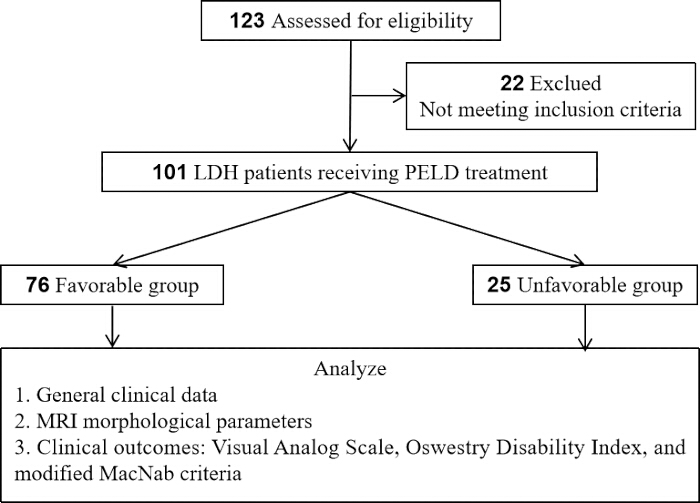
그림 1: 연구 흐름도. 환자 선정 및 그룹화 흐름도. 평가 대상 환자 123명 중, PELD를 시행한 단일 레벨 LDH 환자 101명이 포함되었으며, 1년 수정 MacNab 기준에 따라 양호(Favorable, n = 76) 그룹과 불량(Unfavorable, n = 25) 그룹으로 나누었다. 분석 체계에는 일반 임상 데이터, MRI 파라미터 및 임상 결과(VAS, ODI, MacNab)가 포함되었다. 이 그림의 더 큰 버전을 보시려면 여기를 클릭하십시오.
선정 기준
연령 18세에서 75세 사이; 임상 증상, 신체 검사 및 MRI/CT 소견을 바탕으로 L3/4, L4/5 또는 L5/S1의 단일 레벨 LDH로 진단된 환자; 신경근 압박의 명확한 증상과 징후가 있으며, 최소 6주 이상의 체계적인 보존적 치료(침상 안정, 약물 치료, 물리 치료 등 포함)에 반응이 없는 경우; PELD 수술을 받은 환자; 완전한 임상 데이터가 포함된 수술 후 추적 관찰 기간이 최소 1년 이상인 경우.
제외 기준
다분절 LDH 또는 이전의 요추 수술 이력; 동반된 요추 척추전방전위증, 요추 척추관 협착증, 척수 종양, 척수 감염 또는 척추 골절; 응급 수술이 필요한 마미 증후군의 존재; 정확한 측정을 방해하는 낮은 수술 전 MRI 영상 품질; 수술에 부적합한 중증 동반 질환(예: 심장, 폐, 간 또는 신부전); 추적 관찰 실패 또는 불완전한 임상 데이터.
수술 절차
모든 수술은 동일한 숙련된 척추 외과 전문의 팀에 의해 수행되었습니다. 수술 접근법(추간공 접근법, TF 또는 층간 접근법, IL)은 추간판 탈출증의 형태와 해부학적 제약 요인에 대한 종합적인 평가를 기반으로 결정되었습니다. 구체적으로, TF 접근법은 추간공, 추간공 외측 또는 추궁근(pedicle) 레벨 아래에 위치한 중앙/측방 탈출증에 우선적으로 채택되었습니다. 반대로, Choi 등16이 이전에 기술한 바와 같이, 고도로 이동되었거나 격리된 파편, 특히 L5 추궁근 방향으로 머리 쪽(cephalad)으로 이동한 경우나 장골능(iliac crest)이 높은 환자에게는 IL 접근법이 선택되었습니다. 이러한 차별화된 선택 전략을 통해 수술 경로가 목표 병변과 최적으로 일치하도록 하였습니다.
시술은 정맥 진정 요법의 동반 여부와 관계없이 국소 마취 하에 환자를 복와위로 위치시킨 상태에서 진행되었습니다. C-arm 투시 장비를 사용하여 표적 디스크 공간을 확인하였습니다. 캄빈 삼각(Kambin's triangle) 또는 층간 공간을 통해 척수강 내로 천자침을 진입시킨 후, 가이드와이어를 삽입하고 순차적으로 확장하여 작업 채널을 형성하였습니다. 작업용 내시경(직경 6.9 mm, 시야각 30°)을 삽입하고, 신경근 감압을 위해 직접 시야 하에서 탈출된 추간판 핵 조직을 제거하였습니다. 고주파 프로브를 사용하여 섬유륜 성형술과 지혈을 시행하였습니다. 이후 작업 채널을 제거하고 피부 절개 부위를 봉합하였습니다.
수술 전 DHI가 25% 이하이거나 MRI상 심한 추간공 협착이 나타나는 경우, Kambin's working zone을 확장하고 채널 삽입을 용이하게 하기 위해 집도의의 판단하에 리머(reamer) 또는 고속 드릴을 이용한 추가 추간공 성형술을 시행하였습니다. 다만, 본 연구는 후향적 설계로 인해 이러한 추간공 성형술의 수행 방식이 엄격하게 표준화되지 않았으며 수술 기록지에 체계적으로 기록되지 않았으므로, 본 코호트에서 임상적 결과에 미치는 구체적인 영향에 대한 정량적 분석은 불가능합니다.
관찰 지표
본 연구에서는 T0, T1, T2, T3를 각각 수술 전, 수술 후 1주, 수술 후 3개월, 수술 후 1년으로 정의하였다.
일반 임상 데이터
연령, 성별, 체질량 지수(BMI), 질환 기간, 흡연력, 당뇨병 병력, 고혈압 병력, 수술 레벨 및 수술 접근법을 포함한 환자 데이터를 수집하였다.
MRI 형태학적 파라미터 측정
모든 환자를 대상으로 시상면 T1 강조 영상(T1WI), T2 강조 영상(T2WI) 및 횡단면 T2 강조 영상(T2WI) 시퀀스를 포함한 수술 전 요추 MRI 검사를 시행하였다. 두 명의 숙련된 영상의학과 전문의가 이중 맹검 방식으로 파라미터를 독립적으로 측정하였으며, 분석에는 평균값을 사용하였다. Pfirrmann 등급: 디스크의 신호 강도, 구조, 높이 및 수핵(nucleus pulposus)과 섬유륜(annulus fibrosus) 간의 구별 정도를 바탕으로 시상면 T2 강조 영상에서 1~5등급으로 평가하였다.11. Modic 변화: 척추 종판과 인접 골수의 신호 변화로 평가하며, 없음, type I (T1WI에서 저신호, T2WI에서 고신호), type II (T1WI에서 고신호, T2WI에서 등신호 또는 약간의 고신호), 또는 type III (T1WI와 T2WI 모두에서 저신호)로 분류함12. 추간판 탈출 유형: 축상 T2 강조 영상(T2WI)을 기준으로 팽윤(bulging), 돌출(protrusion), 탈출(extrusion) 또는 격리(sequestration)로 분류함17. 탈출 위치: 중앙, 중앙 주변, 추간공, 또는 극외측으로 분류함4이탈 범위: 시상면 T2 강조 영상(T2WI)에서 탈출된 파편의 상단 또는 하단 경계로부터 디스크 높이까지의 수직 거리로 측정하며, Lee 등의 분류 기준에 따라 분류함.18 이동 없음(0등급), 저등급 이동(<디스크 높이의 50%, 1-2등급) 또는 고도 이동(디스크 높이의 50% 이상, 3-4등급). 척수관 점유율: 최대 탈출 수준의 축상 T2WI에서 (탈출부의 전후경 / 척수관의 전후경)으로 계산함 × 100%19,20DHI: 시상면 T2WI에서 [(전방 디스크 높이 + 후방 디스크 높이) / 2] / [(상위 척추체 전후경 + 하위 척추체 전후경) / 2]로 측정함 × 100%21추간공 면적: ImageJ 소프트웨어를 사용하여 추간공이 최대인 레벨의 축상 T2 강조 영상(axial T2WI)에서 수동으로 구획하고 측정함22,23최대 탈출 면적: 최대 탈출 높이의 축상 T2 강조 영상(T2WI)에서 탈출된 파편의 단면적으로 측정함24.
임상 결과 평가
통증 평가: 요통(VAS-Back) 및 다리 통증(VAS-Leg)에 대해 시각 아날로그 척도(Visual Analog Scale, VAS)를 사용하여 통증 강도를 평가했으며, 점수는 0(통증 없음)에서 10(상상 가능한 최악의 통증)까지로 산정하였다25,26. 기능적 장애: 요추 기능은 Oswestry 장애 지수(Oswestry Disability Index, ODI)를 사용하여 평가하였다27,28. 수정된 MacNab 기준: 1년 추적 관찰 시 결과를 평가하여, 우수(excellent, 증상의 완전한 소실, 정상 활동으로 복귀), 양호(good, 가끔 발생하는 통증, 업무나 일상생활에 지장 없음), 보통(fair, 증상은 개선되었으나 활동에 제한이 있음), 또는 불량(poor, 개선 없음 또는 증상 악화)으로 분류하였다29,30. "우수" 및 "양호" 결과는 양호한 예후로 정의하였고, "보통" 및 "불량" 결과는 불량한 예후로 정의하였다. 수정된 MacNab 기준은 통증 완화, 기능적 회복, 일상 활동로의 복귀를 통합하여 수술 성공 여부를 환자 중심으로 종합적으로 평가하며, 이는 단일 연속 척도보다 임상적 의사 결정에 더 관련성이 높은 영역들이기 때문에 일차 결과 측정 지표로 선택되었다. 또한, MacNab 분류법은 PELD 문헌에서 널리 활용되고 검증되어 왔으므로29,30, 이전 연구들과의 직접적인 비교가 용이하다.
표본 크기 추정
표본 크기 추정은 G*Power 소프트웨어31를 사용하여 수행되었습니다. 회고적 관찰 연구로서, 표본 크기는 연구 기간 동안 선정 및 제외 기준을 충족한 환자의 총 수를 기준으로 결정되었으며, 사후 검정력 분석(post-hoc power analysis)을 통해 그 적절성을 검증하였습니다. 이전의 관련 연구8를 바탕으로, PELD 후 불량 예후의 발생률을 약 25%로 가정하고 디스크 돌출/격리(disc extrusion/sequestration)를 주요 노출 요인으로 설정했을 때, 양측 α = 0.05 및 검정력(1-β) 80%에서 최소 89명의 참여자가 필요했습니다. 최종적으로 본 연구에는 통계적 요구 사항을 충족하는 총 101명의 환자가 포함되었습니다.
통계 분석
통계 분석은 SPSS를 사용하여 수행되었습니다. 연속 변수의 정규성은 Shapiro-Wilk 검정으로 평가하였습니다. 정규 분포를 따르는 데이터는 평균 ± SD로 표시하였으며, 독립 표본 및 대응 표본 t-검정을 사용하여 비교하였습니다. 정규 분포를 따르지 않는 데이터는 중앙값(Q1, Q3)으로 표시하고 Mann-Whitney U 검정을 통해 분석하였습니다. 범주형 변수는 카이제곱 검정을 사용하여 비교하였습니다. Pearson 상관 분석과 다변량 로지스틱 회귀 분석을 적용하였습니다. 모든 검정은 양측 검정으로 수행되었으며, P < 0.05를 통계적으로 유의한 것으로 간주하였습니다.
다변량 로지스틱 회귀 분석을 위해 종속 변수를 양호한 예후 = 1, 불량한 예후 = 0으로 인코딩하였으며, 그 결과 76건의 양성 이벤트가 도출되었다. 최종 모델에 포함되는 공변량의 수는 EPV 원칙을 준수해야 한다.32MRI 파라미터 간의 다중공선성을 평가하기 위해 분산팽창요인(VIF)을 사용하였으며, VIF 값은 <5는 허용 가능한 수준으로 간주됩니다.
결과
환자에 대한 일반 정보
본 연구에는 총 101명의 환자가 포함되었습니다. 표 1에 나타난 바와 같이, 불량 그룹의 환자들은 연령이 유의하게 더 높았으며 (P < 0.05), 질환 기간이 더 길었습니다 (P < 0.05). 성별, BMI, 흡연력, 동반 질환, 수술 레벨 또는 수술 접근법과 관련하여 두 그룹 간에 통계적으로 유의미한 차이는 관찰되지 않았습니다 (P > 0.05).
| 변수 | 양호군 (n = 76) | 불량군 (n = 25) | t/χ²/Z | P |
| 연령 (years) | 45.2 ± 11.8 | 51.6 ± 12.9 | 2.322 | 0.022 |
| 남성 | 42 (55.3) | 16 (64.0) | 0.587 | 0.444 |
| BMI (kg/m2) | 24.4 ± 3.0 | 25.4 ± 3.6 | 1.531 | 0.129 |
| 질병 기간 (months) | 9 (8, 13) | 15 (12, 17) | 4.174 | <0.001 |
| 흡연력 | 18 (23.7) | 9 (36.0) | 1.457 | 0.227 |
| 당뇨병 병력 | 8 (10.5) | 5 (20.0) | 0.779 | 0.337 |
| 고혈압 병력 | 12 (15.8) | 6 (24.0) | 0.396 | 0.529 |
| 수술 분절 | ||||
| L3/4 | 11 (14.5) | 1 (4.0) | ||
| L4/5 | 40 (52.6) | 16 (64.0) | ||
| L5/S1 | 25 (32.9) | 8 (32.0) | 2.18 | 0.336 |
| 수술 접근법 | ||||
| 추간공 접근법 | 59 (77.6) | 19 (76.0) | ||
| 추궁판 간 접근법 | 17 (22.4) | 6 (24.0) | 0.029 | 0.866 |
표 1: 기초 특성[평균±표준편차, n (%), 중앙값(Q1, Q3)]. 예후군별로 층화된 기초 특성. 데이터는 평균 ± SD, n (%), 또는 중앙값 (Q1, Q3)으로 표시됨. Unfavorable 군은 연령이 유의하게 더 높았으며 (P = 0.022), 질병 기간이 더 길었음 (P < 0.001). 다른 변수들은 군 간에 유의미한 차이를 보이지 않음. BMI, 체질량 지수.
수술 전 MRI 형태학적 파라미터 비교
101명 환자의 수술 전 MRI 형태학적 파라미터를 표 2에 나타내었다. Modic 변화의 비율과 추간판 탈출 유형 측면에서 두 그룹 간에 유의미한 차이(P < 0.05)가 있었다. Favorable 그룹의 DHI와 추간공 면적은 Unfavorable 그룹보다 유의하게 높았으며(P < 0.05), Favorable 그룹의 척추관 점유율과 최대 탈출 면적은 Unfavorable 그룹보다 유의하게 낮았다(P < 0.05). Pfirrmann 등급, 탈출 위치 및 이동 등급에서는 통계적으로 유의미한 차이(P > 0.05)가 없었다. 이러한 결과는 위의 파라미터들이 PELD의 수술 후 예후를 예측할 수 있는 잠재적 지표가 될 수 있음을 시사한다.
| 유리한 군 (n = 76) | 불량 그룹 (n = 25) | 제공된 텍스트가 없습니다. 번역할 소스 텍스트를 입력해 주십시오.χ² | P | |
| 피르만 등급(Pfirrmann Grade) | ||||
| I-II | 28(36.8) | 5(20.0) | ||
| III | 32(42.1) | 10(40.0) | ||
| IV-V | 16(21.1) | 10(40.0) | 4.277 | 0.118 |
| 모딕 변화 (Modic Changes) | ||||
| 입력된 텍스트가 없습니다. 번역할 소스 텍스트를 제공해 주세요. | 48(63.2) | 8(32.0) | ||
| 제I형 | 12(15.8) | 3(12.0) | ||
| 제2형 | 14(18.4) | 13(52.0) | ||
| 제III형 | 2(2.6) | 1(4.0) | 11.529 | 0.009 |
| 탈출 유형 | ||||
| 팽윤 | 8(10.5) | 1(4.0) | ||
| 돌출 | 44(57.9) | 7(28.0) | ||
| 압출 | 20(26.3) | 12(48.0) | ||
| 격리 | 4(5.3) | 5(20.0) | 11.605 | 0.009 |
| 탈출 위치 | ||||
| 중앙 | 12(15.8) | 2(8.0) | ||
| 방중심성 | 48(63.2) | 18(72.0) | ||
| 추간공의 | 12(15.8) | 4(16.0) | ||
| 추간공 외 | 4(5.3) | 1(4.0) | 1.11 | 0.775 |
| 이동도 | ||||
| 이동 없음 (0등급) | 25(32.9) | 6(24.0) | ||
| 저등급 이동 (1~2등급) | 32(42.1) | 10(40.0) | ||
| 고등급 이동 (3-4등급) | 19(25.0) | 9(36.0) | 1.326 | 0.515 |
| 척수강 점유율 (%) | 35.6 ± 6.9 | 47.2 ± 8.4 | 6.892 | <0.001 |
| 척추관 점유율 > 40% | 30(39.5) | 14(56.0) | ||
| DHI (%) | 28.5 ± 5.2 | 24.5 ± 4.8 | 3.392 | 0.001 |
| 직접 모발 이식(DHI) > 25% | 58(76.3) | 11(44.0) | ||
| 추공 영역 (cm²²) | 1.1 ± 0.3 | 0.8 ± 0.3 | 4.029 | <0.001 |
| 추간공 부위 > 1 cm² | 49(64.5) | 13(52.0) | ||
| 최대 탈출 면적(mm²²) | 59.8 ± 11.9 | 86.0 ± 15.7 | 8.764 | <0.001 |
| 최대 탈출 부위 > 75 mm² | 21(27.6) | 13(52.0) |
표 2: 수술 전 MRI 형태학적 파라미터 비교 [평균±표준편차, n (%)]. 그룹 간 수술 전 MRI 파라미터를 비교하였다. 데이터는 평균 ± 표준편차 또는 n (%)로 표시하였다. Modic 변화, 탈출 유형, 이동 등급, 척추관 점유율, DHI, 추간공 면적 및 탈출 면적에서 유의미한 차이가 관찰되었으나(모두 P < 0.05), Pfirrmann 등급이나 탈출 위치에서는 차이가 없었다. DHI, 추간판 높이 지수.
임상 결과 지표의 변화
표 3은 수정된 MacNab 기준을 사용하여 1년 추적 관찰 시 평가한 101명 환자의 임상 결과를 나타냅니다. 결과적으로 51명(50.5%)의 환자에게서 우수한 결과가, 25명(24.8%)의 환자에게서 양호한 결과가 나타나, 양호한 군에 총 76명(75.2%)의 환자가 포함되었습니다. 보통의 결과는 16명(15.8%)의 환자에서, 불량한 예후는 9명(8.9%)의 환자에서 관찰되어 불량한 군에 25명(24.8%)의 환자가 포함되었으며, 이는 전반적으로 양호한 임상적 효능을 나타냅니다.
| 결과 등급 | 사례 수 (n) | 백분율 (%) | 임상 결과의 정의 |
| 훌륭합니다 | 51 | 50.50% | 양호한 군 |
| 좋음 | 25 | 24.80% | 양호군 |
| 공정함 | 16 | 15.80% | 불리한 군 |
| 불량한 | 9 | 8.90% | 불리한 군 |
표 3: 수정된 MacNab 기준에 따른 수술 후 1년 임상 효능 평가 수정된 MacNab 기준에 따른 수술 후 1년 결과. Excellent/Good 결과는 양호군(75.2%)으로, Fair/Poor 결과는 불량군(24.8%)으로 분류되었다.
표 4는 수술 후 임상 결과의 동적 변화를 보여줍니다. 수술 전, 요통에 대한 VAS, 하지 통증에 대한 VAS 또는 ODI 점수에서 두 그룹 간에 통계적으로 유의미한 차이가 없었습니다 (P > 0.05). 모든 수술 후 시점에서 두 그룹 모두 수술 전 값과 비교하여 점수가 유의하게 개선되었으며 (그룹 내 P < 0.001), 이는 PELD의 전반적인 효능을 나타냅니다. 그러나 그룹 간 비교 결과, 양호 그룹이 불량 그룹에 비해 모든 수술 후 시점에서 VAS (허리 및 다리) 및 ODI 점수가 유의하게 더 좋게 나타났습니다 (P < 0.001). 또한, 수술 후 1년 시점의 ODI 개선 정도는 불량 그룹보다 양호 그룹에서 유의하게 더 컸습니다. 이러한 결과는 수술 자체가 상당한 개선을 이끌어내지만, 수술 전 MRI 형태학적 특징으로 층화된 환자들은 임상적 회복 궤적에서 지속적인 차이를 보인다는 것을 입증하며, 이는 장기 결과에 대한 수술 전 MRI 특성의 예측 가치를 더욱 뒷받침합니다.
| 시간 | 양호군 (n = 76) | 불량군 (n = 25) | P (군 내) | P (군 간) | |
| VAS-요통 | T0 | 6(6, 7) | 7(6, 7) | - | 0.212 |
| T1 | 3(3, 4) | 4(4, 5) | <0.001 | <0.001 | |
| T2 | 2(1, 2) | 3(2, 4) | <0.001 | <0.001 | |
| T3 | 1(1, 1) | 2(2, 3) | <0.001 | <0.001 | |
| VAS-하지통 | T0 | 8(7, 9) | 8(7, 8) | - | 0.486 |
| T1 | 3(2, 3) | 5(4, 5) | <0.001 | <0.001 | |
| T2 | 2(1, 2) | 3(3, 4) | <0.001 | <0.001 | |
| T3 | 1(1, 1) | 3(2, 3) | <0.001 | <0.001 | |
| ODI(%) | T0 | 68.4 ± 9.6 | 70.2 ± 10.4 | - | 0.408 |
| T1 | 30.1 ± 7.4 | 42.4 ± 8.9 | <0.001 | <0.001 | |
| T2 | 18.7 ± 5.5 | 29.5 ± 6.4 | <0.001 | <0.001 | |
| T3 | 10.1 ± 4.0 | 21.8 ± 5.5 | <0.001 | <0.001 | |
| ODI 변화 점수 | T3-T0 | 58.2 ± 10.9 | 48.5 ± 11.5 | - | <0.001 |
표 4: 수술 전후 임상 결과 지표의 변화 [평균 ± 표준편차, M(Q1, Q3)]. VAS 및 ODI 점수의 종단적 변화. 데이터는 중앙값(Q1, Q3) 또는 평균 ± 표준편차로 표시됨. 두 그룹 모두 수술 후 유의하게 개선되었으나(그룹 내 P < 0.001), Favorable 그룹이 모든 시점에서 일관되게 더 나은 점수를 보임(그룹 간 P < 0.001). 그룹 내 비교는 Wilcoxon 부호 순위 검정을 통해 수행되었으며, 그룹 간 비교는 Mann-Whitney U 검정을 통해 수행됨.
MRI 형태학적 파라미터와 임상 결과 간의 상관관계 분석 표 5 피어슨 상관관계 분석 결과, 척추관 점유율 및 최대 추간판 탈출 면적이 수술 후 요통 VAS, 하지 통증 VAS 및 ODI 점수와 유의미한 정적 상관관계를 보이는 것으로 나타났다(r: 0.375–0.538, P < 0.001). 이는 척수강 압박의 심각도가 높고 추간판 탈출 부피가 클수록 잔류 증상이 더 뚜렷하며 기능적 회복이 더 낮음을 나타냅니다. 반면, DHI와 추간공 면적은 모든 임상 결과 측정치와 유의미한 음의 상관관계를 보였습니다 (r: -0.637 ~ -0.240, P < 0.05). 그중 추간공 면적과 ODI 점수 간의 상관관계는 유의 수준에 근접하였다(P=0.053). 이러한 결과는 보존된 추간판 높이와 적절한 추간공 공간이 수술 후 신경 감압과 증상 완화를 촉진한다는 것을 시사한다. 이러한 상관관계 분석 결과는 MRI 파라미터와 예후 사이의 연관성 강도를 더욱 정량화한다. DHI는 VAS-Back과 가장 강한 상관관계를 보였으며(r = -0.637), 이는 수술 후 기능적 회복에 대한 DHI의 특별한 예측 가치를 강조하며 수술 전 영상 평가를 위한 정량적 근거를 제공한다.
| VAS-허리 | VAS-다리 | ODI | ||||
| MRI 파라미터 | r | P | r | P | r | P |
| 척수강 점유율 | 0.501 | <0.001 | 0.52 | <0.001 | 0.404 | <0.001 |
| DHI | -0.637 | <0.001 | -0.608 | <0.001 | -0.323 | 0.001 |
| 추간공 면적 | -0.24 | 0.016 | -0.298 | 0.002 | -0.193 | 0.053 |
| 최대 탈출 면적 | 0.475 | <0.001 | 0.538 | <0.001 | 0.375 | <0.001 |
표 5: 수술 1년 후 MRI 형태학적 파라미터와 임상 결과 지표 간의 피어슨 상관분석.MRI 파라미터와 1년 후 결과 간의 피어슨 상관관계. 척추관 점유율과 추간판 탈출 면적은 VAS/ODI 점수와 양의 상관관계를 보였으며(r = 0.457–0.653), DHI 및 추간공 면적은 음의 상관관계를 보였다(r = –0.627 ~ –0.311), 모두 P < 0.01.
다변량 로지스틱 회귀 분석
수술 후 1년 시점의 수정된 MacNab 기준을 종속 변수(양호한 예후 = 1, 불량한 예후 = 0)로 사용하여, MRI 형태학적 파라미터(Modic 변화, 추간판 탈출 유형, 이동 범위, 척추관 점유율, DHI, 추간공 면적 및 최대 탈출 면적)의 단변량 분석에서 P < 0.1인 변수들을 다변량 로지스틱 회귀 분석에 포함했다. 추간판 탈출 유형과 최대 탈출 면적 간의 공선성 때문에, 그리고 연속 변수로서의 최대 탈출 면적이 더 많은 정보를 제공하기 때문에, 추간판 탈출 유형 대신 최대 탈출 면적을 모델에 포함했다. 표 6에 나타난 바와 같이, 다변량 보정 후 Modic type II 변화(OR = 0.213, 95% CI: 0.062–0.728, P = 0.014)는 불량한 예후의 독립적인 위험 요인으로 확인된 반면, DHI > 25%(OR = 5.154, 95% CI: 1.622–16.376, P = 0.005)는 양호한 예후의 독립적인 보호 요인이었다. 표 6은 모든 공변량의 VIF가 2 미만임을 보여주며, 이는 유의미한 공선성 문제가 없음을 나타낸다.
| OR의 95% 신뢰구간 | ||||||||
| B | 표준 오차 | 발드 | P | 또는 | 하향 | 상부 | 분산팽창요인 | |
| Modic 변화 1형 | -0.542 | 0.853 | 0.404 | 0.525 | 0.581 | 0.109 | 3.095 | 1.082 |
| 모딕 변화 2형 | -1.547 | 0.627 | 6.082 | 0.014 | 0.213 | 0.062 | 0.728 | |
| Modic 변화 3형 | -0.802 | 1.452 | 0.305 | 0.581 | 0.449 | 0.026 | 7.72 | |
| 저등급 이동 | -0.539 | 0.707 | 0.583 | 0.445 | 0.583 | 0.146 | 2.329 | 1.073 |
| 고등급 전이 | -0.953 | 0.738 | 1.669 | 0.196 | 0.386 | 0.091 | 1.637 | |
| 척수강 점유율 > 40% | -0.813 | 0.563 | 2.084 | 0.149 | 0.444 | 0.147 | 1.337 | 1.016 |
| 직접 모발 이식(DHI) > 25% | 1.64 | 0.59 | 7.729 | 0.005 | 5.154 | 1.622 | 16.376 | 1.026 |
| 추간공 영역 > 1 cm² | 0.84 | 0.582 | 2.084 | 0.149 | 2.316 | 0.741 | 7.245 | 1.047 |
| 최대 탈출 면적 > 75 mm² | -1.153 | 0.614 | 3.526 | 0.06 | 0.316 | 0.095 | 1.052 | 1.154 |
| 상수 | 1.596 | 0.782 | 4.173 | 0.041 | 4.936 | - | - | |
표 6: 수술 후 임상 결과에 대한 다변량 로지스틱 회귀 분석. 양호한 예후 예측 인자에 대한 다변량 로지스틱 회귀 분석 결과이다. Modic type II는 독립적인 위험 요인이었으며(OR = 0.213, P = 0.014), DHI > 25%는 보호 요인이었다(OR = 5.154, P = 0.005). VIF < 2는 공선성이 없음을 나타냈다(Hosmer-Lemeshow P = 0.933). Hosmer-Lemeshow 검정 χ2=3.017, P=0.933, Nagelkerke R2 = 0.353.
다양한 유형의 Modic 변화가 있는 환자의 예후 분석
표 7은 Modic 변화의 유형별로 층화된 환자들의 예후 결과를 보여줍니다. VAS 점수 4–6점은 중등도 통증을 나타내며, ODI 점수 21%–40%는 중등도 장애를 나타냅니다. Modic type II 변화가 있는 환자들은 가장 낮은 양호한 예후 비율(51.9%)과 상대적으로 높은 기능적 장애 비율(33.3%)을 보였습니다.
| 모딕 변화(Modic Changes) | n | 양호한 예후 | 중등도 요통 | 중등도 다리 통증 | 중등도 장애 |
| 번역할 원문 텍스트가 제공되지 않았습니다. 번역이 필요한 텍스트를 입력해 주세요. | 56 | 48(85.7) | 2(3.6) | 1(1.8) | 5(8.9) |
| I | 15 | 12(80.0) | 0 | 0 | 2(13.3) |
| II | 27 | 14(51.9) | 2(7.4) | 2(7.4) | 8(33.3) |
| III | 3 | 2(66.7) | 0 | 0 | 1(33.3) |
| χ²/F | - | 11.529 | 1.608 | 2.668 | 6.617 |
| P | - | 0.009 | 0.658 | 0.446 | 0.085 |
표 7: Modic 변화 유형별 임상 결과 비교 [n (%)].Modic 변화 유형에 따른 임상 결과. 데이터는 n (%)로 표시됨. Modic II 그룹에서 양호한 예후율이 가장 낮았으며(51.9%), 중등도 장애율이 가장 높았음(32.0%, P = 0.048).
데이터 가용성:
본 연구의 결과물을 뒷받침하는 모든 데이터는 본 논문 및 보충 파일 1에 포함되어 있습니다.
보충 파일 1: 원시 데이터. 이 파일을 다운로드하려면 여기를 클릭하십시오.
토론
This retrospective study analyzed 101 patients with single-level LDH who underwent PELD, aiming to investigate the correlation between preoperative MRI morphological features, clinical factors, and 1-year postoperative clinical outcomes. The findings demonstrated that PELD achieved favorable overall clinical efficacy for LDH, with an excellent-to-good rate (favorable group) of 75.2% at the 1-year follow-up. However, 24.8% of patients experienced unfavorable outcomes (unfavorable group). This result is highly consistent with recent multicenter large-sample studies reporting residual symptoms or recurrence rates ranging from 10% to 25% following PELD33,34. By comparing baseline data and morphological features between the two groups, this study confirmed that MRI morphological features, including Modic type II changes, are closely associated with unfavorable clinical outcomes.
To contextualize our findings within the existing body of literature, we summarized recent large-scale clinical series that investigated MRI predictors for outcomes after transforaminal PELD (Table 8)33,34,35. Compared to previous studies that predominantly focused on single parameters such as herniation type or Modic changes in isolation, our study provides a multidimensional morphological evaluation integrating qualitative (Modic subtypes, herniation grade) and quantitative metrics (DHI, foraminal area, and herniation cross-sectional area). This comprehensive approach allows for a more nuanced risk stratification, particularly highlighting the independent protective role of DHI > 25%, which has been underreported in earlier series. Therefore, the main contribution of this study is not only to identify individual MRI predictive factors, but also to establish a systematic and multidimensional preoperative evaluation framework that can accurately predict results and provide actionable guidance for tailoring surgical strategies based on the patient's specific imaging phenotype, including deciding to undergo intervertebral foramen shaping surgery or considering alternative methods.
| Author (Year) | N | MRI Parameters Evaluated | Key Prognostic Findings | Limitations |
| Shen et al. (2019) | 241 | Herniation type, Modic | Extrusion & Modic I as risk factors | No quantitative foraminal/DHI analysis |
| Li et al. (2021) | 582 | Canal occupation ratio | Occupation > 50% predicted poor outcome | Did not analyze Modic subtypes separately |
| Zhu et al. (2022) | 120 | Modic type II | Modic II linked to higher residual back pain | Focused only on Modic changes |
| Present study | 101 | Modic type, DHI, Foraminal area, Herniation area | Modic II (OR=0.213) risk; DHI>25% (OR=5.154) protective | Single-center, no post-op MRI, limited 1-year F/U |
Table 8: Summary of recent clinical series reporting MRI predictors for outcomes after transforaminal PELD. Comparison with recent PELD outcome studies. Unlike prior single-parameter studies, the present study integrates qualitative and quantitative MRI metrics, highlighting DHI > 25% as an independent protective factor. Shen et al. (2019)33; Li et al. (2021)34; Zhu et al. (2022)35.
Before further exploring the predictive value of individual MRI parameters, it is important to validate the clinical relevance of the outcome grouping itself. In our cohort, the MacNab-based classification demonstrated strong consistency with VAS and ODI changes. The favorable group (excellent/good MacNab) showed a mean ODI improvement from 68.4% preoperatively to 10.1% at 1 year (a reduction of 58.3 percentage points), whereas the unfavorable group improved from 70.2% to 21.8% (a reduction of 48.4 percentage points). These improvements substantially exceeded the established minimal clinically important difference (MCID) for ODI in lumbar disc surgery, generally accepted as a 10–15 percentage point reduction36. The marked between-group difference in ODI improvement (58.3% vs. 48.4%) further confirms that the modified MacNab criteria effectively distinguish patients with clinically meaningful functional recovery from those with suboptimal outcomes, reinforcing the validity of this grouping for subsequent prognostic analysis.
Univariate analysis revealed a significant association between disc extrusion/sequestration, maximum herniation area, and unfavorable postoperative outcomes, along with a strong positive correlation between maximum herniation area and postoperative pain and disability scores. These findings indicate that morphological features of disc herniation are closely related to functional recovery following surgery for LDH. From a pathophysiological standpoint, while a sequestered fragment may occasionally undergo spontaneous resorption37, the persistence of suboptimal outcomes following PELD is often rooted in structural pathologies extending beyond the herniated nucleus itself. Annular tears, cartilaginous endplate injuries, posterior ring apophyseal fractures (PRAF), and calcified disc components, all of which may be overlooked or inadequately addressed during standard endoscopic procedures, can impair disc healing, create non-resorbable fragments, and lead to incomplete decompression38,39. Furthermore, it is critical to recognize that the severity of clinical symptoms does not strictly correlate with herniation size40; rather, symptom severity and postoperative recovery are modulated by the local inflammatory response triggered by exposure of the nucleus pulposus to the epidural space. While some studies have identified extrusion/sequestration and fragment weight as predictors of residual symptoms and slower recovery41,42, others have found no direct correlation between herniation size and symptom severity39, reflecting the complex interplay of mechanical compression, anatomical disruption, and chemical radiculopathy. Nevertheless, as an objective baseline parameter, maximum herniation area retains predictive value for postoperative prognosis.
In this study, Modic type II changes were identified as an independent risk factor for unfavorable outcomes (OR = 0.213, P = 0.014), consistent with previous research35,43. While our findings establish a clear statistical association, the underlying mechanisms remain incompletely understood. Based on prior literature, several hypotheses have been proposed. First, Rajasekaran et al.38 demonstrated that endplate damage is the most common anatomical failure mode in lumbar disc herniation, impairing disc nutrition and predisposing to irreversible degeneration. Second, Kjaer et al.43 reported that Modic changes are closely associated with chronic low back pain, suggesting that endplate-derived pain may persist even after adequate nerve root decompression. Third, a meta-analysis by Luo et al.44 (n = 5,446) reported that Modic changes increased the risk of recurrence after PELD by 2.48-fold (OR = 2.48, 95% CI: 1.54–3.99). Additionally, Zhu et al.35 specifically found that patients with Modic type II changes had significantly higher rates of persistent low back pain and recurrence following PELD compared to those without. In summary, our study confirms the prognostic relevance of Modic type II changes as an imaging biomarker, but the mechanistic pathways linking these changes to unfavorable prognosis are multifactorial and not fully elucidated. Future prospective studies incorporating histologic or biochemical analyses are needed to clarify the causal mechanisms.
DHI and foraminal area were significantly reduced in the unfavorable group and showed negative correlations with postoperative outcomes, with DHI demonstrating the strongest correlation with low back pain VAS (r = -0.637). Disc height loss triggers a cascade of anatomical changes, such as facet subluxation, ligamentum flavum buckling, and secondary foraminal stenosis, thereby reducing Kambin's triangle and increasing surgical difficulty45,46. Moreover, the resulting stenosis is osseous or ligamentous in nature; PELD primarily targets soft disc tissue, and without adequate foraminoplasty, removing the herniated fragment alone cannot fully address the stenotic volume47,48. These findings support the routine use of power instruments for foraminoplasty or consideration of alternative approaches when DHI is severely reduced or foraminal stenosis is present.
This study inevitably has certain limitations. First, as a single-center retrospective observational study, inherent selection bias may exist, and the findings may not be representative of broader population characteristics. Second, the follow-up duration was limited to one year. While this captures short- to medium-term clinical outcomes, it is insufficient for evaluating long-term recurrence rates and adjacent segment degeneration, necessitating future studies with 5- to 10-year follow-up periods. Third, all MRI morphological parameters were manually measured on two-dimensional images by two radiologists, with averaged values used for analysis. Despite the double-blind design, subjective measurement errors and variations in image slice selection may have influenced the results. Fourth, we did not routinely perform postoperative MRI to verify the adequacy of decompression. Thus, we cannot definitively exclude the possibility that incomplete resection of occult calcified fragments, missed PRAF, or untreated secondary stenosis contributed to the unfavorable outcomes. Fifth, although two radiologists independently measured all MRI parameters, we did not formally calculate interobserver reliability coefficients (ICC or kappa). This represents a methodological limitation, as reproducibility of imaging measurements cannot be quantified. Sixth, this study did not incorporate potentially confounding factors such as individual history of heavy occupational labor and postoperative rehabilitation adherence into the multivariate model, as these variables are difficult to quantify, which may have had some impact on the findings. Seventh, this study has several additional methodological constraints that should be considered. The single-center retrospective design inherently carries selection bias, and the relatively modest sample size (101 patients) limits the generalizability of our findings. Moreover, the absence of an external validation cohort precludes assessment of the reproducibility and transportability of our proposed multidimensional MRI evaluation model. Finally, there is the issue of selection bias related to surgical indications. Because the same surgical team believes that all patients are suitable candidates for PELD, this may introduce confusion that cannot be fully adjusted in our analysis.
In summary, this study confirms that preoperative MRI features are significantly associated with 1-year clinical outcomes, yet these findings should not be interpreted deterministically. Instead, they serve as a roadmap for surgical decision-making. For patients presenting with Modic type II changes, DHI ≤ 25%, or large calcified/sequestered fragments, the surgeon must recognize the inherent technical limitations of standard transforaminal PELD. In this high-risk phenotype, achieving the endpoint of decompression, which is the ultimate determinant of surgical success, may require assisted foraminal reconstruction, switching to interlayer or UBE approaches, or even open microdissection. Therefore, preoperative MRI empowers the surgeon not merely to predict outcomes, but to critically assess whether the chosen approach can adequately address both the disc fragment and the concomitant secondary stenotic pathology.
Preoperative MRI morphological features, particularly Modic type II changes (independent risk factor) and DHI > 25% (protective factor), are closely linked to 1-year outcomes after PELD. However, the clinical value of these imaging biomarkers lies in their ability to guide proactive and adaptable surgical planning rather than serving as static prognosticators. The pivotal lesson for spine surgeons is to respect the limitations of their chosen approach and to prioritize the achievement of objective decompression endpoints. If standard PELD is unlikely to achieve these endpoints based on preoperative MRI traits (e.g., severe foraminal stenosis, calcified discs), alternative strategies such as UBE or fusion should be considered. Future prospective studies with mandatory post-operative imaging are warranted to validate whether adapting surgical aggressiveness based on these MRI features can effectively convert a predicted “unfavorable” prognosis into a favorable one.
공개 사항
저자들은 어떠한 재정적 이해상충도 없음을 확인합니다.
재료
| 이름 | 회사 | 카탈로그 번호 | 댓글 |
|---|---|---|---|
| C-arm 투시촬영 장치 | Philips | <글꼴 색상="#333333">718133 | 수술 중 표적 디스크 공간의 국소화 |
| G*Power | 하인리히 하이네 대학교ät Dü뒤셀도르프 | 버전 3.1.9.7 | 표본 크기에 대한 사후 검정력 분석을 수행하였습니다. |
| ImageJ 소프트웨어 | 미국 국립보건원(NIH) | 버전 1.54r | 추공 면적 측정 & 탈출 부위 |
| MRI 스캐너 (3.0T Siemens Skyra) | Siemens Healthineers, 독일 에를랑겐 | syngo MR E11 | 수술 전 요추 MRI: 전용 척추 코일을 이용한 시상면 T1 강조 영상(T1WI), T2 강조 영상(T2WI) 및 횡단면 T2 강조 영상(T2WI) 시퀀스. |
| 무선주파수 시스템 | Elliquence | IEC6-SU170 | 수술 중 환상성형술 및 지혈 |
| 사회과학 통계 패키지 (SPSS) | IBM | 버전 25.0 | 모든 통계 분석에 사용되었습니다. |
참고문헌
- Hoy D, et al. The global burden of low back pain: estimates from the Global Burden of Disease 2010 study. Ann Rheum Dis. 2014;73(6):968-974.
- Konstantinou K, Dunn KM. Sciatica: review of epidemiological studies and prevalence estimates. Spine (Phila Pa 1976). 2008;33(22):2464-2472.
- Lan W, Cui H, Zhou D, Xiao X, Xiong H. Advances in minimally invasive surgical techniques for lumbar disc herniation: a comprehensive review. Front Surg. 2025;12.
- Yeung AT, Tsou PM. Posterolateral endoscopic excision for lumbar disc herniation: surgical technique, outcome, and complications in 307 consecutive cases. Spine (Phila Pa 1976). 2002;27(7):722-731.
- Ruetten S, Komp M, Merk H, Godolias G. Full-endoscopic interlaminar and transforaminal lumbar discectomy versus conventional microsurgical technique: a prospective, randomized, controlled study. Spine (Phila Pa 1976). 2008;33(9):931-939.
- Pan M, et al. Percutaneous endoscopic lumbar discectomy: indications and complications. Pain Physician. 2020;23(1):49-56.
- Choi KC, Kim JS, Park CK. Percutaneous endoscopic lumbar discectomy as an alternative to open lumbar microdiscectomy for large lumbar disc herniation. Pain Physician. 2016;19(2)-E300.
- Yao Y, et al. Risk factors for recurrent herniation after percutaneous endoscopic lumbar discectomy. World Neurosurg. 2017;100:1-6.
- Yin S, et al. Prevalence of recurrent herniation following percutaneous endoscopic lumbar discectomy: a meta-analysis. Pain Physician. 2018;21(4):337-350.
- Krishnan A, et al. End-points of decompression of in lumbar transforaminal endoscopic spine surgery: a narrative review of objective and subjective criteria to prevent failures. J Minim Invasive Spine Surg Tech. 2022;7(1):68-83.
- Pfirrmann CW, Metzdorf A, Zanetti M, Hodler J, Boos N. Magnetic resonance classification of lumbar intervertebral disc degeneration. Spine (Phila Pa 1976). 2001;26(17):1873-1878.
- Modic MT, Steinberg PM, Ross JS, Masaryk TJ, Carter JR. Degenerative disk disease: assessment of changes in vertebral body marrow with MR imaging. Radiology. 1988;166(1 Pt 1):193-199.
- Choi G, et al. Percutaneous endoscopic lumbar herniectomy for high-grade down-migrated L4-L5 disc through an L5-S1 interlaminar approach: a technical note. Minim Invasive Neurosurg. 2010;53(3):147-152.
- Guo Y, D X, Liu M, Liu D, Wang J. Robotic-assisted systems in minimally invasive neurosurgical techniques: a review. Neurosci Res Clin Pract. 2025;1(1):27-40.
- Wen B, Zhang G, Zhan C, Chen C, Yi H. The 2024 revision of the Declaration of Helsinki: a modern ethical framework for medical research. Postgrad Med J. 2025;101(1194):371-382.
- Choi KC, Kim JS, Lee DC, Park CK. Percutaneous endoscopic lumbar discectomy: minimally invasive technique for multiple episodes of lumbar disc herniation. BMC Musculoskelet Disord. 2017;18.
- Fardon DF, et al. Lumbar disc nomenclature: version 2.0: recommendations of the combined task forces of the North American Spine Society, the American Society of Spine Radiology and the American Society of Neuroradiology. Spine J. 2014;14(11):2525-2545.
- Lee S, et al. Percutaneous endoscopic lumbar discectomy for migrated disc herniation: classification of disc migration and surgical approaches. Eur Spine J. 2007;16(3):431-437.
- Kalidindi KKV, Sath S, Vishwakarma G, Chhabra HS. Magnetic resonance imaging findings in intervertebral disc herniation: comparison of canal compromise and canal size in patients with and without cauda equina syndrome. Surg Neurol Int. 2020;11:171.
- Korse NS, Kruit MC, Peul WC, Vleggeert-Lankamp CLA. Lumbar spinal canal MRI diameter is smaller in herniated disc cauda equina syndrome patients. PLoS One. 2017;12(10).
- Shiwen L, et al. MRI changes and clinical results of low energy semiconductor percutaneous laser disc decompression (LS-PLDD) for lumbar disc herniation in adolescents. Lasers Med Sci. 2025;40(1):481.
- Lee CK, Rauschning W, Glenn W. Lateral lumbar spinal canal stenosis: classification, pathologic anatomy and surgical decompression. Spine (Phila Pa 1976). 1988;13(3):313-320.
- Inufusa A, et al. Anatomic changes of the spinal canal and intervertebral foramen associated with flexion-extension movement. Spine (Phila Pa 1976). 1996;21(21):2412-2420.
- Yuan S, Tang Q, Wang X, Kong K. Significance of spinal canal and dural sac dimensions in predicting treatment of lumbar disc herniation. Acta Orthop Belg. 2014;80(4):575-581.
- Huskisson EC. Measurement of pain. Lancet. 1974;2(7889):1127-1131.
- Aun C, Lam YM, Collett B. Evaluation of the use of visual analogue scale in Chinese patients. Pain. 1986;25(2):215-221.
- Fairbank JC, Pynsent PB. The Oswestry Disability Index. Spine (Phila Pa 1976). 2000;25(22):2940-2952.
- Liu H, Tao H, Luo Z. Validation of the simplified Chinese version of the Oswestry Disability Index. Spine (Phila Pa 1976). 2009;34(11):1211-1216.
- Macnab I. Negative disc exploration: an analysis of the causes of nerve-root involvement in sixty-eight patients. J Bone Joint Surg Am. 1971;53(5):891-903.
- Gugliotta M, et al. Surgical versus conservative treatment for lumbar disc herniation: a prospective cohort study. BMJ Open. 2016;6(12).
- Kim N, Fischer AH, Dyring-Andersen B, Rosner B, Okoye GA. Research techniques made simple: choosing appropriate statistical methods for clinical research. J Invest Dermatol. 2017;137(10)-e178.
- Peduzzi P, Concato J, Kemper E, Holford TR, Feinstein AR. A simulation study of the number of events per variable in logistic regression analysis. J Clin Epidemiol. 1996;49(12):1373-1379.
- Shen Z, et al. Predictors for poor outcomes after percutaneous endoscopic lumbar discectomy: a retrospective study of 241 patients. World Neurosurg. 2019;126-e431.
- Li Y, et al. Adjuvant surgical decision-making system for lumbar intervertebral disc herniation after percutaneous endoscopic lumber discectomy: a retrospective nonlinear multiple logistic regression prediction model based on a large sample. Spine J. 2021;21(12):2035-2048.
- Zhu H, et al. Percutaneous endoscopic lumbar discectomy for lumbar disc herniation with Type II Modic changes. World Neurosurg. 2022;164-e149.
- Copay AG, et al. Minimum clinically important difference: current trends in the orthopaedic literature, Part II lower extremity—a systematic review. JBJS Rev. 2018;6.
- Krishnan P. Spontaneous complete absorption of a large prolapsed lumbar intervertebral disc with extrusion and cranial migration. J Neurosci Rural Pract. 2017;8(2):307-309.
- Rajasekaran S, Bajaj N, Tubaki V, Kanna RM, Shetty AP. ISSLS Prize winner: the anatomy of failure in lumbar disc herniation: an in vivo, multimodal, prospective study of 181 subjects. Spine (Phila Pa 1976). 2013;38(17):1491-1500.
- Dunsmuir RA, Nisar S, Cruickshank JA, Loughenbury PR. No correlation identified between the proportional size of a prolapsed intravertebral disc with disability or leg pain. Bone Joint J. 2022;104-B(6):715-720.
- Sriri H, et al. Postoperative chemical radiculitis mimicking recurrent disc herniation: a case report. Cureus. 2026;18(4).
- Yu Y, et al. Prognostic factors for residual symptoms following percutaneous endoscopic lumbar discectomy. Neurosurg Rev. 2024;47(1).
- Mariajoseph FP, et al. Relationship between herniated intervertebral disc fragment weight and pain in lumbar microdiscectomy patients. J Clin Neurosci. 2022;102:75-79.
- Kjaer P, Korsholm L, Bendix T, Sorensen JS, Leboeuf-Yde C. Modic changes and their associations with clinical findings. Eur Spine J. 2006;15(9):1312-1319.
- Luo M, et al. Risk factors for lumbar disc herniation recurrence after percutaneous endoscopic lumbar discectomy: a meta-analysis of 58 cohort studies. Neurosurg Rev. 2023;46(1):159.
- Yusof MI, Salim AA, Johari J, Rajagopal AR. Determination of the entry point for lower lumbar intradiscal procedure using transforaminal technique: cross-sectional study using magnetic resonance imaging. Spine Surg Relat Res. 2022;6(6):689-695.
- Sinha MB, Rachel John M, Abraham Poonuraparampil J, Prasad Sinha H. Dimensions of bony Kambin’s triangle pertaining to endoscopic procedure. Med J Armed Forces India. 2024;80-S134.
- Choi KC, et al. Advanced Techniques of Endoscopic Lumbar Spine Surgery. Springer Singapore; 2020:103-113.
- Nurmukhametov R, et al. Exploring pathways for pain relief in treatment and management of lumbar foraminal stenosis: a review of the literature. Brain Sci. 2024;14(8):740.