This study investigated high-risk human papillomavirus infection among women undergoing cervical screening. It demonstrated a substantial burden of infection, predominance of HPV16, and significant associations with age and behavioral risk factors.
연구 논문
High-risk Human Papillomavirus Genotypes in Women Undergoing Cervical Screening in Leshan, China: Real-time and Nested Multiplex PCR
이 논문에서
요약
초록
High-risk human papillomavirus (HR-HPV) infection is the principal etiological factor in cervical precancer and cervical cancer. Regional surveillance of HR-HPV prevalence and genotype distribution is important because circulating genotypes may vary according to geography, age structure, screening access, and vaccination coverage. This retrospective cross-sectional study evaluated HR-HPV infection among women undergoing routine cervical cancer screening at Leshan Maternal and Child Health Hospital, Leshan, Sichuan, China, between January 2022 and December 2024. A total of 5,000 eligible women aged 25–60 years were included. Cervical specimens were collected from the transformation zone using a sterile cytobrush and preserved in liquid-based cytology medium. Primary HR-HPV detection was performed using the Roche Cobas 4800 HPV assay, which detects HPV16 and HPV18 individually and detects 12 additional high-risk genotypes as a pooled result. Samples positive for pooled HR-HPV were further analyzed by nested multiplex PCR to identify additional genotypes. Cytology and histopathology results, where available, were reviewed and correlated with HPV detection results. Among 5,000 screened women, 612 were HR-HPV positive, corresponding to an overall prevalence of 12.2%. Single-genotype infections accounted for 65.7% of HPV-positive cases, while multiple-genotype infections accounted for 34.3%. HPV16 was the most frequent genotype, followed by HPV31, HPV56, HPV66, and HPV51. HR-HPV prevalence was highest in women aged 25–30 years and declined with increasing age. Multivariate analysis showed that age below 35 years, multiple sexual partners, smoking, and lack of prior screening were associated with higher odds of HR-HPV positivity. These findings describe the regional epidemiology of HR-HPV infection in a cervical screening cohort from Leshan and support continued genotype surveillance, while conclusions regarding genotype replacement and vaccine impact should be interpreted with caution due to incomplete vaccination history data.
서론
Despite substantial advances in cervical cancer screening, human papillomavirus (HPV) vaccination, and clinical management, cervical cancer remains a major global public health challenge. It is the fourth most common cancer among women worldwide, with approximately 660,000 new cases and 350,000 deaths reported annually1. The burden of disease remains disproportionately high in low- and middle-income countries, where access to organized screening programs and HPV vaccination is often limited. Persistent infection with high-risk human papillomavirus (HR-HPV) genotypes is recognized as the principal etiological factor in the development of cervical intraepithelial neoplasia and invasive cervical cancer. Consequently, understanding the epidemiology, genotype distribution, and clinical correlates of HR-HPV infection is essential for optimizing prevention strategies and reducing disease burden2,3,4.
Human papillomaviruses are small, non-enveloped, double-stranded DNA viruses belonging to the family Papillomaviridae. More than 200 HPV genotypes have been identified, of which at least 14 are classified as high-risk because of their established oncogenic potential. These include HPV16, HPV18, HPV31, HPV33, HPV35, HPV39, HPV45, HPV51, HPV52, HPV56, HPV58, HPV59, HPV66, and HPV68. HPV16 and HPV18 alone are responsible for approximately 70% of cervical cancer cases worldwide. The oncogenic activity of HR-HPV is primarily mediated by the viral E6 and E7 proteins, which disrupt key tumor-suppressor pathways by promoting the degradation of p53 and the inactivation of the retinoblastoma protein, thereby facilitating uncontrolled cellular proliferation and genomic instability5,6,7.
Most HPV infections are transient and are cleared spontaneously within 12–24 months. However, persistent infection with HR-HPV genotypes is a major risk factor for the development of high-grade cervical lesions and invasive cervical cancer. The likelihood of viral persistence and disease progression is influenced by multiple factors, including host immune status, viral characteristics, co-infections, and behavioral risk factors. Accordingly, the characterization of genotype-specific distribution patterns and their association with demographic and clinical variables provides valuable information for risk stratification and disease prevention8,9,10.
Over the past decade, cervical cancer prevention strategies have evolved considerably with the introduction of prophylactic HPV vaccination and the adoption of primary HPV testing as a screening approach. Currently available vaccines protect against the most clinically important oncogenic genotypes, with the nonavalent vaccine extending coverage to additional high-risk types beyond HPV16 and HPV18. Although vaccination programs have substantially reduced the prevalence of vaccine-covered genotypes in many populations, emerging epidemiological studies continue to report a notable contribution of non-vaccine HR-HPV genotypes, including HPV35, HPV56, and HPV66, to the overall burden of infection. Ongoing surveillance of circulating HPV genotypes, therefore, remains important for monitoring epidemiological trends, evaluating vaccination programs, and informing future prevention strategies11.
Despite increasing implementation of HPV-based screening and vaccination programs, contemporary data describing HR-HPV prevalence and genotype distribution remain limited in Leshan, Sichuan, China. Regional epidemiological data are needed to support local screening policies, guide genotype surveillance efforts, and identify demographic and behavioral factors associated with HR-HPV infection. Therefore, the present study aimed to determine the prevalence, genotype distribution, and selected clinical correlates of HR-HPV infection among women undergoing routine cervical cancer screening at Leshan Maternal and Child Health Hospital between January 2022 and December 2024.
프로토콜
This study was conducted in accordance with the ethical principles of the Declaration of Helsinki and institutional guidelines for retrospective human-subject research. Ethical approval was obtained from the Ethics Committee of Leshan Maternal and Child Health Hospital, Leshan, Sichuan, China (Approval No. LM/2022/45BM-120). Because this retrospective study used de-identified clinical, cytological, histopathological, and laboratory records, the Ethics Committee waived the requirement for written informed consent. The chemicals, equipment, and tools used in the protocol are listed in the Table of Materials.
1. Study population and sampling frame
The study population comprised women aged 25–60 years who underwent routine cervical cancer screening during the study period and had adequate cervical specimens available for HR-HPV testing. After application of the predefined inclusion and exclusion criteria, 5,000 eligible screening records were included in the final analysis. Only one screening episode per participant was analyzed. When multiple screening records were available for the same woman during the study period, the first adequate screening sample was retained, and subsequent records were excluded to prevent duplication. Records were excluded if cervical specimens were inadequate, demographic information was incomplete, HPV test results remained invalid after repeat testing, or insufficient residual material was available for extended genotyping.
Women were eligible for inclusion if they underwent routine cervical cancer screening at Leshan Maternal and Child Health Hospital, were between 25 and 60 years of age, had complete demographic and clinical information available, provided an adequate cervical specimen for HR-HPV DNA testing, and had a valid HPV result following initial or repeat testing. Women were excluded if they had a history of cervical cancer, previous hysterectomy, current pregnancy, treatment for cervical intraepithelial neoplasia within the preceding 12 months, an inadequate cervical specimen, a duplicate screening record, or missing clinical or laboratory data required for analysis.
2. Data collection and clinical variables
Data were extracted from electronic medical records and laboratory information systems using a standardized data abstraction sheet. Extracted variables included age, menopausal status, marital status, parity, age at first sexual intercourse, number of lifetime sexual partners, smoking status, hormonal contraceptive use, history of sexually transmitted infection, prior cervical screening history, cytology result, colposcopy result, where available, histopathology diagnosis where biopsy was performed, HR-HPV result, genotype result, and year of testing.
All data were anonymized before analysis. Patient identifiers, including name, registration number, telephone number, and address, were removed before the dataset was transferred for statistical analysis. The final dataset was stored in password-protected files accessible only to authorized study investigators.
3. Cervical specimen collection and processing
Cervical specimens were collected by trained gynecologists during speculum examination (Figure 1). After visualizing the cervix, a sterile cytobrush was inserted into the cervical canal and rotated at the transformation zone to obtain ectocervical and endocervical cells. The brush head was immediately rinsed into a vial containing PreservCyt liquid-based cytology transport medium. Each vial was labeled with a unique study code and transported to the molecular laboratory within 24 h.
Samples were maintained at 2–8 °C before processing. Samples were rejected if they were unlabeled or mislabeled, leaked during transport, visibly contaminated, stored outside acceptable temperature conditions, had insufficient volume, or failed sample adequacy criteria. Sample adequacy was assessed using internal control amplification and laboratory cellularity criteria.
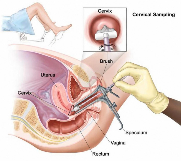
Figure 1: Conceptual workflow of cervical sample collection and processing. Visualization of the cervix during gynecological examination, followed by collection of cervical cells from the transformation zone using a cytobrush. Please click here to view a larger version of this figure.
4. Cytology prodedure and interpretation
Liquid-based cytology slides were prepared from cervical specimens according to the manufacturer’s protocol and institutional laboratory procedures. Slides were stained using the Papanicolaou staining method and examined by trained cytopathologists. Cytological findings were classified using the Bethesda System into negative for intraepithelial lesion or malignancy; atypical squamous cells of undetermined significance; low-grade squamous intraepithelial lesion; high-grade squamous intraepithelial lesion; and other categories, where applicable.
A second cytopathologist reviewed abnormal or equivocal slides. Discrepant interpretations were resolved by consensus review. Cytology results were linked with HPV results using anonymized study codes.
5. Histopathology procedure and interpretation
Women with abnormal cytology, positive high-risk HPV results requiring further evaluation, or clinically indicated colposcopic findings underwent cervical biopsy according to routine clinical practice. Biopsy specimens were fixed in 10% neutral buffered formalin, processed routinely, embedded in paraffin, sectioned, and stained with hematoxylin and eosin. Histopathological diagnoses were classified as benign/negative, cervical intraepithelial neoplasia grade 1, cervical intraepithelial neoplasia grade 2, cervical intraepithelial neoplasia grade 3, or invasive carcinoma, where applicable.
All histopathology slides were reviewed by qualified pathologists. A second pathologist reviewed equivocal or high-grade lesions, and the final diagnosis was established by consensus. Histopathology results were correlated with cytology and HPV genotype results for participants with available biopsy data.
6. Primary HR-HPV detection
Primary HR-HPV DNA testing was performed using a clinically validated real-time PCR assay that individually detected HPV16 and HPV18 and reported 12 additional high-risk genotypes as a pooled result. DNA extraction, amplification, and detection were performed according to the manufacturer's instructions. The assay targeted the L1 region of the HPV genome and incorporated β-globin as an internal control for specimen adequacy and amplification validity. Each run included internal controls and external positive and negative controls. Samples with invalid internal control results were repeated. Samples that remained invalid after repeat testing were excluded from the final analysis. The overall laboratory workflow for HR-HPV detection and extended genotyping is illustrated in Figure 2.
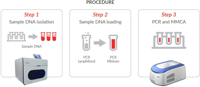
Figure 2: Laboratory workflow for HR-HPV detection and extended genotyping. Schematic overview of primary HR-HPV detection followed by nested multiplex PCR for extended genotype identification. Mix 1 included HPV31, HPV45, and HPV59; Mix 2 included HPV33, HPV58, HPV52, HPV56, and HPV35; and Mix 3 included HPV68, HPV39, HPV51, and HPV66. Amplicon identification was performed using a 50-bp molecular weight ladder. Please click here to view a larger version of this figure.
For extended HR-HPV genotyping, genotype-specific nested multiplex PCR assays were performed using previously validated primers. Detailed primer sequences, expected amplicon sizes, and multiplex reaction allocations are summarized in Supplementary Table 1.
7. Agarose gel electrophoresis and band interpretation
Nested multiplex PCR products were separated on 2% agarose gels containing nucleic acid stain and electrophoresed in 1× Tris–Borate–EDTA buffer at 100 V for 45 min. A 50-bp molecular weight ladder was included in each run to estimate amplicon size. Bands were visualized using a gel documentation system under ultraviolet illumination in accordance with laboratory safety procedures. Genotype assignment was based on the presence of a clearly identifiable band at the expected amplicon size for the corresponding genotype-specific primer pair. Samples producing faint, non-specific, overlapping, or discordant bands were retested. Unresolved results following repeat testing were classified as indeterminate and excluded from genotype-specific analyses.
8. Quality control
Quality-control procedures were applied throughout sample collection, molecular testing, data extraction, and statistical analysis. Sample adequacy was verified using internal control amplification. Each PCR batch included positive, negative, and no-template controls. 10% of the samples were randomly selected for repeat testing to assess reproducibility. Repeat testing was considered concordant when the same HR-HPV positivity status and genotype interpretation were obtained. Discordant results were repeated, reviewed by a senior laboratory scientist, and finalized only after consensus interpretation.
Data entry was independently checked by two investigators. Inconsistencies between clinical records, laboratory files, and analysis sheets were resolved by reviewing the original source records.
9. Statistical analysis
Continuous variables were summarized as mean ± standard deviation or median with interquartile range, depending on distribution. Categorical variables were summarized as frequencies and percentages. HR-HPV prevalence was calculated as the number of HR-HPV-positive women divided by the total number of eligible screened women.
Categorical variables were compared using the chi-square test or Fisher’s exact test, as appropriate. Continuous variables were compared using the independent-samples t-test or the Mann–Whitney U test, depending on normality. Variables with clinical relevance and variables showing p < 0.10 in univariate analysis were considered for inclusion in multivariate logistic regression. Adjusted odds ratios, 95% confidence intervals, and p-values were reported. Missing data were not imputed; complete-case analysis was performed for regression models.
Age-stratified analyses were performed using predefined groups: 25–30, 31–40, 41–50, and 51–60 years. Temporal trends were evaluated by comparing annual HR-HPV prevalence across 2022, 2023, and 2024. Genotype-specific frequencies were reviewed descriptively because genotype-specific annual datasets were not analyzed as formal temporal trend outcomes. A p-value < 0.05 was considered statistically significant.
결과
Baseline characteristics of the study population
A total of 5,000 eligible women who underwent cervical cancer screening between January 2022 and December 2024 were included in the final analysis. The mean age of the study population was 38.7 ± 8.9 years. The largest proportion of participants was in the 31–40-year age group (1,730/5,000, 34.6%), followed by the 41–50-year age group (1,490/5,000, 29.8%), the 25–30-year age group (1,070/5,000, 21.4%), and the 51–60-year age group (710/5,000, 14.2%). Most participants were multiparous (3,115/5,000, 62.3%), whereas 1,885 women (37.7%) were nulliparous. Prior cervical screening had been documented in 2,435 women (48.7%), while 2,565 women (51.3%) had no recorded screening history. Smoking was reported by 1,125 women (22.5%), and 910 women (18.2%) reported more than one lifetime sexual partner. Detailed baseline demographic and clinical characteristics are presented in Table 1.
| Variable | n (%) |
| Age, years, mean ± SD | 38.7 ± 8.9 |
| 25–30 years | 1,070 (21.4) |
| 31–40 years | 1,730 (34.6) |
| 41–50 years | 1,490 (29.8) |
| 51–60 years | 710 (14.2) |
| Multiparous | 3,115 (62.3) |
| Nulliparous | 1,885 (37.7) |
| Prior cervical screening | 2,435 (48.7) |
| No prior cervical screening | 2,565 (51.3) |
| Smoking | 1,125 (22.5) |
| More than one lifetime sexual partner | 910 (18.2) |
Table 1: Baseline characteristics of the study population. Demographic and clinical characteristics of women included in the cervical screening cohort.
Prevalence of high-risk Human Papillomavirus infection
Among the 5,000 screened women, 612 tested positive for HR-HPV, corresponding to an overall prevalence of 12.2%. Single-genotype infections were identified in 402 of the 612 HR-HPV-positive women (65.7%), whereas multiple-genotype infections were detected in 210 women (34.3%). Among the multiple infections, double infections were most common (148/612, 24.2%), followed by triple infections (49/612, 8.0%) and quadruple infections (13/612, 2.1%). The distribution of single and multiple infections is shown in Figure 3.
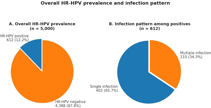
Figure 3: Distribution of single and multiple HR-HPV infections. (A) Overall prevalence of HR-HPV infection among the screened study population (n = 5,000). (B) Distribution of single-genotype and multiple-genotype HR-HPV infections among HR-HPV-positive women (n = 612). Values are presented as n (%). Please click here to view a larger version of this figure.
Genotype distribution of high-risk Human Papillomavirus
Genotype analysis was performed for all HR-HPV-positive women (n = 612). HPV16 was the most frequently detected genotype (18.6%), followed by HPV31 (14.2%), HPV56 (12.9%), HPV66 (11.8%), and HPV51 (9.6%). Additional detected genotypes included HPV52, HPV58, HPV45, HPV18, HPV68, HPV59, HPV35, HPV39, and HPV33. Genotypes covered by the nonavalent HPV vaccine accounted for 61.4% of detected infections, whereas non-vaccine high-risk genotypes accounted for 38.6%. Detailed genotype frequencies are presented in Table 2 and Figure 4.
| Genotype | n (%) |
| HPV16 | 114 (18.6) |
| HPV31 | 87 (14.2) |
| HPV56 | 79 (12.9) |
| HPV66 | 72 (11.8) |
| HPV51 | 59 (9.6) |
| HPV52 | 53 (8.7) |
| HPV58 | 46 (7.5) |
| HPV45 | 42 (6.9) |
| HPV18 | 40 (6.5) |
| HPV68 | 36 (5.9) |
| HPV59 | 34 (5.6) |
| HPV35 | 29 (4.7) |
| HPV39 | 27 (4.4) |
| HPV33 | 24 (3.9) |
Table 2: Distribution of HR-HPV genotypes among positive cases. Frequency and percentage of individual HR-HPV genotypes detected among HPV-positive women.
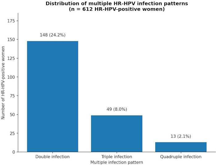
Figure 4: Distribution of HR-HPV genotypes among positive cases. Frequency and proportion of individual HR-HPV genotypes detected among HPV-positive women. Please click here to view a larger version of this figure.
Age-specific distribution of HR-HPV infection
HR-HPV prevalence varied across age groups and was highest among women aged 25–30 years. Prevalence declined progressively with increasing age and was lowest among women aged 51–60 years. Age-specific prevalence data, including denominators and HR-HPV-positive counts, are presented in Table 3 and Figure 5.
| Age group | Screened women, n | HR-HPV positive, n (%) |
| 25–30 years | 1,070 | 202 (18.9) |
| 31–40 years | 1,730 | 252 (14.6) |
| 41–50 years | 1,490 | 146 (9.8) |
| 51–60 years | 710 | n (6.3) |
Table 3: Age-specific prevalence of HR-HPV infection. Prevalence of HR-HPV infection across predefined age groups.
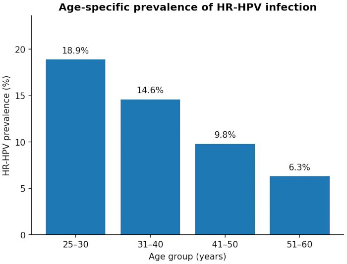
Figure 5: Age-specific prevalence of HR-HPV infection. Distribution of HR-HPV positivity across predefined age groups. Please click here to view a larger version of this figure.
Association between HR-HPV infection and cytological findings
HR-HPV positivity increased with the severity of cytological abnormalities. Women with atypical squamous cells of undetermined significance, low-grade squamous intraepithelial lesions, and high-grade squamous intraepithelial lesions demonstrated higher rates of HR-HPV positivity than women with negative cytology. Detailed cytological categories and corresponding HR-HPV positivity rates are presented in Table 4 and Figures 6, 7.
| Cytology | HR-HPV positive, n (%) |
| Normal cytology | 234 (38.2) |
| ASC-US | 132 (21.6) |
| LSIL | 152 (24.8) |
| HSIL | 94 (15.4) |
| Total | 612 (100.0) |
Table 4: HR-HPV infection according to cytological findings. Distribution of HR-HPV positivity according to Bethesda cytology categories.
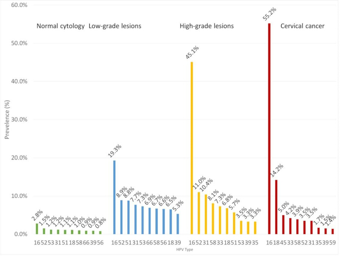
Figure 6: HR-HPV genotype distribution according to cytology category. Distribution of HR-HPV genotypes across cytological categories, including negative cytology, atypical squamous cells of undetermined significance, low-grade squamous intraepithelial lesion, and high-grade squamous intraepithelial lesion. Please click here to view a larger version of this figure.
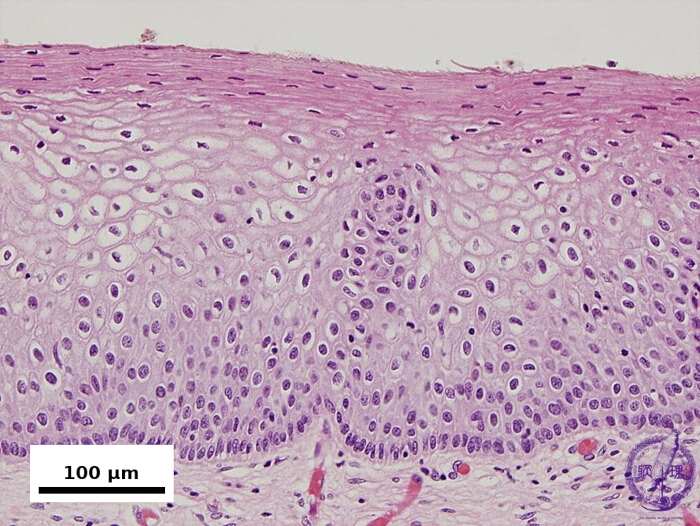
Figure 7: Correlation between cytological and histopathological findings. Representative hematoxylin and eosin-stained cervical biopsy section demonstrating a high-grade squamous intraepithelial lesion. Scale bar = 100 µm. Please click here to view a larger version of this figure.
Descriptive correlation between HPV testing, cytology, and histopathology
HR-HPV positivity showed a descriptive association with increasing severity of cytological abnormalities. HPV-positive cases were more frequently observed among women with abnormal cytology, and HPV16 and HPV18 were more commonly detected in women with high-grade cytological and histopathological findings. Histopathological evaluation was available only for clinically indicated cases; therefore, these observations should be interpreted descriptively. Formal concordance analyses between PCR, cytology, and histopathology were not performed.
Clinical correlates and risk factors
Multivariate logistic regression analysis identified age below 35 years, multiple sexual partners, smoking, and lack of prior cervical screening as factors associated with HR-HPV positivity. Adjusted odds ratios, 95% confidence intervals, and p-values for the final model are presented in Table 5 and Figure 8.
| Variable | Adjusted OR | 95% CI | p-value |
| Age <35 years | 2.14 | 1.76–2.60 | <0.001 |
| Multiple lifetime sexual partners | 2.89 | 2.21–3.78 | <0.001 |
| Smoking | 1.52 | 1.18–1.96 | 0.002 |
Table 5: Multivariate analysis of risk factors associated with HR-HPV infection. Adjusted odds ratios, 95% confidence intervals, and p-values for variables associated with HR-HPV positivity.
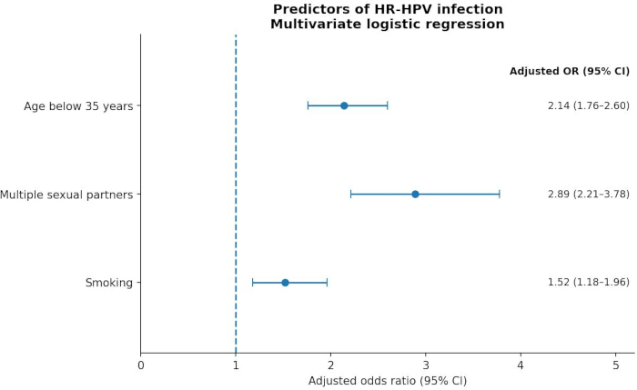
Figure 8: Multivariate logistic regression analysis of predictors of HR-HPV infection. Forest plot showing adjusted odds ratios and 95% confidence intervals for independent predictors of HR-HPV positivity. The vertical reference line indicates an odds ratio of 1.0. Please click here to view a larger version of this figure.
Temporal trends in HR-HPV infection
Annual HR-HPV prevalence remained relatively stable throughout the study period from 2022–2024, with only minor year-to-year variation. Because genotype-specific annual datasets and complete vaccination-history data were not available for all participants, genotype-specific temporal patterns were evaluated descriptively only. Consequently, no definitive conclusions regarding increasing or decreasing genotype-specific trends can be drawn from the present dataset. Year-wise HR-HPV prevalence is presented in Table 6.
| Year | Screened women, n | HR-HPV positive, n (%) |
| 2022 | 1,600 | 189 (11.8) |
| 2023 | 1,700 | 214 (12.6) |
| 2024 | 1,700 | 209 (12.3) |
| Total | 5,000 | 612 (12.2) |
Table 6: Year-wise distribution of HR-HPV infection. Annual prevalence of HR-HPV infection during the study period from 2022–2024.Overall, HR-HPV infection was detected in 12.2% of screened women. HPV16 was the predominant genotype, and HR-HPV positivity was associated with younger age, multiple sexual partners, smoking, and lack of prior cervical screening.
Overall, HR-HPV infection was detected in 12.2% of screened women. HPV16 was the predominant genotype, and HR-HPV positivity was associated with younger age, multiple sexual partners, smoking, and lack of prior cervical screening.
DATA AVAILABILITY:
The de-identified dataset supporting the findings of this study is provided as Supplementary File 1 and includes demographic variables, HR-HPV test results, genotype assignments, cytological findings, histopathological diagnoses (where available), and the data underlying the statistical analyses. All personal identifiers were removed prior to data sharing to protect participant confidentiality.
Supplementary Table 1: Primer sequences, expected amplicon sizes, and multiplex mixture allocation for nested multiplex PCR. Primer sequences, expected amplicon sizes, and multiplex PCR mixture assignments used for extended HR-HPV genotyping. Please click here to download this file.
Supplementary File 1: The de-identified dataset supporting the findings of this study. Please click here to download this file.
토론
This study provides a comprehensive assessment of the prevalence, genotype distribution, and selected clinical correlates of high-risk human papillomavirus (HR-HPV) infection among 5,000 women undergoing routine cervical cancer screening in Leshan, Sichuan, China. The overall HR-HPV prevalence of 12.2% indicates a substantial burden of infection within this screening population and is broadly consistent with rates reported in recent epidemiological studies from Asia and other regions, although differences in population characteristics, screening practices, and vaccination coverage may contribute to regional variation12,13,14. The predominance of single-genotype infections, together with the substantial proportion of multiple-genotype infections, highlights the heterogeneous nature of HR-HPV infection within the study population15,16,17,18. Although the clinical significance of multiple-genotype infections remains incompletely understood, previous studies have suggested that co-infections may influence viral persistence, host immune responses, and progression to cervical lesions19,20,21,22,23. Because the present study employed a cross-sectional design, the independent contribution of multiple infections to disease progression could not be determined.
In addition to providing regional epidemiological data, this study describes a standardized workflow for cervical specimen collection, HR-HPV detection, extended genotyping, and interpretation of results. The protocol integrates cytobrush-based sample collection, liquid-based specimen preservation, primary HR-HPV testing, nested multiplex PCR for extended genotype characterization, and quality-control procedures designed to support reproducibility. By documenting these methodological steps in detail, the study provides a framework that may facilitate implementation of HPV surveillance and molecular screening workflows in comparable laboratory settings.
HPV16 was the most frequently detected genotype, followed by HPV31, HPV56, HPV66, and HPV51. Consistent with previous reports, HPV16 was more frequently associated with higher-grade cytological and histopathological abnormalities than other genotypes22,23,24,25,26. Non-vaccine HR-HPV genotypes, including HPV35, HPV56, and HPV66, also accounted for a substantial proportion of detected infections27,28,29. However, because individual vaccination histories were not fully available, the observed genotype distribution should be interpreted with caution. The present findings do not permit conclusions regarding vaccine-driven genotype replacement and are more appropriately viewed as descriptive regional surveillance data. Continued monitoring of genotype distribution remains important for evaluating long-term epidemiological patterns and informing future prevention strategies30,31,32,33,34.
Multivariate analysis identified younger age, multiple sexual partners, smoking, and lack of prior cervical screening as factors associated with HR-HPV positivity. These findings are consistent with established risk factors for HPV acquisition and persistence reported in previous studies35,36,37,38. Smoking has been associated with impaired local immune responses and increased susceptibility to persistent HPV infection39,40,41, whereas behavioral factors may increase opportunities for viral exposure. The association between the absence of prior screening and HR-HPV positivity further emphasizes the importance of regular cervical cancer screening programs for early detection and risk reduction. Temporal evaluation showed a relatively stable overall HR-HPV prevalence over the three-year study period. Although minor variation in genotype frequencies was observed descriptively, genotype-specific temporal trends could not be evaluated conclusively because annual genotype-specific datasets and complete vaccination-history information were unavailable. Consequently, the present study does not provide sufficient evidence to support conclusions regarding genotype replacement or sustained genotype-specific epidemiological shifts41,42.
Several strengths enhance the value of this study. These include the large screening population, comprehensive genotype characterization, integration of clinical and demographic variables, and use of standardized molecular diagnostic methods for HPV detection and genotyping. The inclusion of cytological and histopathological information also provides clinically relevant context for interpreting HPV infection patterns. Nevertheless, several limitations should be acknowledged. The retrospective cross-sectional design precludes assessment of HPV persistence, clearance, reinfection, and causal progression to cervical disease. The hospital-based study population may not fully represent the broader female population of Leshan or Sichuan Province. In addition, incomplete vaccination history data limited the evaluation of vaccine impact, and behavioral variables obtained from clinical records may have been affected by reporting bias or incomplete documentation. Histopathological data were available only for clinically indicated biopsy cases, introducing the possibility of verification bias. Finally, temporal observations were based on a relatively short three-year study period and should therefore be interpreted cautiously.
Overall, this study provides contemporary evidence regarding the epidemiology of HR-HPV infection in a large cervical screening cohort from Leshan, Sichuan, China. The findings demonstrate a measurable burden of HR-HPV infection, a diverse distribution of genotypes, and significant associations between HR-HPV positivity and selected demographic, behavioral, and screening-related factors. These data support the continued use of HPV-based screening strategies and underscore the importance of ongoing regional surveillance to inform cervical cancer prevention programs and public health policy.
Conclusion:
This retrospective cross-sectional study characterized the prevalence, genotype distribution, and selected clinical correlates of HR-HPV infection among women undergoing cervical cancer screening in Leshan, Sichuan, China. Among 5,000 screened women, the overall HR-HPV prevalence was 12.2%. HPV16 was the most frequently detected genotype, followed by HPV31, HPV56, HPV66, and HPV51. Single-genotype infections were more common than multiple-genotype infections, although multiple infections were observed in a substantial proportion of HR-HPV-positive women. HR-HPV positivity was associated with younger age, multiple sexual partners, smoking, and lack of prior cervical screening.
These findings highlight the value of continued regional HPV surveillance and genotype monitoring within cervical cancer screening programs. However, the cross-sectional design, incomplete vaccination history data, and limited follow-up preclude drawing conclusions about HPV persistence, vaccine effectiveness, genotype replacement, or long-term clinical outcomes. Future prospective studies incorporating longitudinal follow-up, comprehensive vaccination histories, and expanded cytology-histopathology correlation are needed to further clarify the epidemiological and clinical significance of emerging HR-HPV genotype patterns.
공개 사항
The authors declare that they have no competing financial interests, conflicts of interest, or other disclosures relevant to this study.
감사의 글
The authors would like to thank the staff of the Department of Gynecology, Department of Pathology, and Molecular Diagnostic Laboratory of Leshan Maternal and Child Health Hospital for their assistance with specimen processing, laboratory testing, data management, and quality-control procedures. The authors also acknowledge all healthcare personnel involved in cervical cancer screening services during the study period.
재료
| 이름 | 회사 | 카탈로그 번호 | 댓글 |
|---|---|---|---|
| Agarose | Thermo Fisher Scientific | R0491 | Preparation of 2% agarose gels for electrophoresis |
| Automated Coverslipper | Sakura Finetek | Tissue-Tek Film Coverslipper | Coverslipping of cytology and histopathology slides |
| β-globin Internal Control Reagents | Roche Diagnostics | Included with assay | Verification of specimen adequacy and PCR validity |
| Cervical Cytobrush | CooperSurgical | Rovers Cervex-Brush | Collection of cervical specimens |
| Cobas 4800 HPV Test Kit | Roche Diagnostics | 5880715190 | Primary HR-HPV detection assay |
| Cobas x 480 Instrument | Roche Diagnostics | Cobas x480 | Automated DNA extraction |
| Cobas z 480 Analyzer | Roche Diagnostics | Cobas z480 | Real-time PCR amplification and detection |
| Colposcope | Olympus Corporation | OCS-500 Series | Colposcopic evaluation of abnormal cervical findings |
| Coverslips | Thermo Fisher Scientific | 12-545M | Cytology and histopathology slide preparation |
| DNA Molecular Weight Marker (50 bp Ladder) | Thermo Fisher Scientific | SM0373 | Determination of PCR amplicon size |
| Ethanol, Absolute | Merck | 100983 | Histological and cytological processing |
| Formalin, Neutral Buffered 10% | Sigma-Aldrich | HT501128 | Fixation of cervical biopsy specimens |
| Gel Documentation System | Bio-Rad Laboratories | Gel Doc EZ System | Visualization and documentation of PCR products |
| Hematoxylin Solution | Sigma-Aldrich | H3136 | Histopathological staining |
| Histology Embedding System | Leica Biosystems | EG1150 | Paraffin embedding of tissue specimens |
| Light Microscope | Olympus Corporation | BX53 | Cytology and histopathology examination |
| Liquid-Based Cytology Medium (PreservCyt) | Hologic | 70098-002 | Preservation and transport of cervical samples |
| Microcentrifuge Tubes (1.5 mL) | Eppendorf | 30120086 | PCR reaction preparation and sample handling |
| Microtome | Leica Biosystems | RM2235 | Sectioning of paraffin-embedded tissue blocks |
| Microscope Slides | Thermo Fisher Scientific | J1800AMNZ | Cytology and histopathology evaluation |
| Multiplex PCR Master Mix | Qiagen | 206143 | Nested multiplex PCR amplification |
| Nuclease-Free Water | Thermo Fisher Scientific | AM9937 | Preparation of PCR reactions |
| Paraffin Wax | Leica Biosystems | 39601095 | Tissue embedding |
| Papanicolaou Stain Kit | Merck | 1.01646 | Cytological staining and evaluation |
| Pathology Tissue Processor | Leica Biosystems | TP1020 | Automated tissue processing |
| PCR Primer Sets for HPV31, HPV33, HPV35, HPV39, HPV45, HPV51, HPV52, HPV56, HPV58, HPV59, HPV66, and HPV68 | Integrated DNA Technologies (IDT) | Custom synthesis | Extended HR-HPV genotyping |
| PCR Tubes (0.2 mL) | Eppendorf | 30124359 | PCR amplification reactions |
| Refrigerated Centrifuge | Eppendorf | 5810R | Sample preparation and processing |
| R Statistical Software v4.3.1 | R Foundation for Statistical Computing | Version 4.3.1 | Statistical analysis and data visualization |
| Roche Cobas 4800 HPV Assay Software | Roche Diagnostics | Included with system | HPV result interpretation and reporting |
| SPSS Statistical Software v26.0 | IBM Corporation | Version 26 | Statistical analysis |
| Thermal Cycler | Bio-Rad Laboratories | T100 Thermal Cycler | Nested multiplex PCR amplification |
| Tris-Borate-EDTA (TBE) Buffer | Thermo Fisher Scientific | BP1333 | Agarose gel electrophoresis |
| Ultraviolet Transilluminator | Bio-Rad Laboratories | Universal Hood III | Visualization of nucleic acid bands |
| Xylene | Merck | 108298 | Histological slide preparation and deparaffinization |
참고문헌
- Van Doorslaer K et al. ICTV virus taxonomy profile: Papillomaviridae. J Gen Virol. 2018;99(8):989-990.
- Tsakogiannis D et al. Nucleotide polymorphisms of the human papillomavirus 16 E1 gene. Arch Virol. 2014;159(1):51-63.
- Tsakogiannis D et al. Molecular and phylogenetic analysis of the HPV16 E4 gene in cervical lesions from women in Greece. Arch Virol. 2012;157(9):1729-1739.
- Bernard HU et al. Classification of papillomaviruses (PVs) based on 189 PV types and proposal of taxonomic amendments. Virology. 2010;401(1):70-79.
- Bernard HU, Calleja-Macias IE, Dunn ST. Genome variation of human papillomavirus types: Phylogenetic and medical implications. Int J Cancer. 2006;118(5):1071-1076.
- International Agency for Research on Cancer. Human papillomaviruses. IARC Monogr Eval Carcinog Risks Hum. 2007;90:1-636.
- Li Y, Xu C. Human papillomavirus-related cancers. Adv Exp Med Biol. 2017;1018:23-34.
- Martinez-Zapien D et al. Structure of the E6/E6AP/p53 complex required for HPV-mediated degradation of p53. Nature. 2016;529(7587):541-545.
- Shimada M et al. The human papillomavirus E6 protein targets apoptosis-inducing factor (AIF) for degradation. Sci Rep. 2020;10(1):14195.
- Aarthy M, Kumar D, Giri R, Singh SK. E7 oncoprotein of human papillomavirus: Structural dynamics and inhibitor screening study. Gene. 2018;658:159-177.
- Zhang Y et al. Roles of human papillomavirus in cancers: Oncogenic mechanisms and clinical use. Signal Transduct Target Ther. 2025;10(1):44.
- Bletsa G et al. Genetic variability of the HPV16 early genes and LCR: Present and future perspectives. Expert Rev Mol Med. 2021;23.
- Zygouras I et al. Human papillomavirus 16 DNA methylation patterns and investigation of integration status in head and neck cancer cases. Int J Mol Sci. 2023;24(18):14593.
- Bray F et al. Global cancer statistics 2022: GLOBOCAN estimates of incidence and mortality worldwide for 36 cancers in 185 countries. CA Cancer J Clin. 2024;74(4):229-263.
- Tsakogiannis D et al. Mutation profile of HPV16 L1 and L2 genes in different geographic areas. Viruses. 2023;15(1):141.
- Ronco G et al. Efficacy of HPV-based screening for prevention of invasive cervical cancer: Follow-up of four European randomised controlled trials. Lancet. 2014;383(9916):524-532.
- Lew JB et al. Primary HPV testing versus cytology-based cervical screening in women in Australia vaccinated for HPV and unvaccinated: Effectiveness and economic assessment for the National Cervical Screening Program. Lancet Public Health. 2017;2(2):e96-e107.
- Ostrbenk Valencak A et al. Clinically validated HPV assays offer comparable long-term safety in primary cervical cancer screening: A 9-year follow-up of a population-based screening cohort. Int J Cancer. 2025;156(4):788-801.
- Tsakogiannis D, Gartzonika C, Levidiotou-Stefanou S, Markoulatos P. Molecular approaches for HPV genotyping and HPV-DNA physical status. Expert Rev Mol Med. 2017;19:e1.
- Fontham ETH et al. Cervical cancer screening for individuals at average risk: 2020 guideline update from the American Cancer Society. CA Cancer J Clin. 2020;70(5):321-346.
- Perkins RB et al. 2019 ASCCP risk-based management consensus guidelines for abnormal cervical cancer screening tests and cancer precursors. J Low Genit Tract Dis. 2020;24(2):102-131.
- Lin S et al. Worldwide trends in cervical cancer incidence and mortality, with predictions for the next 15 years. Cancer. 2021;127(21):4030-4039.
- Du J, Ahrlund-Richter A, Nasman A, Dalianis T. Human papilloma virus (HPV) prevalence upon HPV vaccination in Swedish youth: A review based on findings from 2008-2018 and perspectives on cancer prevention. Arch Gynecol Obstet. 2021;303(2):329-335.
- Oyervides-Munoz MA et al. Multiple HPV infections and viral load association in persistent cervical lesions in Mexican women. Viruses. 2020;12(4):380.
- Zhang W et al. Prevalence and genotype distribution of human papillomavirus infections in Beijing, China between 2016 and 2020. Virol J. 2023;20(1):11.
- Sotlar K et al. Detection and typing of human papillomavirus by E6 nested multiplex PCR. J Clin Microbiol. 2004;42(7):3176-3184.
- Malagon T, Franco EL, Tejada R, Vaccarella S. Epidemiology of HPV-associated cancers past, present and future: Towards prevention and elimination. Nat Rev Clin Oncol. 2024;21(8):522-538.
- Singh D et al. Global estimates of incidence and mortality of cervical cancer in 2020: A baseline analysis of the WHO Global Cervical Cancer Elimination Initiative. Lancet Glob Health. 2023;11(2).
- Arroyo Muhr LS et al. Concomitant human papillomavirus vaccination and screening for elimination of HPV and cervical cancer. Nat Commun. 2024;15(1):3679.
- Kutz JM et al. Barriers and facilitators of HPV vaccination in Sub-Saharan Africa: A systematic review. BMC Public Health. 2023;23(1):974.
- Wendland EM et al. Prevalence of HPV infection among sexually active adolescents and young adults in Brazil: The POP-Brazil Study. Sci Rep. 2020;10(1):4920.
- Alarcon-Romero LDC et al. Prevalence and distribution of human papillomavirus genotypes (1997-2019) and their association with cervical cancer and precursor lesions in women from Southern Mexico. Cancer Control. 2022;29:10732748221103331.
- Bhattacharya M, Reiter PL, McRee AL. Nativity status and genital HPV infection among adults in the U.S. Hum Vaccin Immunother. 2019;15(8):1897-1903.
- Hu JP et al. Prevalence and genotype distribution of human papillomavirus infection among 66,000 women from 2014 to 2023 in the plateau region of Southwest China. Virol J. 2024;21(1):176.
- Bruni L et al. Human papillomavirus and related diseases in the world. ICO/IARC Information Centre on HPV and Cancer. Summary Report. 2023:1-376. Available from: https://www.hpvcentre.net/statistics/reports/XWX.pdf. Accessed June 24, 2026.
- Agorastos T et al. Epidemiology of HPV infection and current status of cervical cancer prevention in Greece: Final results of the LYSISTRATA cross-sectional study. Eur J Cancer Prev. 2014;23(5):425-431.
- Agorastos T et al. Prevalence and distribution of high-risk human papillomavirus in Greece. Eur J Cancer Prev. 2009;18(6):504-509.
- Argyri E et al. A cross-sectional study of HPV type prevalence according to age and cytology. BMC Infect Dis. 2013;13(1):53.
- Tsiodras S et al. Molecular epidemiology of HPV infection using a clinical array methodology in 2952 women in Greece. Clin Microbiol Infect. 2011;17(8):1185-1188.
- Panotopoulou E et al. Prevalence of human papillomavirus types 6, 11, 16, 18, 31, and 33 in a cohort of Greek women. J Med Virol. 2007;79(11):1898-1905.
- Salazar KL et al. Multiple human papilloma virus infections and their impact on the development of high-risk cervical lesions. Acta Cytol. 2015;59(5):391-398.
- Bruno MT, Scalia G, Cassaro N, Boemi S. Multiple HPV16 infection with two strains: A possible marker of neoplastic progression. BMC Cancer. 2020;20(1):444.