본 연구는 5xFAD 마우스 모델에서 Qi-fu-yin (QFY) 치료의 효과를 평가합니다. 연구 결과, QFY 투여는 학습 및 기억 능력의 향상, 뇌 내 Aβ 침착 및 세포 노화의 감소, 그리고 대뇌 피질의 시냅스 구조 손상 완화와 관련이 있는 것으로 나타났습니다.
연구 논문
5xFAD 마우스 뇌의 인지 기능 장애 개선 및 세포 노화 감소에 미치는 Qi-fu-yin의 효과
* These authors contributed equally
이 논문에서
요약
초록
전통 중의학 처방인 Qi-fu-yin (QFY)의 기전은 아직 완전히 밝혀지지 않았으나, 알츠하이머병 (AD)에서 치료 가능성을 보여주었습니다. 본 연구는 5xFAD 마우스에서 QFY 치료와 인지 장애 사이의 연관성을 조사하는 것을 목표로 하였습니다. 우리는 5xFAD 마우스 뇌 내의 Amyloid-β protein 1-42 (Aβ1-42), Aβ1-40, Growth-associated protein 43 (GAP-43), Synaptophysin (SYN) 및 Postsynaptic density protein 95 (PSD-95) 수치를 평가하기 위해 악력, 보행, 군집 둥지 만들기, 셔틀 박스, 모리스 수중 미로 검사 및 ELISA를 사용하였습니다. 피질 내의 노화 관련 분비 표현형 (SASP) 수치를 정량화하기 위해 Luminex 사이토카인 분석이 사용되었습니다. 동물 뇌의 노화 관련 β-galactosidase (SA-β-Gal) 양, Aβ 침착 및 수지상 돌기 가시의 양은 각각 SA-β-Gal 염색, 면역조직화학 염색 및 Golgi-Cox 염색으로 측정하였습니다. 면역형광 검사 결과, QFY 치료는 5xFAD 마우스의 피질 Aβ 플라크 부담을 극적으로 감소시키고 p21 발현을 낮추었습니다. 5xFAD 마우스에서 QFY 요법은 학습 및 기억 능력을 현저히 향상시키고, 노화를 완화하며, 마우스 피질 내 GAP-43, PSD-95 및 SYN 단백질의 양을 증가시켰습니다. QFY 치료 후, 뇌 내 Aβ 플라크와 SA-β-Gal 활성뿐만 아니라 해마 내 Aβ1-42 및 Aβ1-42/Aβ1-40 수치가 유의하게 감소하였습니다. 5xFAD 마우스의 피질에서 IL-1α, IL-1β, IL-6, IL-17A 및 IFN-γ 수치가 상당히 감소하였습니다. PCA 및 Pearson 상관관계 분석 결과, SASP 사이토카인과 시냅스 단백질 수치 사이에 강한 음의 상관관계가 있음이 입증되었습니다. 이러한 결과는 QFY 치료가 5xFAD 마우스의 시냅스 구조 손상을 완화함과 동시에 노화 관련 인지 저하 및 세포 노화 표현형의 개선과 연관이 있음을 시사합니다. 본 연구 결과는 알츠하이머병의 잠재적 치료 후보로서 QFY에 대한 추가 연구의 근거를 제공합니다.
서론
Alzheimer's disease (AD)1 is the most prevalent form of dementia, characterized by amyloid-β protein (Aβ), hyperphosphorylated tau proteins (P-Tau), synaptic loss, and chronic inflammation. Currently available medications for AD have a number of side effects and are unable to stop the disease's progression2,3,4,5.
Senescence, which manifests as organism-wide disruptions across cellular and interstitial microenvironments rather than localized organ-specific alterations, is a key risk factor in AD pathogenesis. This systemic dysregulation distinguishes AD progression from single tissue-pathology models by fundamentally changing homeostatic mechanisms across biological systems6,7. Although cellular senescence initially serves as a homeostatic regulatory mechanism, the accumulation of senescent cells eventually causes progressive tissue degradation and persistent inflammatory cascades, fundamentally disrupting physiological homeostasis8. The ensuing inflammation may harm synapses and impair cognitive function9,10.
Cellular senescence is a complex cellular stress response characterized by telomere shortening, permanent cell-cycle arrest, and altered secretory activity11. Additional hallmarks include enlarged, flattened cell morphology, positive senescence-associated β-galactosidase (SA-β-Gal) staining, lipofuscin accumulation, and secretion of SASP factors12. Memory formation is affected by the notable decline in the levels of proteins associated with synaptic plasticity of axons and dendrites, which occurs with cellular senescence13,14. Because synaptic plasticity is essential for learning and memory, disruptions in synaptic proteins contribute to cognitive impairment. These proteins include GAP-43, SYN, and PSD-95. Undoubtedly, synaptic function is affected by reductions in dendritic numbers and functional abnormalities in the brains of the elderly15.
QFY is a traditional Chinese medicine formula derived from Jing Yue Quan Shu. It is composed of Panax ginseng C.A.Mey (Araliaceae), Polygala tenuifolia Willd (Polygalaceae), Rehmannia glutinosa (Gaertn.) Libosch. ex Fisch. & C. A. Mey (Orobanchaceae), Angelica sinensis (Oliv.) Diels (Apiaceae), Glycyrrhiza uralensis Fisch. ex DC. (Fabaceae), Atractylodes macrocephala Koidz (Asteraceae), Ziziphus jujuba Mill. var. spinosa (Bunge) Hu ex H.F. Chou (Rhamnaceae). Research has demonstrated that QFY significantly ameliorates cognitive dysfunction in rats with diabetes-induced brain injury16. Our previous study found that QFY could improve cognitive learning and memory functions impairment in 5xFAD and APP/PS1 mice. In addition, QFY demonstrated high safety in non-clinical studies, and no obvious acute, subacute, or long-term toxic reactions were observed17,18,19. Analysis of the clinical therapeutic effect of QFY showed that AD patients experienced a significant increase in their capacity for activities of daily living and MMSE scores after QFY treatment20. Following three consecutive days of QFY administration in male Sprague-Dawley rats, ten prototype QFY-derived components, including butylidenephthalide, butylphthalide, 20(S)-ginsenoside Rh1, 20(R)-ginsenoside Rh1, and zingibroside R1, were detected in cerebrospinal fluid21,22. However, the therapeutic mechanism of QFY in AD is still unclear. In this study, we investigated the association between QFY treatment, cellular senescence, and AD-related pathological changes in 5xFAD mice.
Although previous studies have demonstrated the neuroprotective effects of QFY in AD models, the specific involvement of cellular senescence in mediating these effects remains unexplored. Moreover, whether QFY confers cognitive benefits through senescence‑associated synaptic preservation, rather than solely through amyloid‑β reduction, has not been addressed. The present study was therefore designed to investigate whether QFY treatment attenuates cellular senescence and whether this attenuation is associated with improved synaptic plasticity in 5xFAD mice.
프로토콜
모든 동물 실험은 산동중의약대학교 실험동물복지윤리위원회의 승인을 받았습니다(SDUTCM20221101001).
약물
QFY 과립 건조 크림 분말은 Lunan Pharmaceutical Houpu 연구개발 센터에서 제조 및 테스트되었습니다. Donepezil과 Memantine은 Shanghai Yuanye Technology에서 구입하였습니다. 본 연구에는 초기 체중 범위가 19–27 g인 수컷 5xFAD 마우스를 사용하였습니다. QFY 투여량은 임상 인간 용량인 43 g/d에서 도출되었으며, 이는 QFY 과립 건조 크림 분말로 환산 시 16.297 g/d에 해당합니다. 본 실험의 중간 용량은 임상 인간 용량을 기준으로 계산되었습니다 (16.297 g/70 kg x 9.1 = 2.11861 g/kg/d). 고용량은 중간 용량의 2배(4.24 g/kg/d)로, 저용량은 중간 용량의 절반(1.06 g/kg/d)으로 설정하였습니다. 모든 약물 용액은 체중 10 g당 0.1 mL의 부피로 투여되었습니다.
양성 대조군으로는 donepezil과 memantine hydrochloride의 병용 투여를 실시하였습니다. 투여 용량은 현재의 임상 적용 용량(각각 10 mg/d 및 20 mg/d)과 본 연구팀의 이전 데이터를 참고하여 결정하였으며, 그 결과 donepezil 1.0 mg/kg/d와 memantine 2.8 mg/kg/d를 병용하여 총 3.8 mg/kg/d의 용량으로 설정하였습니다. donepezil-memantine 혼합액의 최종 농도는 체중 10 g당 0.1 mL의 투여 부피로 나누어 3.8 mg/kg으로 계산하여 0.38 mg/mL로 조제하였습니다.
저용량, 중용량 및 고용량의 QFY 용액을 다음과 같이 준비하였습니다: 5.3 g, 10.6 g 및 21.2 g의 QFY 건조 분말을 각각 별도의 100 mL 비커에 넣고, 30 mL의 정제수를 가하여 충분히 교반하며 혼합한 후, 메스실린더로 옮겨 정제수로 최종 부피를 50 mL로 맞추어 최종 질량 농도를 각각 0.106 g/mL, 0.212 g/mL 및 0.424 g/mL가 되게 하였습니다. 양성 대조군의 경우, 5 mg의 donepezil과 14 mg의 memantine을 정제수에 녹여 최종 부피를 50 mL로 조정하였습니다. 각 그룹의 동물에게 해당 약물 용액을 1일 1회 경구 투여하였습니다.
동물
5xFAD 형질전환 마우스 [B6. Cg-Tg (APPSwFlLon, PSEN1* M146L* L286V)6799Vas/Mmjax]를 실험실에서 번식시켰다. 모든 동물은 산동중의약대학교의 SPF 환경에서 온도 22–24 oC, 습도 50–60%, 12시간 명암 주기로 유지되었다. 동물에게는 먹이와 물을 상시 제공하였다.
생후 2.5개월 된 수컷 5xFAD 마우스를 무작위로 처리하지 않은 5xFAD군, donepezil 및 memantine 투여군, 저용량 QFY군, 중용량 QFY군, 고용량 QFY군의 다섯 그룹으로 나누었습니다. 연령이 일치하는 수컷 야생형(WT) 형제 마우스를 WT 대조군으로 설정하였습니다. 처치는 220일 동안 매일 1회 실시하였습니다. (그림 1).
행동 테스트
악력 테스트
마우스의 전지 골격근 강도는 악력 측정기를 사용하여 측정하였다23. 실험 동안 측정 장치는 수평으로 배치하여 마우스가 테이블 표면과 평행을 유지하도록 하였다. 장치의 전원을 켠 후, 마우스를 악력 플레이트 위에 조심스럽게 놓았다. 마우스가 플레이트를 단단히 잡으면, 마우스가 잡고 있던 것을 놓을 때까지 꼬리를 일정한 힘으로 뒤로 당겼다. 이때 측정된 최대 악력을 기록하였다. 이 측정 과정을 3회 반복하였으며, 이 값들의 평균치를 마우스의 악력으로 결정하였다.
보행 테스트
CatWalk XT를 사용하여 마우스의 보행 규칙성을 측정하였다. 실험은 훈련 기간과 테스트 기간으로 나누어 진행하였다24. 실험 하루 전, 실험 쥐들을 실험실로 옮겨 새로운 환경에 적응하도록 하였다. 훈련 기간 동안 마우스들을 시작점에 두고 통로를 따라 끝점까지 달리게 하였다. 각 마우스는 하루에 세 번씩 훈련을 받았다. 3일간의 훈련 후(각 마우스가 통로를 빠르게 통과할 수 있을 때까지), 마우스들을 다시 케이지로 돌려보냈다. 테스트 기간은 훈련 기간과 동일하게 진행되었으며, 컴퓨터 소프트웨어가 각 이동 궤적을 자동으로 분석하였다. 궤적이 적합한 경우에는 녹색 점이 표시되었고, 부적합한 경우에는 주황색 점이 표시되었으며, 각 마우스당 최소 세 개의 녹색 점이 나타난 경우를 적합한 것으로 간주하여 CatWalkXT 10.0 소프트웨어로 분석하였다.
셔틀 박스 테스트
마우스의 조건부 능동 회피 능력을 평가하기 위해 셔틀 박스 테스트를 사용하였다.25장치는 방음실 내에 설치된 셔틀 박스(18 cm x 18 cm)로 구성되었다. 실험은 4일간의 학습 기간과 이후 1일간의 테스트 기간으로 구성되었습니다. 매 학습일마다 마우스는 훈련 시작 전 셔틀 박스 내에서 120초간의 적응 시간을 가졌다. 각 시행은 6초간의 조건 자극(70dB, 1000Hz 톤과 500lux 광원의 결합)으로 시작되었으며, 이어서 그리드 바닥을 통해 3초간 0.3mA의 족부 전기 충격이 가해졌고, 12초의 시행 간 간격으로 마무리되었다. 마우스가 조건 자극 중에 반대편 구획으로 이동하면 전기 충격을 생략하고 이를 능동 회피로 기록하였다. 마우스가 조건 자극 중에는 이동하지 않았으나 족부 전기 충격 중에 이동한 경우에는 이를 수동 회피 반응. 만약 생쥐가 시험 전 과정 동안 가로질러 가지 않았다면, 이는 다음과 같이 기록되었습니다. 회피 실패. 훈련은 하루 30회씩 4일 연속으로 반복되었다. 테스트 기간(5일 차) 동안 다른 모든 매개변수는 학습 기간과 동일하게 유지하되, 족부 전기 충격은 생략하였다. 학습 및 기억의 일차적 측정 지표로 능동적 회피 반응 횟수를 기록하였다.
모리스 수중 미로
실험 동물의 공간 학습 및 기억 능력을 평가하기 위해 Morris 수중 미로 검사를 사용하였다26. 실험은 직경 120 cm, 깊이 50 cm의 수조에서 수행되었다. 수조는 탁한 흰색 물로 채웠으며, 온도는 21 °C로 유지하였다. 직경 10 cm의 플랫폼을 사분면 중 하나에서 수면 아래 1 cm 지점에 설치하였다. 마우스가 공간적 지표로 삼을 수 있도록 수조 주변에 시각적 단서를 배치하였다. 학습 과정은 4일 연속으로 진행되었으며, 매일의 세션은 세 개의 사분면 중 하나에서 시작되었다. 시작 위치는 네 개의 방위점 사이에서 유사 랜덤 방식으로 변경하였다. 각 시행은 동물이 플랫폼에 성공적으로 도달했을 때 종료되었다. 최대 시행 시간은 60 s로 설정하였으며, 만약 동물이 이 시간 내에 플랫폼을 찾지 못하면 플랫폼까지 조심스럽게 안내하였다. 플랫폼에 도달한 후, 동물에게 10 s 동안 짧은 휴식 시간을 준 뒤 홈 케이지로 돌려보냈다. 학습 기간이 종료된 후 24 h 뒤에 테스트 기간을 시작하였다. 5일 차에 마우스를 플랫폼의 반대편 사분면에서 배치하였다. 테스트 기간의 탈출 잠복기, 표적 사분면에서의 이동 시간, 그리고 원래의 수중 플랫폼 구역을 통과한 횟수를 기록하였다.
군락 중첩 시험
실험 동물의 일상 활동은 군집 둥지 만들기 시험(colony nesting test)을 통해 평가되었다.27실험은 그룹당 3마리의 동물을 대상으로 24시간 동안 진행되었다. 약물 투여 당일, 둥지 만들기를 위해 6g의 부드러운 종이를 깐 새 케이지를 준비하였으며, 6시간, 12시간, 24시간 시점에 사진을 촬영하였다. 실험 종료 시, 종이가 케이지 전체에 흩어져 있고 씹은 흔적이 없는 경우 1점으로 처리하였다. 종이 조각들이 케이지 한쪽으로 모여 있으나 느슨하고, 형성된 둥지가 없으며 뚜렷한 찢김이나 접힘이 없는 경우 2점으로 처리하였다. 종이 조각들이 모여 접혀 형성된 둥지 형태를 띠고 있으나 상대적으로 평평하고 뚜렷한 찢김이 없는 경우 3점으로 처리하였다. 종이가 광범위하게 찢어져 깊은 둥지를 형성한 경우 4점을 부여하였다. 실험 종료 시, 남은 찢어지지 않은 조직 (무게가 다음의 기준인 종이 파편으로 정의됨) >(잘게 찢기거나 둥지에 포함되지 않은 0.1 g의 시료를) 조심스럽게 수집하여 풍건시킨 후, 전자 저울(정밀도: 0.01 g)을 사용하여 무게를 측정하였다.
샘플링 및 시료 수집
WT, 5xFAD 및 QFY (2.12 g/kg/d) 그룹에서만 샘플을 수집하였습니다. CO2 질식사 후 머리를 절단하고, 우뇌 반구를 빠르게 열어 해마와 피질을 분리한 다음, 인산완충생리식염수(PBS)로 헹궈 액체 질소가 담긴 1.5 mL 냉동 튜브에 넣었습니다. 이는 생화학적 분석을 위해 -80 oC에서 보관되었습니다. 좌뇌 반구는 4% 파라포름알데히드에 24시간 동안 침전시켜 고정하고, 절대 에탄올에 침전시켜 탈수한 후, 파라핀 왁스에 포매하였습니다. 이후 5 µm 두께로 절편을 만들어 면역조직화학적 검출을 위해 파라핀을 제거하였습니다.
효소결합 면역흡착 분석법 (ELISA)
뇌 조직 내 표적 단백질 수준을 정량화하기 위해 ELISA를 수행하였습니다. 해마 균질액 내 Aβ1–40 및 Aβ1–42 함량은 상용 키트를 사용하여 측정하였으며, 피질 내 GAP-43, PSD-95 및 SYN 수준은 Jiangsu Enzyme Immunoassay Industry에서 제공하는 키트를 사용하여 결정하였습니다. 모든 과정은 제조사의 프로토콜을 엄격히 준수하여 수행되었습니다. 표준물질 시리즈는 키트 지침에 따라 연속 희석하여 준비하였습니다. 마이크로플레이트 배치는 공시료 웰, 표준물질 웰, 5xFAD 샘플 웰, QFY 처리(2.12 g/kg/d) 샘플 웰 및 야생형 웰을 포함하였습니다. 로딩 전, 모든 샘플은 제공된 희석액으로 2배 희석하였습니다. 시약은 피펫 팁을 웰 바닥과 수직이 되도록 유지하여 첨가한 후, 충분히 혼합되도록 부드럽게 흔들어 주었습니다.
플레이트를 밀봉한 후, 37 °C에서 30분 동안 초기 배양을 수행하였습니다. 이 과정 동안 농축 스톡을 초순수로 1:29 비율(농축액 1 mL당 물 29 mL)로 희석하여 세척 완충액을 준비하였습니다. 배양이 끝난 후, 밀봉 필름을 제거하고 액체를 버린 뒤 플레이트를 닦아 건조시켰습니다. 그 후 각 웰에 200 µL의 세척 완충액을 채우고 30초 동안 방치한 뒤 비워냈으며, 이 과정을 5회 반복하였습니다. 이어서 공백 시료를 제외한 모든 웰에 효소 표지 시약(50 µL)을 첨가하고, 플레이트를 다시 밀봉하여 37 °C에서 30분 동안 배양한 후, 이전과 동일하게 5회 세척 단계를 수행하였습니다.
발색을 위해 각 웰에 50 µL의 chromogen solution A를 첨가한 후 50 µL의 chromogen solution B를 첨가하였으며, 각 첨가 단계마다 가볍게 혼합하였다. 플레이트를 37 °C의 암소에서 10분 동안 배양하였다. 웰당 50 µL의 stop solution을 첨가하여 효소 반응을 종결시켰으며, 이에 따라 청색이 즉시 황색으로 변했다. 마이크로플레이트 리더를 사용하여 450 nm에서 흡광도(광학 밀도)를 측정하였고, 공백 웰을 대조군으로 사용하였다. 모든 측정값은 반응 종결 후 15분 이내에 얻었다.
Luminex
SASP 인자의 피질 수준은 Bio‑Plex Pro Mouse Cytokine 23‑plex Assay 키트를 사용하여 측정하였으며, 모든 절차는 제조사의 지침에 따라 수행되었습니다. 시약은 4 °C 보관소에서 꺼내어 사용 전 30분 동안 실온에서 평형을 맞추었습니다. 키트 프로토콜에 따라 표준 스톡을 연속 희석하여 하나의 블랭크 웰을 포함한 7점 표준 곡선을 생성하였습니다. 자성 비드는 미리 혼합하여 권장 작동 농도로 희석하였고, 검출 항체는 작동 용액으로 준비하였으며, 세척 버퍼 농축액은 탈이온수로 1x로 희석하였습니다. PE‑streptavidin 접합체 또한 제공된 희석 버퍼를 사용하여 1x로 희석하였습니다.
분석을 위해, 플레이트 배치도에 따라 지정된 웰에 50 µL의 비드 현탁액, 표준 물질, 품질 관리 시료 및 희석된 조직 샘플(5.6 µg/µL)을 순차적으로 첨가하였다. 그 후 플레이트를 밀봉하고 800 rpm으로 궤도 진탕하며 4 °C에서 하룻밤 동안 배양하였다. 배양 후, 웰당 200 µL의 세척 완충액으로 비드를 3회 세척하였다. 이어서, 준비된 검출 항체 50 µL를 각 웰에 첨가하고, 플레이트를 800 rpm으로 진탕하며 실온에서 1시간 동안 배양하였다. 다시 3회의 세척 과정을 거친 후, PE‑streptavidin 작동 용액 50 µL를 넣고 동일한 진탕 조건의 실온에서 30분간 추가로 배양하였다. 마지막 세척 단계(200 µL 세척 완충액으로 3회)를 수행한 후, 각 웰에 150 µL의 세척 완충액을 첨가하고 실온에서 800 rpm으로 2분간 진탕시킨 다음 Bio‑Plex 기기로 판독하였다.
SA-β-Gal 염색 및 면역조직화학 염색
SA-β-Gal 염색은 Senescence β-Galactosidase Staining Kit의 설명서에 따라 수행되었습니다. SA-β-Gal 염색 후, 절편을 citrate buffer (pH 6.0)에서 가열하여 항원 회복 과정을 거쳤습니다. 내인성 peroxidase 활성을 차단하기 위해 절편을 peroxidase 차단 용액에 넣고 실온에서 10 min 동안 배양하였습니다. 그 후, 절편을 rabbit monoclonal anti‑Aβ₁-₄₂ 항체 (1:400)와 함께 4 °C에서 하룻밤 동안 배양하였습니다. 세척 후, 절편을 anti‑rabbit/mouse IgG 2차 항체와 함께 실온에서 20 min 동안 배양하였습니다. 추가 세척 후, DAB를 사용하여 2 min 동안 면역 반응성을 시각화하였습니다. 마지막으로, 절편을 탈수 및 투명화 처리한 후 중성 수지로 봉입하여 현미경으로 관찰하였습니다.
면역형광법
면역형광 염색을 위해, 파라핀 절편을 자일렌으로 탈파라핀화(3회 x 15분)하고 단계별 에탄올 시리즈(100%, 95%, 75%; 각 2회 x 5분)를 통해 재수화하였다. 항원 회복은 절편을 1x sodium citrate buffer에 넣고 95 °C에서 30분 동안 가열한 후, 서서히 상온까지 냉각시켜 수행하였다. Tween 20이 포함된 인산염 완충 식염수(PBST)로 3회 세척(각 5분)한 후, 0.3% Triton X‑100이 포함된 10% normal goat serum으로 상온에서 90분 동안 투과성 부여 및 블로킹을 진행하였다. 이후 절편을 p21/CIP1/CDKN1A (1:200) 및 Aβ₁‑₄₂ (1:400)에 대한 1차 항체와 함께 37 °C에서 2시간 동안 반응시켰다. PBST로 4회 세척(각 5분)한 후, 해당 종에 맞는 Alexa Fluor-접합 2차 항체(1:500)와 함께 37 °C의 암실에서 2시간 동안 반응시켰다. 추가 세척(PBST로 4회 x 5분) 후, DAPI가 포함된 항퇴색 마운팅 용액으로 절편을 마운팅하고 형광 현미경을 사용하여 이미징하였다.
골지-콕스 염색법
Golgi‑Cox 염색은 제조사의 지침에 따라 상용 키트를 사용하여 수행되었습니다. 용액 A와 B는 실험 24시간 전에 혼합하였습니다. 마우스를 안락사시킨 후, 뇌 조직을 빠르게 적출하여 PBS로 세척하고 용액 A와 B의 혼합액에 침지시켰습니다. 조직을 암실 내 상온에서 6시간 동안 보관한 후, 침전 용액을 새 용액으로 교체하고 암실 내 상온에서 2주 동안 추가로 배양하였습니다(뇌 조직당 최소 2 mL의 침전 용액 사용). 그 후 조직을 용액 C로 옮겨 24시간 동안 배양한 다음, 다시 새 용액 C로 교체하여 암실 내 상온에서 7일 동안 추가로 배양하였습니다. 뇌 조직은 바이브로톰(vibratome)을 사용하여 100 µm 두께로 절편화하였습니다. 절편을 젤라틴 코팅 슬라이드에 마운트하고 상온에서 3일 동안 건조시켰습니다. 슬라이드를 증류수로 두 번 세척(각 5분)한 후, 용액 D, 용액 E 및 증류수의 혼합액(1:1:2 비율)에 15분 동안 침지시켰습니다. 증류수로 두 번 더 세척(각 5분)한 후, 단계별 에탄올 용액(50%, 75%, 95%; 각 5분)으로 절편을 탈수시켰으며, 이어서 절대 에탄올로 4회(각 4분) 및 자일렌으로 3회(각 5분) 세척하였습니다. 마지막으로 마운팅 매질을 사용하여 절편에 커버슬립을 덮었습니다. 현미경으로 이미지를 캡처하였으며, ImageJ를 사용하여 수지상 돌기 가시 밀도를 정량화하였습니다.
통계 분석
모든 결과는 평균 ± 표준편차(SD)로 표기하였다. 데이터 분석 및 그래프 작성에는 GraphPad Prism 8.1.0을 사용하였다. 두 그룹 간의 비교에는 Student’s t-test를 사용하였다. 다중 그룹 비교의 경우 일원 분산 분석(one-way ANOVA)을 실시하였으며, Dunnett's test를 통해 사후 쌍별 비교를 수행하였다. 통계적 유의성은 p < 0.05로 설정하였다.
다변량 및 상관 분석
WT, 5xFAD 및 QFY (2.12 g/kg/day) 그룹(n = 그룹당 8) 간의 SASP 인자(IL-1α, IL-1β, IL-6, IL-17A, IFN-γ, CCL3, CCL4 및 CCL5)의 그룹별 클러스터링을 시각화하기 위해 GraphPad Prism 8.0.1을 사용하여 주성분 분석(PCA)을 수행하였다. 스코어 플롯은 처음 두 개의 주성분을 기반으로 생성되었다. PCA 수행 전 데이터 변환은 적용하지 않았다. SASP 인자와 시냅스 단백질(PSD-95, GAP-43 및 SYN)뿐만 아니라 Aβ₁₋₄₀, Aβ₁₋₄₂, 그리고 Aβ₁₋₄₂/Aβ₁₋₄₀ 비율 간의 선형 관계를 평가하기 위해 피어슨 상관 분석을 실시하였다. 분석은 GraphPad Prism을 사용하여 수행되었으며, 양측 검정 유의 수준은 p < 0.05로 설정하였다. 연관성의 강도와 방향을 시각화하기 위해 상관 행렬과 히트맵을 생성하였다.
결과
Administration of QFY enhances learning and memory in 5xFAD mice
After QFY administration, a shuttle box test was conducted to assess the conditional active avoidance response of mice (Figure 2A–C). The results showed the number of active avoidances on days 1-4 (Figure 2A, p < 0.05) and the area under the curve (AUC) of active avoidance (Figure 2B, p < 0.01) during the learning period, and the number of active avoidances (Figure 2C, p < 0.05) during the test period were significantly reduced in 5xFAD mice compared with WT mice. The administration of donepezil and memantine significantly increased the number of active avoidances on day 2 during the learning period in 5xFAD mice (Figure 2A, p < 0.05). The administration of QFY significantly increased the number of active avoidances on days 3 and 4 (Figure 2A, p < 0.05), and the AUC of active avoidance (Figure 2B, p < 0.01 in QFY high-dose group) during the learning period, and the number of active avoidances (Figure 2C, p < 0.05 in QFY low-dose group) during the test period. This data indicates that the ability to perform an active avoidance response was impaired in 5xFAD mice, whereas QFY administration improved it.
We used the Morris water maze to evaluate spatial learning and memory in mice. The results showed (Figure 2D–I) that the escape latency on the first day (Figure 2D, p < 0.05) and AUC of escape latency (Figure 2E, p < 0.01) during learning period, escape latency in testing period (Figure 2F, p < 0.05) were obviously increased in 5xFAD mice compared with WT mice, the number of platform crossings (Figure 2H, p < 0.05) was reduced significantly in 5xFAD mice compared with WT mice. The administration of donepezil and memantine significantly reduced the AUC of escape latency during the learning period (Figure 2E, p < 0.05) and during the testing period (Figure 2F, p < 0.01). The treatment of QFY significantly reduced the AUC of escape latency during the learning period (Figure 2E, p < 0.05 in QFY low-dose and high-dose groups, p < 0.01 in QFY medium-dose group) and escape latency (Figure 2F, p < 0.01 in three QFY groups) during the testing period, and also prolonged the time in the target quadrant (Figure 2G, p < 0.05 in low-dose QFY group), increased the number of crossings (Figure 2H, p < 0.05 in low-dose QFY), evidently. These results indicate spatial learning and memory deficits in 5xFAD mice and that QFY treatment ameliorated these deficits.
Administration of QFY alleviates aging in 5xFAD mice
After QFY administration, gait, grip strength, and colony nesting tests were used to evaluate mouse aging. The results (Figure 3A) showed that, compared with WT mice, run duration, run maximum variation, and the step sequence regularity index decreased significantly in 5xFAD mice (p < 0.05). Run average speed (Figure 3A, p < 0.05), right forelimb (RF) stand index (Figure 3A, p < 0.01), left hindlimb (LH) stand index (Figure 3A, p < 0.05), RF max contact (Figure 3A, p < 0.01), right hindlimb (RH) swing speed (Figure 3A, p < 0.05), LH swing speed (Figure 3A, p < 0.01) and duty cycle (FIGURE 3A, p < 0.05) in 5xFAD was significant increased. The treatment of donepezil and memantine increased RH, stand index left forelimb (LF) stand index (Figure 3A, p < 0.05) and decreased LH duty cycle (Figure 3A, p < 0.01), also reduced RH initial dual stance (Figure 3A, p < 0.05) and LH terminal dual stance (Figure 3A, p < 0.01) of 5xFAD mice. The administration of QFY reduced LH duty cycle (Figure 3A, p < 0.05 in QFY low- and p < 0.01 in medium-dose group), RH initial dual stance (Figure 3A, p < 0.05 in QFY low-dose and high-dose group, p < 0.01 in QFY medium-dose group) and LH terminal dual stance (Figure 3A, p < 0.05 in QFY low-dose group, p < 0.01, in QFY medium- and high-dose group), also increased the run duration (Figure 3A, p < 0.05 in QFY low-and medium-dose group), run maximum variation (Figure 3A, p < 0.05 in QFY low- and medium-dose group), step sequence regularity index (Figure 3A, p < 0.01 in QFY low- and medium-dose group), LF stand index (Figure 3A, p < 0.05 in QFY low-dose group) and LH stand index (Figure 3A, p < 0.05 in QFY low-dose group) of 5xFAD mice. These results indicate that 5xFAD mice exhibited an abnormal gait, which QFY treatment restored.
After QFY administration, a grip-strength test was conducted to assess forelimb strength. The experimental data (Figure 3B) show that the forelimb strength of the 5xFAD mice was significantly weaker than that of WT mice (p < 0.01). The administration of donepezil and memantine (Figure 3B, p < 0.01), QFY (Figure 3B, p < 0.01, in QFY low-dose and high-dose groups) significantly increased the grip strength of the 5xFAD mice. These studies suggest that forelimb strength in 5xFAD mice has been enhanced by QFY treatment.
The results of colony nesting test showed (Figure 3C–G) that the nesting score of the 5xFAD mice were significantly lower than the WT mice at 6 h (Figure 3C, p < 0.05), 12 h and 24 h after the start of colony nesting (Figure 3DE, p < 0.01), and the untorn tissue weight was significantly higher of 5xFAD mice (Figure 3F, p < 0.01). Administration with donepezil and memantine just decreases the untorn tissue weight (Figure 3F, p < 0.01). The treatment with QFY increased nesting scores at 12 h (Figure 3D; QFY p < 0.05 in the QFY medium-dose group) and 24 h (Figure 3E; QFY p < 0.05 in the QFY low- and medium-dose groups). Moreover, the untorn tissue weight was significantly lower in the QFY low- and medium-dose groups (Figure 3F, p < 0.01) and in the QFY high-dose group (Figure 3F, p < 0.05). These results indicated that QFY treatment enhanced daily activity in 5xFAD mice.
Treatment of QFY reduces the synaptic structure damage in the cerebral cortex of 5xFAD mice
After QFY treatment, the cerebral cortex of mice was collected to measure the protein levels of GAP-43, PSD-95, and SYN by ELISA. The results (Figure 4A–C) showed that the contents of GAP-43 (Figure 4A, p < 0.05), PSD-95 (Figure 4B, p < 0.01), and SYN (Figure 4C, p < 0.01) in the cerebral cortex of the 5xFAD mice were significantly lower than those in WT mice. The protein content of GAP-43 (Figure 4A, p < 0.05), PSD-95 (Figure 4B, p < 0.01), and SYN (Figure 4C, p < 0.01) significantly increased after the treatment of QFY. The results of Golgi-Cox staining (Figure 4D) showed that the length of dendrites in the cerebral cortex of the 5xFAD mice was shorter (Figure 4G, p < 0.05) than that of WT mice. The treatment with QFY tended to increase the length, number, and density of dendritic spines.
Treatment of QFY reduced Aβ deposition and cellular senescence in the brains of 5xFAD mice
Aβ deposition and cellular senescence play important roles in AD pathogenesis. We used a combination of SA-β-Gal staining and Aβ immunohistochemistry to assess co-localization between SA-β-Gal and Aβ plaques. The result showed there was a large amount of SA-β-Gal aggregation and Aβ plaque deposition in the cerebral cortex of 5xFAD mice (Figure 5A), while only a few SA-β-Gal aggregations and Aβ plaque depositions were found after QFY treatment (Figure 5A). In addition, there was co-localization of SA-β-Gal and Aβ plaque.
Immunofluorescence assays revealed that 5xFAD mice brains were heavily laden with both Aβ plaques and p21-positive cells (Figure 5B,C). Administration of QFY significantly curtailed cortical Aβ plaque and attenuated p21 expression in these animals (Figure 5B,C). The results of ELISA showed that compared with WT mice, the content of Aβ1-42 and the ratio of Aβ1-42/ AΒ1-40 was significantly increased in the hippocampus of 5xFAD mice (Figure 5D,F, p < 0.01), while the treatment of QFY reduced them obviously (Figure 5D, p < 0.01; Figure 5F, p < 0.05, respectively).
Senescent cells typically secrete large amounts of SASP factors. The results of Luminex analysis showed the contents of IL-1α (Figure 5G, p < 0.05), IL-1β (Figure 5H, p < 0.01), IL-17A (Figure 5J, p < 0.05), CCL3 (Figure 5K, p < 0.01), CCL4 (Figure 5L, p < 0.01), CCL5 (Figure 5M, p < 0.01) and IFN-γ (Figure 5N, p < 0.01) were significantly increased in cortex of 5xFAD mice. QFY treatment decreased the contents of IL-1α (Figure 5G, p < 0.05), IL-1β (Figure 5H, p < 0.05), IL-6 (Figure 5I, p < 0.05), IL-17A (Figure 5J, p < 0.05), and IFN-γ (Figure 5N, p < 0.01) in the cortex significantly.
These data indicate that cellular senescence appeared in the brain of 5xFAD mice, while the treatment of QFY reduced cellular senescence in 5xFAD mice.
Effects of QFY ameliorating cognitive impairment and reducing the aging of brain cells in 5xFAD mice correlated with synaptic damage
Principal component analysis (PCA) was performed to assess the overall SASP expression profiles (IL‑1α, IL‑1β, IL‑6, IL‑17A, CCL3, CCL4, CCL5, and IFN‑γ) among WT, 5xFAD, and QFY‑treated groups (Figure 6A). The first two principal components accounted for 44.11% and 23.46% of the total variance, respectively. As shown in the PCA score plot (Figure 6A), the WT group was clearly separated from the 5xFAD group. Notably, the QFY‑treated group showed an intermediate position, clustering more closely with the WT group than with the 5xFAD group, suggesting partial normalization of the SASP profile after QFY treatment. Statistical analysis of PC1 scores (Figure 6B) revealed that the 5xFAD group exhibited significantly higher PC1 values than the WT group. PC2 scores (Figure 6C) also showed significant differences between the 5xFAD and WT groups. Loading plot analysis (Figure 6D) indicated that IL‑1α, IL‑1β, and IFN‑γ were the major contributors to PC1, whereas IL‑6 and CCL3 contributed more substantially to PC2.
We performed Pearson correlation analyses between IL-1α, IL-1β, and IFN-γ and the contents of PSD-95, GAP-43, and SYN in the cortex, as well as the contents of Aβ1-40 and Aβ1-42 and the ratio of Aβ1-42/Aβ1-40 in the hippocampus, and developed heatmaps to visualize the interplay between synaptic-associated protein and SASP function. IL-1α, IL-1β, and IFN-γ levels positively correlated with the contents of Aβ1-40 and Aβ1-42 and the ratio of Aβ1-42/Aβ1-40 (Figure 6E, p < 0.05), and negatively correlated with the content of SYN protein. The levels of IL-1α and IFN-γ were negatively correlated with the content of GAP-43 protein, and IL-1β was negatively correlated with PSD-95 protein (Figure 6E, p < 0.05). These data suggest that IL-1α, IL-1β, and IFN-γ are major contributors to synaptic dysfunction.
Data Availability:
All raw data supporting the findings of this study have been deposited in Figshare and are accessible via the private reviewer link: https://doi.org/10.6084/m9.figshare.33208029.
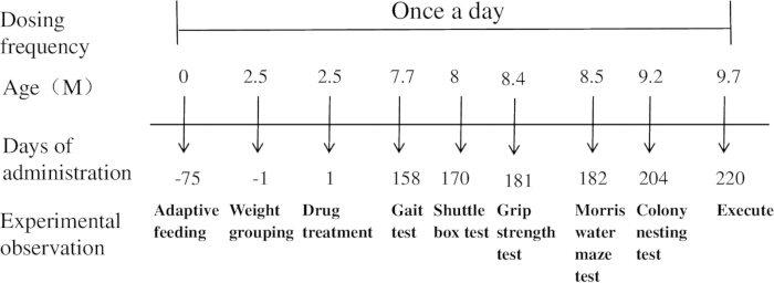
Figure 1: Schematic diagram of the experimental procedure. Please click here to view a larger version of this figure.
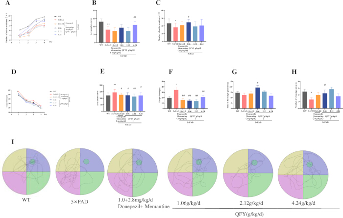
Figure 2: QFY administration improves learning and memory performance in 5xFAD mice. (A–C) Shuttle box test: (A) Number of active avoidance responses during the learning period. (B) Area under the curve (AUC) for active avoidance responses during the learning period. (C) Number of active avoidance responses during the test period. (D–I) Morris water maze test: (D) Escape latency during the learning period. (E) AUC for escape latency during the learning period. (F) Escape latency during the test period. (G) Time spent in the target quadrant. (H) Number of platform crossings. (I) Movement trajectories during the test period. Mean ± SD, n = 11–20 per group, with exact n indicated in each bar. * = p < 0.05, vs WT, Student`s t-test; # = p < 0.05, ## = p < 0.01, vs 5xFAD; One-way ANOVA followed by Dunnett's multiple comparisons test. Please click here to view a larger version of this figure.
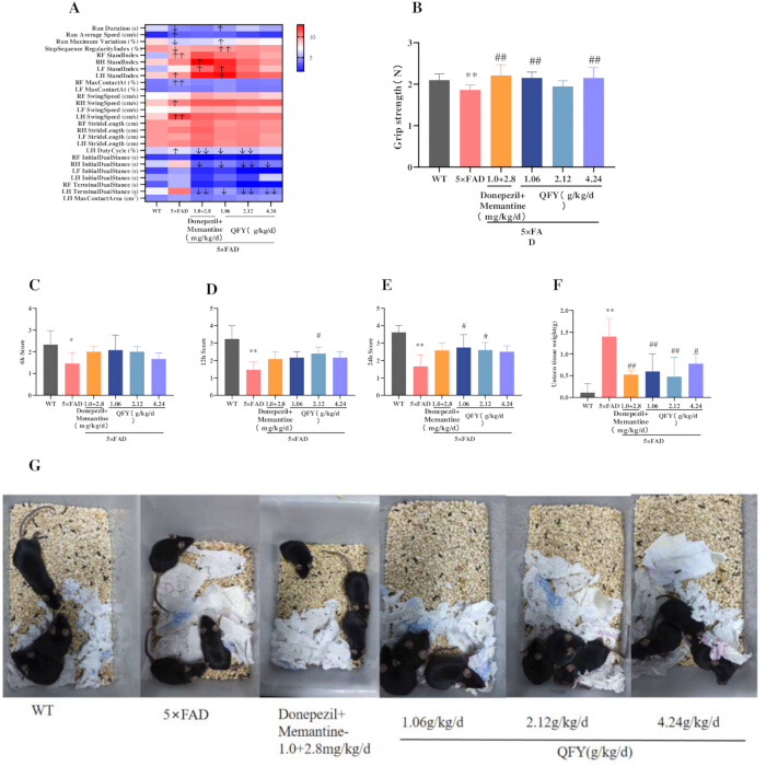
Figure 3: Effects of QFY administration on aging-associated functional measures in 5xFAD mice. (A) Gait heat map. The color scale is based on row‑wise Z‑score normalization, where red indicates values above the group mean, blue indicates values below the group mean, and white represents the mean level. (B) Grip strength test. (C–G) Colony nesting test: (C) Nesting score 6 h, (D) Nesting score at 12 h. (E) Nesting score at 24 h. (F) Untorn tissue weight. (G) Representative images of nests at 24 h. Mean ± SD, n = 3 per group, with exact n indicated in each bar. * = p < 0.05, ** = p < 0.01, vs WT, Student`s t-test; # = p < 0.05, ## = p < 0.01, vs 5x FAD, One-way ANOVA followed by Dunnett's multiple comparisons test. Please click here to view a larger version of this figure.
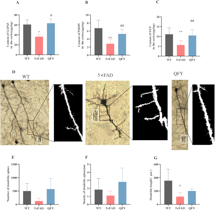
Figure 4: QFY attenuates synaptic structural alterations in the cerebral cortex of 5xFAD mice. (A) Content of GAP-43 in the cortex. (B) Content of PSD-95 in the cortex. (C) Content of SYN in the cortex. (D) Golgi-Cox staining. (E) Number of dendritic spines. (F) Density of dendritic spines. (G) Dendritic length. Mean ± SD, n = 3–14 per group, with exact n indicated in each bar. * = p < 0.05, ** = p < 0.01, vs WT, Student`s t-test; # = p < 0.05, ## = p < 0.01, vs 5xFAD, Student`s t-test. Please click here to view a larger version of this figure.
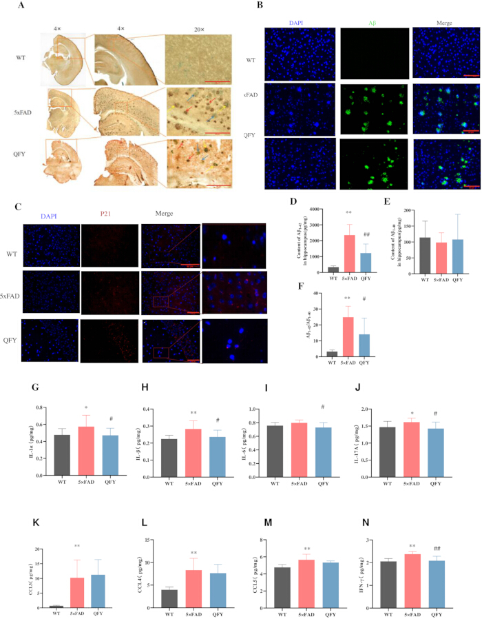
Figure 5: QFY reduced Aβ deposition and cell senescence in the brains of 5xFAD mice. (A) Combined SA-β-Gal staining and Aβ immunohistochemistry, SA-β-Gal (green, blue arrow), Aβ (brown, red arrow), SA-β-Gal + Aβ (yellow arrow), n = 3, Scale bar = 10 µm, 20x objective. (B) Immunofluorescence images showing Aβ in the cerebral cortex after treatment of QFY, n = 3, Scale bar = 50 µm, 40x objective. (C) Immunofluorescence images showing p21in the cerebral cortex after treatment of QFY. n = 1, Scale bar = 50 µm, 40x objective. (D–F) ELISA. (D) Content of Aβ1-42 in the hippocampus. (E) Content of Aβ1-40 in the hippocampus. (F) Aβ1-42/ Aβ1-40. (G–N) Luminex cytokine analysis. (G) Content of IL-1α in the cortex. (H) Content of IL-1β in the cortex. (I) Content of IL-6 in the cortex. (J) Content of IL-17A in the cortex. (K) Content of CCL3 in the cortex. (L) Content of CCL4 in the cortex. (M) Content of CCL5 in the cortex. (N) Content of IFN-γ in the cortex. Mean ± SD, n = 8–14 per group, with exact n indicated in each bar. * = p < 0.05, ** = p < 0.01, vs WT, Student’s t-test; # = p < 0.05 ## = p < 0.01, vs 5xFAD, Student’s t-test. Please click here to view a larger version of this figure.
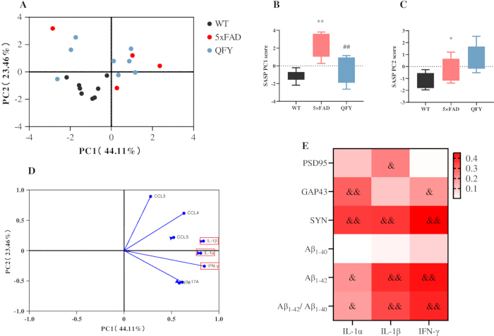
Figure 6: Principal component and correlation analyses of SASP factors, synaptic proteins, and Aβ levels in 5xFAD mice following QFY treatment. (A–D) Principal component analysis (PCA) of SASP factors. (A) PCA score plot. (B) PC1 scores. (C) PC2 scores. (D) PCA loading plot. (E) Heatmap generated from the correlation analysis between the SASP and PSD-95, GAP-43, SYN, Aβ1-40, Aβ1-42, and the ratio of Aβ1-42/Aβ1-40. Colors closer to red indicate higher R-values, while colors closer to white indicate lower R-values. Mean ± SD; n = 8 per group. * = p < 0.05, ** = p < 0.01,vs WT; ## = p < 0.01, vs 5xFAD, & = p < 0.05, && = p < 0.01, indicates a significant correlation. Pearson correlation analysis was used to assess associations between variables. Please click here to view a larger version of this figure.
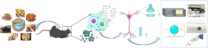
Figure 7: Proposed model of the effects of QFY on age-related cognitive decline and cellular senescence-associated phenotypes in 5xFAD mice. Please click here to view a larger version of this figure.
토론
신경 시냅스 가소성은 인지 기능에서 핵심적인 역할을 합니다. AD 환자의 뇌에서는 Aβ 침전물이 여러 영역으로 확산되어 시냅스 구조 손상, 흥분성 시냅스 소실 및 인지 기능 저하를 직접적으로 유발합니다28. GAP-43(시냅스 전 막 단백질)29, PSD-95(시냅스 후 막에 위치한 스캐폴드 단백질)30, 그리고 SYN(시냅스 소포 관련 인린화 단백질)31은 시냅스 구조 가소성과 AD의 병태생리에서 중요한 역할을 합니다32. 본 연구에서는 QFY 처리가 5xFAD 마우스의 뇌에서 수지상 돌기 가시의 수와 GAP-43, PSD-95 및 SYN의 단백질 수준을 증가시킴으로써 학습 및 기억 능력을 향상시키고, Aβ 플라크 부하를 감소시키며, 시냅스 구조를 회복시킨다는 것을 확인하였습니다.
노화는 AD 발생의 결정적인 위험 요인이며 Aβ 생성을 촉진합니다. 노화의 특징으로는 근력 감소, 보행 불균형, 그리고 기본적인 일상생활 수행 능력의 저하가 포함됩니다33,34. SA-β-Gal로 특징지어지는 세포 노화는 노화 및 AD와 같은 연령 관련 질환의 발병과 연관이 있는 것으로 보고되었습니다11,35. 세포 노화의 선택적 제거는 건강 수명을 연장하고 연령 관련 질환을 지연시킵니다36. 유전적 또는 약리학적 방법을 통한 노화 세포 제거는 AD 모델 마우스에서 Aβ 침착을 완화하고 학습 및 기억 능력을 향상시켰습니다37. Dasatinib와 Quercetin (D+Q) 병용 요법은 희소돌기아교세포 전구세포의 노화를 감소시키고, 신경염증을 완화하며, Aβ 플라크와 SA-β-Gal 활성을 줄여 APP/PS1 마우스의 Aβ 축적과 인지 장애를 예방합니다37. Memantine 치료는 SAMP8 마우스 뇌의 SA-β-Gal 활성을 유의하게 감소시키고 공간 학습 및 기억 능력을 향상시켰으며, memantine과 donepezil의 병용 투여에서도 동일한 효과가 관찰되었습니다38. 본 연구에서는 QFY 치료가 5xFAD 마우스 뇌의 SA-β-Gal 염색으로 나타난 세포 노화를 완화함으로써, 특히 보행을 개선하고 전지 근력을 강화하며 기본적인 일상생활 수행 능력을 향상시켰음을 확인하였습니다.
AD 환자와 동물 모델 모두에서 노화된 성상교세포, 미세아교세포, 혈관내피세포 및 뉴런이 관찰됩니다39,40. 케모카인과 염증성 사이토카인을 포함하는 SASP의 분비는 세포 노화의 특징적인 현상이며, 과도한 SASP 분비는 만성 염증을 유발하여 조직 손상과 세포 노화로 이어질 수 있습니다41. IL-1α, IL-1β, IL-6, IL-8, IFN-γ, CCL2, CCL3, CCL4, CCL5, CXCL1 등을 포함한 SASP의 지속적인 분비는 만성 염증, 정상 세포의 파라크린 노화, 세포 대사 및 후성유전적 조절의 변화, 그리고 Aβ의 지속적인 생성을 촉진합니다42,43,44,45. AD 환자와 AD 모델의 뇌에서는 IL-1α, IL-1β, IL-6, IL-17A 및 IFN-γ의 수치가 유의하게 상향 조절됩니다46. 연구에 따르면 D+Q는 IL-1α, IFN-γ를 포함한 염증 인자의 분비를 억제하고 APP/PS1 마우스에서 Aβ 플라크와 SA-β-Gal 활성을 감소시킬 수 있음이 밝혀졌습니다37. 또한 QFY가 IL-1α, IL-1β, IL-6, IL-17A 및 IFN-γ를 포함한 여러 SASP 인자의 발현을 감소시켜 노화 관련 염증 표현형을 완화한다는 연구 결과가 있었습니다.
p21은 cyclin-dependent kinase 억제제인 CIP/KIP 가족에 속하며, cyclin-CDK2/4 복합체의 근접 제동 장치로 작용하여 세포 스트레스, DNA 손상 및 노화가 발생했을 때 G1/G2 정지를 유도합니다. p21 수치의 지속적인 상승은 일반적으로 안정적인 노화 프로그램으로의 진입으로 해석됩니다47,48. 최근 데이터에 따르면 5xFAD 마우스는 비정상적으로 높은 뇌 내 p21 부하를 가지며, 이는 D+Q 병용 투여 시 현저히 감소하는 것으로 나타났습니다49. 이와 일관되게, 본 연구에서는 QFY 투여가 이들 동물에서 피질 Aβ 플라크를 유의하게 감소시키고 p21 발현을 완화했음을 확인하였습니다.
종합적으로, 이러한 결과는 QFY 처리가 5xFAD 마우스의 연령 관련 인지 기능 저하 및 세포 노화 표현형의 개선과 더불어 시냅스 구조 손상의 완화와 연관이 있음을 시사합니다(그림 7).
본 연구의 몇 가지 한계점을 인정해야 합니다. 첫째, 수컷 5xFAD 마우스만을 사용했으므로, 관찰된 효과가 성별에 따라 다른지는 아직 확인되지 않았습니다. 둘째, 현재의 데이터는 감쇠된 노화 관련 표현형과 인지 기능 개선 사이의 강한 연관성을 보여주지만, 인과 관계를 확립하기 위한 직접적인 기전적 개입 실험은 수행하지 않았습니다. 셋째, 종 간의 차이와 임상 약동학 데이터의 부족으로 인해 본 연구 결과의 인간 AD에 대한 중개적 관련성에는 한계가 있습니다. 이러한 발견을 확장하기 위해서는 암컷 동물을 포함하고, 노화 특이적 개입 및 잘 설계된 임상 시험을 통합하는 향후 연구가 필요합니다.
공개 사항
저자들은 선언할 이해관계의 충돌이 없습니다.
감사의 글
본 연구는 중국 국가자연과학재단(82205078, 82374062), 산둥성 자연과학재단(ZR2026MS1293, ZR2021QH157), 산둥성 중의약 과학기술 프로젝트(M20251730), 중국 시짱 자치구 과학기술 프로젝트(XZ202601ZY0167) 및 산둥성 기술혁신지도 프로그램(중앙정부 지도 지방 과학기술 발전 기금)(YDZX2023137)의 지원을 받았으며, 'Class 1 신규 중의약으로서의 Tongli 장용성 캡슐의 개발 및 산업화 연구'라는 제목의 프로젝트를 통해 수행되었습니다.
재료
| 이름 | 회사 | 카탈로그 번호 | 댓글 |
|---|---|---|---|
| 약물 | |||
| 도네페질 | 상하이 원예 테크놀로지(Shanghai Yuanye Technology) | lot.J15HB188725 | |
| 메만틴 | Shanghai Yuanye Technology | 로트 번호 J21HB189391 | |
| 기부음 | 루난 제약 후푸 연구개발 센터 | 로트 번호 2209001 | |
| 행동 테스트 | |||
| CatWalk XT | Noldus | CatWalkXT 10.0 소프트웨어 | |
| 악력계 | Bileadbiosci | BLD-ZL-20 | |
| 모리스 수중 미로 | Shanghai Xinruan Information Technology Co., Ltd | 120cm | |
| 셔틀 박스 | 상하이 신루안 정보기술 유한공사 (Shanghai Xinruan Information Technology Co., Ltd) | XR-XC404 | |
| 항체 | |||
| Anti-Aβ1-42 항체 | Abcam | Cat# ab201061 | |
| Anti-p21/CIP1/CDKN1A 항체 | Novus Biologicals | 카탈로그 번호 NBP3-15661 | |
| 분석 키트 | |||
| Aβ1-40 ELISA 키트 | Thermo Fisher Scientific | 제품번호 KHB3482 | |
| Aβ1-42 ELISA 키트 | Thermo Fisher Scientific | 제품 번호 KHB3442 | |
| Bio-Plex Pro Mouse Cytokine 23-plex Assay 키트 | R&D 시스템즈 | 카탈로그 번호 LXLBM23-1 | |
| GAP-43 ELISA 키트 | 강소성 효소면역측정 산업 | 카탈로그 번호 MM-47506M1 | |
| Golgi-Cox 염색 키트 | BeiJing Biolead Biology Sci & Tech Co | 카탈로그 번호 PK401 | |
| PSD-95 ELISA 키트 | 강쑤성 효소면역측정 산업 | 카탈로그 번호 MM-45953M1 | |
| 세포 노화 β-갈락토시다아제 염색 키트 | Beyotime | 카탈로그 번호 C0602 | |
| 시냅토피신 (SYN) ELISA 키트 | 강쑤성 효소면역측정 산업 | 카탈로그 번호 MM-46504M1 | |
| 화학 물질 & 시약 | |||
| 항토끼 및 항마우스 IgG (2차 항체) | 시작 단계 | 카탈로그 번호 S0C1001 | |
| DAB 발색제 | 시작 도구 | 제품 번호 S0C1001 | |
| 소프트웨어 | |||
| CatWalk XT | Noldus Information Technology | 버전 10.0 | |
| ImageJ | 미국 국립보건원(NIH) | 버전 1.53 |
참고문헌
- Joe E, Ringman JM. Cognitive symptoms of Alzheimer's disease: clinical management and prevention. BMJ. 2019;367:l6217.
- Scheff SW, et al. Synaptic alterations in CA1 in mild Alzheimer disease and mild cognitive impairment. Neurology. 2007;68(18):1501-8.
- Muralidar S, et al. Role of tau protein in Alzheimer's disease: the prime pathological player. Int J Biol Macromol. 2020;163:1599-617.
- Hardy JA, Higgins GA. Alzheimer's disease: the amyloid cascade hypothesis. Science. 1992;256(5054):184-5.
- Calsolaro V, Edison P. Neuroinflammation in Alzheimer's disease: current evidence and future directions. Alzheimers Dement. 2016;12(6):719-32.
- Campisi J. Senescent cells, tumor suppression, and organismal aging: good citizens, bad neighbors. Cell. 2005;120(4):513-22.
- Yao R. An in-silico assessment suggests the potential effects of goji (fruit of Lycium barbarum L.) against aging-related diseases. Food Med Homol. 2025;2(2):9420036.
- Campisi J, d'Adda di Fagagna F. Cellular senescence: when bad things happen to good cells. Nat Rev Mol Cell Biol. 2007;8(9):729-40.
- Salminen A, Kaarniranta K, Kauppinen A. ER stress activates immunosuppressive network: implications for aging and Alzheimer's disease. J Mol Med (Berl). 2020;98(5):633-50.
- Mirabella F, et al. Prenatal interleukin 6 elevation increases glutamatergic synapse density and disrupts hippocampal connectivity in offspring. Immunity. 2021;54(11):2611-31.e8.
- Baker DJ, Petersen RC. Cellular senescence in brain aging and neurodegenerative diseases: evidence and perspectives. J Clin Invest. 2018;128(4):1208-16.
- Lucas V, Cavadas C, Aveleira CA, Hook V. Cellular senescence: from mechanisms to current biomarkers and senotherapies. Pharmacol Rev. 2023;75(4):675-713.
- Bloss EB, et al. Evidence for reduced experience-dependent dendritic spine plasticity in the aging prefrontal cortex. J Neurosci. 2011;31(21):7831-9.
- Ye XS, et al. The food and medicine homologous Chinese medicine from Leguminosae species: a comprehensive review on bioactive constituents with neuroprotective effects on nervous system. Food Med Homol. 2025;2(2):9420033.
- Dickstein DL, Weaver CM, Luebke JI, Hof PR. Dendritic spine changes associated with normal aging. Neuroscience. 2013;251:21-32.
- Wang SY, et al. Qifu-Yin attenuates AGEs-induced Alzheimer-like pathophysiological changes through the RAGE/NF-kappaB pathway. Chin J Nat Med. 2014;12(12):920-8.
- Yang X, et al. Qi-fu-yin attenuated cognitive disorders in 5xFAD mice of Alzheimer's disease animal model by regulating immunity. Front Neurol. 2023;14:1183764.
- Xiao QY, et al. Effects of Qi-Fu-Yin on aging of APP/PS1 transgenic mice by regulating the intestinal microbiome. Front Cell Infect Microbiol. 2023;12:1048513.
- Chen K, et al. Safety and toxicity of Qi-Fu Yin, a classical traditional Chinese medicine prescription: a nonclinical research. J Appl Toxicol. 2025;45(9):1811-38.
- Wang L, Qiao P, Yue L, Sun R. Is Qi Fu Yin effective in clinical treatment of dementia? A meta-analysis of 697 patients. Medicine (Baltimore). 2021;100(5):e24526.
- Li H, et al. Identification of chemical components of Qi-Fu-Yin and its prototype components and metabolites in rat plasma and cerebrospinal fluid via UPLC-Q-TOF-MS. Evid Based Complement Alternat Med. 2021;2021:1995766.
- Lei X, et al. Integrating network pharmacology and component analysis to study the potential mechanisms of Qi-Fu-Yin Decoction in treating Alzheimer's disease. Drug Des Devel Ther. 2023;17:2841-58. doi:10.2147/DDDT.S402624.
- Takeshita H, et al. Modified forelimb grip strength test detects aging-associated physiological decline in skeletal muscle function in male mice. Sci Rep. 2017;7:42323.
- Caggiano V, et al. Midbrain circuits that set locomotor speed and gait selection. Nature. 2018;553(7689):455-60.
- Ehret B, et al. Population-level coding of avoidance learning in medial prefrontal cortex. Nat Neurosci. 2024;27(9):1805-15.
- Curdt N, et al. Search strategy analysis of Tg4-42 Alzheimer mice in the Morris water maze reveals early spatial navigation deficits. Sci Rep. 2022;12:5451.
- Deacon RMJ. Assessing nest building in mice. Nat Protoc. 2006;1(3):1117-9.
- Tu S, Okamoto SI, Lipton SA, Xu H. Oligomeric Aβ-induced synaptic dysfunction in Alzheimer's disease. Mol Neurodegener. 2014;9:48.
- Benowitz LI, Routtenberg A. GAP-43: an intrinsic determinant of neuronal development and plasticity. Trends Neurosci. 1997;20(2):84-91.
- Dore K, et al. PSD-95 protects synapses from β-amyloid. Cell Rep. 2021;35(9):109194.
- Kokotos AC, et al. Synaptophysin sustains presynaptic performance by preserving vesicular synaptobrevin-II levels. J Neurochem. 2019;151(1):28-37.
- Martin SB, et al. Synaptophysin and Synaptojanin-1 in Down syndrome are differentially affected by Alzheimer's disease. J Alzheimers Dis. 2014;42(3):767-75.
- Namioka N, et al. Oxidative stress and inflammation are associated with physical frailty in patients with Alzheimer's disease. Geriatr Gerontol Int. 2016;17(6):913-8.
- Kojima G, Liljas A, Iliffe S, Walters K. Prevalence of frailty in mild to moderate Alzheimer's disease: a systematic review and meta-analysis. Curr Alzheimer Res. 2017;14(12):1256-63.
- Dimri GP, et al. A biomarker that identifies senescent human cells in culture and in aging skin in vivo. Proc Natl Acad Sci U S A. 1995;92(20):9363-7.
- Baker DJ, et al. Naturally occurring p16Ink4a-positive cells shorten healthy lifespan. Nature. 2016;530(7589):184-9.
- Zhang P, et al. Senolytic therapy alleviates Abeta-associated oligodendrocyte progenitor cell senescence and cognitive deficits in an Alzheimer's disease model. Nat Neurosci. 2019;22(5):719-28.
- Ota H, Ogawa S, Ouchi Y, Akishita M. Protective effects of NMDA receptor antagonist, memantine, against senescence of PC12 cells: a possible role of nNOS and combined effects with donepezil. Exp Gerontol. 2015;72:109-16.
- Herdy JR, et al. Increased post-mitotic senescence in aged human neurons is a pathological feature of Alzheimer's disease. Cell Stem Cell. 2022;29(12):1637-52.e6.
- Hu Y, et al. Replicative senescence dictates the emergence of disease-associated microglia and contributes to Abeta pathology. Cell Rep. 2021;35(10):109228.
- Tchkonia T, et al. Cellular senescence and the senescent secretory phenotype: therapeutic opportunities. J Clin Invest. 2013;123(3):966-72.
- Guerrero A, De Strooper B, Arancibia-Cárcamo IL. Cellular senescence at the crossroads of inflammation and Alzheimer's disease. Trends Neurosci. 2021;44(9):714-27.
- Acosta JC, et al. A complex secretory program orchestrated by the inflammasome controls paracrine senescence. Nat Cell Biol. 2013;15(8):978-90.
- Wang B, Han J, Elisseeff JH, Demaria M. The senescence-associated secretory phenotype and its physiological and pathological implications. Nat Rev Mol Cell Biol. 2024;25(12):958-78.
- Gaikwad S, Senapati S, Haque MA, Kayed R. Senescence, brain inflammation, and oligomeric tau drive cognitive decline in Alzheimer's disease: evidence from clinical and preclinical studies. Alzheimers Dement. 2024;20(1):709-27.
- Si Z, Sun L, Wang X. Evidence and perspectives of cell senescence in neurodegenerative diseases. Biomed Pharmacother. 2021;137:111327.
- Chen X, et al. Senescence-like changes induced by expression of p21Waf1/Cip1 in NIH3T3 cell line. Cell Res. 2002;12(3-4):229-33.
- Engeland K. Cell cycle regulation: p53-p21-RB signaling. Cell Death Differ. 2022;29(5):946-60.
- Choi I, et al. Autophagy enables microglia to engage amyloid plaques and prevents microglial senescence. Nat Cell Biol. 2023;25(7):963-74.