This study evaluates the associations of p53 and Ki-67 immunohistochemistry with endometrial polyp recurrence and explores their incremental discriminative power beyond clinical features in women with metabolic syndrome.
연구 논문
p53 Immunophenotype and Clinical Features Associated with Endometrial Polyp Recurrence in Women with Metabolic Syndrome: A Retrospective Cohort Study
* These authors contributed equally
이 논문에서
요약
초록
Endometrial polyp recurrence after hysteroscopic resection remains clinically relevant in women with metabolic syndrome. In this retrospective cohort study, we evaluated the associations of a prespecified study-defined diffuse-strong p53 immunohistochemical phenotype, Ki-67 expression, and selected clinical characteristics with 24-month recurrence and explored the incremental discrimination provided by the IHC markers. The analytic cohort included 180 women with metabolic syndrome (MetS) who underwent hysteroscopic resection of histologically confirmed endometrial polyps (EPs) and had ascertainable recurrence status through 24 months. Associations with recurrence were assessed using a prespecified multivariable logistic model and a supportive multivariable Cox model, while exploratory prediction performance was evaluated using repeated stratified cross-validation. During 24 months of follow-up, 58 of 180 women (32.2%) had documented recurrence. Diffuse-strong p53 phenotype (adjusted odds ratio (aOR) 2.74, 95% CI 1.32–5.68), multiple polyps (aOR 2.74, 95% CI 1.25–6.02), AUB (aOR 2.29, 95% CI 1.12–4.65), and endometrial thickness (aOR 1.25 per mm, 95% CI 1.05–1.48) were independently associated with recurrence. Ki-67 ≥ 10% showed a directionally positive but statistically uncertain adjusted association (aOR 1.85, 95% CI 0.88–3.90). Supportive Cox analysis showed associations of diffuse-strong p53 phenotype, multiple polyps, AUB, and endometrial thickness with time to recurrence, whereas the estimate for Ki-67 remained statistically uncertain. The integrated model showed modestly higher repeated cross-validated discrimination than the clinical-only model (AUC 0.734 vs. 0.679; ΔAUC 0.056, 95% CI 0.006–0.105). Diffuse-strong p53 phenotype and selected clinical characteristics were associated with recurrence, whereas the contribution of Ki-67 remained exploratory; the modest incremental discrimination of the integrated model requires external validation before clinical application.
서론
EPs are among the most frequently encountered benign intrauterine lesions, particularly in perimenopausal and postmenopausal women. Their reported prevalence varies from 7% to 34% depending on diagnostic modality and population characteristics1. Although hysteroscopic polypectomy is the standard treatment and usually improves symptoms, recurrence remains common, with reported 2-year rates ranging from about 10% to more than 30%2. Because recurrence rates vary substantially across clinical populations, factors associated with postoperative recurrence warrant further investigation.
Clinical assessment of EPs commonly considers patient-level metabolic, hormonal, and lesion-related characteristics when estimating the likelihood of adverse endometrial findings or recurrence3. MetS—central adiposity, hypertension, dyslipidemia, and impaired glucose tolerance—has been linked to endometrial pathology. MetS has been associated with several forms of endometrial pathology, but its specific relationship with recurrence of histologically benign EPs remains incompletely defined4. Proposed pathways include insulin resistance with hyperinsulinemia, chronic low-grade inflammation, and increased estrogen bioavailability from peripheral aromatization in adipose tissue5. Together, these disturbances might promote persistent endometrial proliferation and alter tissue homeostasis, contributing to polyp formation and regrowth.
Local uterine factors also appear to influence recurrence risk. Larger lesions, greater endometrial thickness, and polyp multiplicity have been consistently associated with recurrence across cohorts6. Chronic endometritis (CE)—defined histologically by stromal plasma cell infiltration—has similarly been implicated and is clinically relevant because it may be amenable to treatment; persistent endometrial inflammation could impair normal remodeling and increase susceptibility to recurrent disease7. Taken together, these observations support a multifactorial model of EP recurrence in which systemic exposures and local endometrial conditions converge.
These observations have prompted interest in whether tissue immunohistochemical markers provide additional information beyond conventional clinical characteristics. Ki-67, a nuclear antigen expressed throughout the active phases of the cell cycle, is widely used to quantify proliferative activity. Higher Ki-67 labeling indices have been linked to a more proliferative phenotype across a spectrum of endometrial lesions, spanning benign changes to premalignant and malignant pathology8. In EPs, higher Ki-67 expression has been associated with symptomatic presentation, whereas its independent relationship with postoperative recurrence remains less clearly established9. Meanwhile, p53 is a central regulator of cell-cycle arrest, DNA-damage response, and apoptosis. In endometrial carcinoma, p53 immunohistochemistry (IHC) is interpreted using pattern-based approaches within contemporary molecular classification frameworks10. These carcinoma-specific approaches should not be directly extrapolated to histologically benign polyps. Strong p53-immunoreactive foci have nevertheless been described in morphologically benign endometrial glands and endometrial polyps, including p53-signature-type lesions11. These observations provide a rationale for studying the p53 immunophenotype in benign polyps, although its molecular basis and relationship to postoperative recurrence remain uncertain.
However, the associations of p53 and Ki-67 immunophenotypes with EP recurrence in women with MetS, and their potential incremental contribution beyond clinical characteristics, remain poorly characterized. Existing risk models for recurrence often rely mainly on clinical or structural features and show only moderate discrimination.
Therefore, this study evaluated whether the prespecified study-defined diffuse-strong p53 immunohistochemical phenotype and Ki-67 expression, together with clinical variables, were associated with postoperative EP recurrence over 2 years in women with MetS. Clinical-only and integrated clinical-plus-IHC models were further compared in an exploratory analysis to assess whether adding p53 and Ki-67 provided incremental discrimination beyond clinical characteristics.
프로토콜
Ethics approval and consent to participate
The current study was approved by the Ethics Committee of Cangzhou Hospital of Integrated Traditional Chinese and Western Medicine, Hebei Province, China (Approval No. CZX2023-KZ-256). The approval covered retrospective medical record review, pathology review, and supplemental follow-up contact. Because the study primarily used retrospectively collected clinical and pathological data, the requirement for written informed consent for record review was waived; verbal agreement was obtained before structured telephone follow-up when such contact was required. All extracted study data were de-identified before analysis and were accessible only to authorized members of the study team.
Details of the materials, reagents, antibodies, equipment, and software used in this study are provided in the Table of Materials.
Study design and setting
This single-center retrospective observational cohort study was conducted at Cangzhou Hospital of Integrated Traditional Chinese and Western Medicine, Cangzhou, Hebei Province, China. Index hysteroscopic procedures were performed between January 1, 2021, and December 31, 2023, and recurrence outcomes were ascertained through December 31, 2025, to provide 24 months of postoperative follow-up. The primary objective was to evaluate the associations of the prespecified p53 immunohistochemical phenotype, Ki-67 expression, and selected clinical characteristics with 24-month postoperative recurrence of EPs in women with MetS.
Study population
Women with MetS who underwent hysteroscopic resection of histologically confirmed EPs at Cangzhou Hospital of Integrated Traditional Chinese and Western Medicine between January 1, 2021, and December 31, 2023, were eligible. The analytic cohort comprised women with the clinical, imaging, operative, pathological, and follow-up information required for the prespecified analyses and with ascertainable recurrence status through 24 months. Recurrence outcomes were ascertained up to December 31, 2025.
Women were diagnosed with MetS according to the National Cholesterol Education Program Adult Treatment Panel III (NCEP ATP III) criteria, which require ≥3 of the following components: waist circumference ≥ 88 cm, blood pressure ≥ 130/85 mmHg, fasting glucose ≥ 100 mg/dL, triglycerides ≥ 150 mg/dL, and high-density lipoprotein (HDL)-cholesterol < 50 mg/dL12. The NCEP ATP III definition was selected because it represents the standard diagnostic framework used in clinical practice at our institution and aligns with the thresholds routinely measured in our electronic medical record system.
Data were extracted from the hospital electronic medical record, laboratory information system, and pathology archives. Trained gynecologists abstracted standardized clinical variables, laboratory indices, imaging measures, and immunohistochemical results using a prespecified data-collection template. Postoperative surveillance was scheduled at approximately 3, 6, 12, 18, and 24 months and included clinical assessment and transvaginal ultrasonography, with repeat hysteroscopy performed when clinically indicated; structured telephone contact was used only to supplement follow-up status and did not by itself establish recurrence.
Exclusion criteria were a history of endometrial hyperplasia, atypical hyperplasia, or carcinoma; prior hysterectomy or uterine malignancy; intrauterine device use at the time of surgery; incomplete resection of the index polyp; systemic autoimmune or chronic inflammatory disease; use of hormonal therapy (including tamoxifen or hormone replacement therapy) during follow-up; and insufficient follow-up information. No participant in the final analytic cohort received Zhuyang Huazheng Formula during postoperative follow-up. Women who received hormonal therapy, tamoxifen, or hormone-replacement therapy during follow-up were excluded according to the prespecified eligibility criteria, and no such exposure remained in the analytic cohort. No other protocolized pharmacologic treatment specifically intended to prevent endometrial polyp recurrence was administered as part of the study. Of 236 potentially eligible women, 56 were excluded for prespecified reasons, leaving 180 women in the final analytic cohort; the complete patient flow and reason-specific exclusions are shown in Figure 1 and Supplementary Table 1A–C. Transvaginal ultrasonography was used to measure endometrial thickness and the maximum diameter of the largest polyp. Operative records captured polyp number (single vs. multiple) and location (fundal vs. non-fundal). Histopathology confirmed EPs and excluded hyperplasia or malignancy.
IHC was performed on formalin-fixed, paraffin-embedded endometrial tissue sections to assess p53 and Ki-67 expression. The prespecified primary p53 exposure was the diffuse-strong immunohistochemical phenotype, which was classified according to the detailed criteria described under IHC for p53 and Ki-67. Ki-67 expression was assessed according to the immunohistochemical scoring procedure described in the same subsection, using ≥10% as the prespecified primary operational threshold.
All hysteroscopic procedures were performed by experienced gynecological surgeons in the same department, in accordance with standardized protocols. Polyps were removed under direct hysteroscopic visualization together with the visible attachment or base, and complete removal of the index lesion was confirmed by inspection of the uterine cavity before completion of the procedure. Data were extracted independently by two trained gynecologists using a standardized form, and any discrepancies were resolved by consensus.
Data collection and baseline variables
Data were abstracted independently by two trained investigators using a standardized case-report template. Any discrepancies were reconciled by consensus after review of the source records to maximize accuracy and completeness.
Clinical and laboratory data
Baseline demographics and clinical characteristics included age, body mass index, menopausal status, systolic blood pressure, history of previous hysteroscopic polypectomy, and presenting symptoms, with abnormal uterine bleeding (AUB) recorded systematically. AUB encompassed abnormal bleeding patterns in premenopausal women and any postmenopausal bleeding in postmenopausal women; menopausal status was therefore included explicitly in the prespecified multivariable adjustment set. Menstrual-cycle phase at ultrasonography, hormone testing, and tissue collection was not consistently documented in premenopausal women and was therefore not included as a model covariate. Laboratory indices comprised fasting plasma glucose, triglycerides, HDL-cholesterol, hemoglobin concentration, estradiol, and follicle-stimulating hormone. Unless otherwise specified, measurements were obtained at the index procedure or within 2 weeks before surgery.
Imaging and operative findings
Transvaginal ultrasonography was used to measure endometrial thickness and the maximum diameter of the largest polyp. Operative records were reviewed to ascertain polyp number (single vs. multiple) and anatomical location (fundal vs. non-fundal).
Histopathology and chronic endometritis
Histopathological assessment confirmed the diagnosis of EPs and excluded endometrial hyperplasia or malignancy. CE was defined as at least five CD138-positive stromal plasma cells across 10 non-overlapping high-power fields, with plasma cells identified in the stromal compartment and epithelial CD138 staining excluded from the count. The final CE classification was based on review of stromal plasma-cell morphology together with CD138 immunoreactivity, and disagreements between pathologists were resolved by consensus.
IHC for p53 and Ki-67
p53 and Ki-67 immunophenotypes were assessed by IHC on formalin-fixed, paraffin-embedded tissue sections. For the primary analysis, the prespecified p53 exposure was a study-defined diffuse-strong immunohistochemical phenotype, defined as strong (3+) nuclear staining involving at least 50% of lesional glandular epithelial nuclei. The ≥50% cutoff was a prespecified study-specific operational threshold and was not intended to reproduce the WHO/ICCR p53-abnormal molecular-classification algorithm for endometrial carcinoma. Accordingly, we use the descriptive term ‘diffuse-strong p53 phenotype’ throughout rather than assigning a WHO/ICCR p53-abnormal molecular class. Other p53 staining patterns were recorded separately as wild-type/heterogeneous, null, or cytoplasmic; null and cytoplasmic patterns were not combined with the primary diffuse-strong exposure and were grouped with it only in the prespecified expanded abnormal-pattern sensitivity analysis. Because TP53 sequencing was not performed, none of these immunohistochemical categories was interpreted as molecular confirmation of TP53 mutation. Ki-67 was scored manually as the percentage of positively stained glandular epithelial nuclei in hotspot areas and was categorized using the prespecified primary threshold of ≥10% versus <10%.
Specimens, staining protocol, and antibodies. IHC was performed on 4-µm sections. Primary antibodies were p53 clone DO-7 at a dilution of 1:200 and Ki-67 clone MIB-1 at a dilution of 1:150. Antigen retrieval was performed using citrate buffer (pH 6.0) at 95 °C for 20 min. Detection used an EnVision detection system with DAB chromogen, followed by hematoxylin counterstaining. All staining was completed in the same pathology laboratory under an identical standard operating procedure.
Controls and quality control
Appropriate positive and negative controls were included in each run, including a known p53-positive tissue and tonsil as a proliferative control for Ki-67. Negative controls were processed in parallel, omitting the primary antibody. When staining was performed in multiple batches, a reference control slide was included in each batch to monitor staining intensity and background. To minimize inter-run variability, sections were processed under the same fixation and embedding workflow and stained with the same antigen retrieval and detection protocol.
Interpretation and scoring. p53 immunoreactivity was evaluated in endometrial glandular epithelial cells. Cases without the diffuse-strong phenotype were classified separately as wild-type/heterogeneous, null, or cytoplasmic according to their staining pattern. Diffuse-strong staining was retained as the primary p53 exposure, whereas null and cytoplasmic patterns were included only in the prespecified sensitivity analysis of any abnormal p53 phenotype. Ki-67 expression was quantified across three non-overlapping hotspot areas at ×400 magnification, with at least 500 lesional glandular epithelial nuclei evaluated per case; the labeling index was calculated as the percentage of positively stained nuclei among all evaluated nuclei and was categorized as high (≥10%) or low (<10%).
Slide review and reproducibility
All IHC slides were independently evaluated by two gynecologic pathologists blinded to clinical outcomes. Interobserver agreement was assessed separately using Cohen’s kappa, with κ values of 0.84 for p53 classification, 0.81 for Ki-67 classification, and 0.79 for chronic endometritis; discrepant cases were reviewed jointly to reach consensus. Operational definitions, prevalence, and interobserver agreement for the pathological variables are summarized in Supplementary Table 2. Image acquisition and selection for illustrative figures. Representative immunohistochemical fields were selected to illustrate high and low Ki-67 expression and diffuse-strong and non-diffuse-strong p53 staining. Case classification was based on whole-slide assessment using prespecified scoring criteria, and image selection was not based on recurrence status. Representative high and low Ki-67 expression patterns are shown in Figure 2A,B, respectively, and representative diffuse-strong and non-diffuse-strong p53 staining patterns are shown in Figure 2C,D, respectively.
Outcome definition
The primary outcome of interest was the recurrence of EPs within 24 months following hysteroscopic polypectomy. Recurrence was defined as a newly documented intracavitary polyp after complete removal of the index lesion, identified by transvaginal ultrasonography or repeat hysteroscopy; when repeat hysteroscopy was performed, histopathological confirmation was obtained. Telephone follow-up alone was not considered sufficient evidence of recurrence. Because the primary outcome definition did not impose a minimum postprocedural interval, a prespecified sensitivity analysis excluded recurrences documented within the first 3 months to assess the potential influence of residual rather than newly recurrent lesions. Recurrence time was calculated in months from the date of the index hysteroscopic resection to the first documented evidence of recurrence. Patients without evidence of recurrence at the last follow-up were censored at 24 months. To ensure accuracy, recurrence events were adjudicated by two independent gynecologists blinded to baseline molecular marker status. Discrepancies were resolved by consensus review.
Statistical analyses
Continuous variables were first assessed for normality using the Shapiro–Wilk test and for homogeneity of variances using Levene’s test. Variables with a normal distribution are expressed as the mean ± standard deviation and compared using the student’s t-test, whereas non-normally distributed variables are presented as the median with interquartile range and compared using the Mann–Whitney U test. Categorical variables were summarized as counts and percentages and compared using the χ2 test or Fisher’s exact test, as appropriate. Within the final analytic cohort, no values were missing for the outcome or the 10 variables included in the prespecified primary multivariable model; therefore, no statistical imputation was performed. Two-sided p < 0.05 was considered statistically significant.
Multivariable logistic regression analysis
We used multivariable logistic regression to identify factors independently associated with recurrence within 24 months, reporting adjusted odds ratios (aORs) with 95% confidence intervals (CIs). Predictors specified a priori on clinical and pathological grounds rather than selected according to univariable statistical significance. The primary multivariable model simultaneously included diffuse-strong p53 phenotype, Ki-67 ≥10%, multiple polyps, AUB, endometrial thickness, age, menopausal status, BMI, previous hysteroscopic polypectomy, and chronic endometritis. Given the 58 recurrence events, model complexity was deliberately restricted to these 10 prespecified predictors, and stepwise selection, interaction terms, and subgroup-specific models were not used. Collinearity was evaluated using variance inflation factors, and all predictors in the final prespecified model had VIF values below 2; linearity of continuous predictors on the log-odds scale was assessed using the Box–Tidwell method.
Kaplan–Meier survival analysis
Recurrence time data were used to perform a Kaplan–Meier survival analysis to assess recurrence-free survival among patients over a 24-month follow-up period. Kaplan–Meier curves and log-rank tests were used descriptively to compare recurrence-free survival according to diffuse-strong p53 phenotype, Ki-67 ≥10%, multiple polyps, and AUB.
Cox proportional hazards regression
As a supportive time-to-event analysis, a multivariable Cox proportional-hazards model used the same prespecified covariate set as the primary logistic model and reported adjusted hazard ratios with 95% confidence intervals. The proportional hazards assumption was evaluated using Schoenfeld residuals. Kaplan–Meier analyses and log-rank tests were retained as unadjusted descriptive analyses and were not interpreted as confirmation of the adjusted logistic regression findings.
Construction of predictive models and sensitivity analyses
Prediction modeling was exploratory, and model performance was estimated using repeated stratified 5-fold cross-validation with 100 repeats rather than a single random training–testing split; all discrimination, calibration, and prediction-error estimates were derived from out-of-fold predictions. Four prespecified exploratory models were evaluated: a p53-only model, a Ki-67-only model, a clinical-only model, and an integrated model combining the clinical predictors with p53 and Ki-67; full model specifications and coefficients are provided in Supplementary Table 3A.
Discrimination was summarized using cross-validated area under the receiver operating characteristic curve estimates with 95% confidence intervals, and overall prediction error was additionally summarized using the Brier score. The difference in AUC between the integrated and clinical-only models was evaluated in paired resampling iterations, and the resulting distribution was used to estimate the confidence interval and two-sided p-value for ΔAUC. Calibration was assessed using calibration intercepts and slopes, and decision-curve analysis was performed as an exploratory assessment of potential net benefit. Sensitivity analyses evaluated alternative Ki-67 thresholds of 5% and 15%, an alternative p53 definition combining diffuse-strong, null, and cytoplasmic phenotypes as any abnormal p53 phenotype, and exclusion of recurrences documented within 3 months after the index procedure. All statistical analyses were conducted using R software.
결과
Baseline characteristics of included patients
Among 236 potentially eligible women, 56 were excluded for prespecified reasons, and 180 constituted the final analytic cohort; 58 women (32.2%) had documented recurrence within 24 months, and 122 (67.8%) remained recurrence-free. None of the 180 women in the final analytic cohort received Zhuyang Huazheng Formula during postoperative follow-up; hormonal therapy, tamoxifen, and hormone-replacement therapy were absent from the analytic cohort by design, as detailed in Supplementary Table 1A–C. Of the 58 recurrences, 45 (77.6%) were confirmed by repeat hysteroscopy with histopathological assessment, and 13 (22.4%) were documented by transvaginal ultrasonography without repeat hysteroscopy. Five recurrence events occurred within 3 months of the index procedure and were addressed separately in the sensitivity analysis.
In unadjusted comparisons, women with recurrence had greater endometrial thickness, larger maximum polyp diameter, and higher hemoglobin concentrations than women without recurrence. Endometrial thickness was 9.18 ± 2.34 versus 7.97 ± 2.30 mm (p = 0.001), maximum polyp diameter was 1.71 ± 0.50 versus 1.55 ± 0.53 cm (p = 0.043), and hemoglobin concentration was 129.22 ± 13.74 versus 122.50 ± 17.04 g/L (p = 0.005) in the recurrence and non-recurrence groups, respectively. Other continuous baseline characteristics did not differ materially between recurrence groups, as detailed in Table 1. The recurrence time among those with relapses was 10.03 ± 4.62 months.
In unadjusted categorical comparisons, recurrence was more frequent among women with multiple polyps (44.8% vs. 23.8%; p = 0.007), diffuse-strong p53 phenotype (53.4% vs. 29.5%; p = 0.003), Ki-67 ≥10% (46.6% vs. 24.6%; p = 0.005), and AUB (56.9% vs. 33.6%; p = 0.005). These unadjusted group differences are summarized in Table 1; Figure 3 presents the corresponding adjusted associations from the prespecified multivariable model.
Multivariable predictors of EP recurrence
Multivariable logistic regression was performed to identify factors independently associated with 24-month recurrence after hysteroscopic polypectomy in women with MetS. The adjusted odds ratios (aORs) and 95% confidence intervals (CIs) are presented in Table 2. Multicollinearity was assessed using variance inflation factors, and all included predictors demonstrated VIF values below 2, indicating no evidence of problematic collinearity.
In the prespecified multivariable model, diffuse-strong p53 phenotype, multiple polyps, AUB, and endometrial thickness remained independently associated with 24-month recurrence. Diffuse-strong p53 phenotype was associated with higher odds of postoperative recurrence (aOR 2.74, 95% CI 1.32–5.68; p = 0.007). Multiple polyps (aOR 2.74, 95% CI 1.25–6.02; p = 0.012) and AUB (aOR 2.29, 95% CI 1.12–4.65; p = 0.023) were also independently associated with recurrence. Endometrial thickness retained a continuous association, with each 1-mm increase corresponding to a 25% increase in the odds of recurrence (aOR 1.25, 95% CI 1.05–1.48; p = 0.010).
Other variables provided supportive but less definitive signals. Ki-67 ≥10% showed a directionally positive but statistically uncertain adjusted association with recurrence (aOR 1.85, 95% CI 0.88–3.90; p = 0.107). The complete adjusted estimates for all 10 prespecified covariates are presented in Table 2 and Figure 3. No statistically significant adjusted association was observed for Ki-67 ≥10% or the remaining prespecified covariates.
Time-to-recurrence analyses
Given that recurrence time was available in our dataset, Kaplan–Meier survival analysis with log-rank testing was further applied to visualize recurrence-free survival across key categorical predictors. The Kaplan–Meier curves are presented in Figure 4, with numbers at risk displayed below each panel. The unadjusted Kaplan–Meier curves for diffuse-strong p53 phenotype and Ki-67 ≥10% are shown in Figure 4A,B, respectively. Kaplan–Meier curves for multiple polyps and AUB are shown in Figure 4C,D, respectively. These unadjusted Kaplan–Meier comparisons were descriptive and were interpreted separately from the adjusted regression analyses. In the supportive multivariable Cox model using the same prespecified covariate set as the primary logistic model, diffuse-strong p53 phenotype (HR 2.27, 95% CI 1.32–3.89; p = 0.003), multiple polyps (HR 2.27, 95% CI 1.28–4.03; p = 0.005), AUB (HR 1.86, 95% CI 1.08–3.21; p = 0.026), and endometrial thickness (HR 1.20 per mm, 95% CI 1.07–1.35; p = 0.002) were associated with shorter recurrence-free survival. The association for Ki-67 ≥10% remained directionally positive but did not meet the conventional threshold for statistical significance (HR 1.69, 95% CI 0.99–2.91; p = 0.056).
Exploratory prediction analyses
As an exploratory analysis, p53-only, Ki-67-only, clinical-only, and integrated clinical-plus-IHC models were compared using repeated stratified cross-validation. In full-cohort descriptive 2×2 analyses, diffuse-strong p53 had a sensitivity of 53.4% and specificity of 70.5%, whereas Ki-67 ≥10% had a sensitivity of 46.6% and specificity of 75.4%; corresponding cell counts and confidence intervals are reported in Supplementary Table 3B.
The clinical-only model had a repeated cross-validated AUC of 0.679 (95% CI 0.590–0.762). The integrated model had a repeated cross-validated AUC of 0.734 (95% CI 0.649–0.810) (Figure 5). The integrated model showed a statistically detectable but modest increase in discrimination relative to the clinical-only model (ΔAUC 0.056, 95% CI 0.006–0.105; p = 0.027); the Brier score was 0.190 for the integrated model and 0.197 for the clinical-only model. Calibration slopes were 0.95 for the clinical model and 0.98 for the integrated model, with corresponding calibration intercepts of 0.00 and 0.01. Calibration plots and exploratory decision-curve analysis are shown in Supplementary Figure 1A and Supplementary Figure 1B, respectively.
Sensitivity analyses
Sensitivity analyses examined alternative Ki-67 thresholds of ≥5% and ≥15%, an alternative definition of any abnormal p53 phenotype, and exclusion of recurrence events documented within 3 months after the index procedure. Alternative Ki-67 thresholds yielded directionally similar but statistically uncertain adjusted associations: aOR 1.58 (95% CI 0.76–3.30) for ≥5% and aOR 2.15 (95% CI 0.83–5.57) for ≥15% (Supplementary Table 4). When diffuse-strong, null, and cytoplasmic staining were combined under the prespecified expanded abnormal-pattern p53 sensitivity definition, the adjusted association with recurrence remained positive (aOR 3.55, 95% CI 1.70–7.41). After exclusion of the five recurrences documented within 3 months of the index procedure, the associations remained directionally stable for diffuse-strong p53 (aOR 2.66, 95% CI 1.26–5.60) and Ki-67 ≥10% (aOR 1.79, 95% CI 0.82–3.90) in the remaining 175 women.
DATA AVAILABILITY:
The de-identified participant-level analytic dataset underlying the reported analyses is provided in Supplementary File, together with a variable dictionary used for the prespecified and sensitivity analyses.
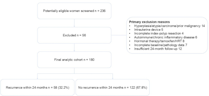
Figure 1: Participant selection and analytic cohort. Flow diagram showing participant screening, exclusions, final cohort selection, and 24-month recurrence status. Abbreviations: HRT = hormone replacement therapy. Please click here to view a larger version of this figure.
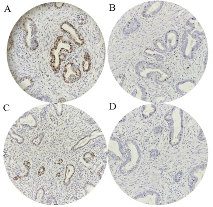
Figure 2: Representative p53 and Ki-67 immunohistochemical staining patterns in endometrial polyps. (A) High Ki-67 expression (≥10%). (B) Low Ki-67 expression (<10%). (C) Diffuse-strong p53 nuclear staining. (D) Non-diffuse-strong p53 staining. Abbreviations: IHC = immunohistochemistry. Please click here to view a larger version of this figure.
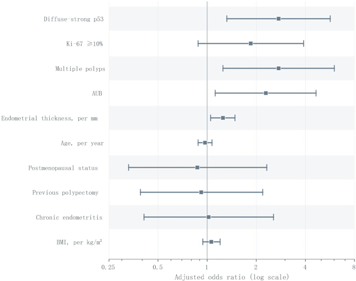
Figure 3: Adjusted associations with 24-month endometrial polyp recurrence. Forest plot showing adjusted odds ratios and 95% confidence intervals for the 10 prespecified predictors in the multivariable logistic regression model. The vertical reference line indicates an odds ratio of 1.0. Abbreviations: AUB = abnormal uterine bleeding; BMI = body mass index; CI = confidence interval. Please click here to view a larger version of this figure.
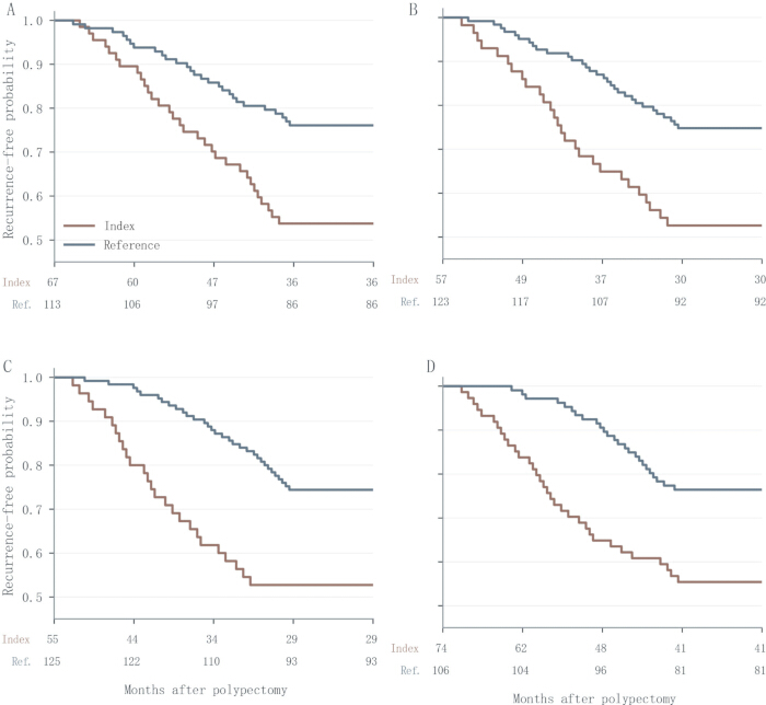
Figure 4: Kaplan–Meier curves for recurrence-free survival according to selected baseline characteristics. Kaplan–Meier curves showing recurrence-free survival according to (A) diffuse-strong versus non-diffuse-strong p53 phenotype, (B) Ki-67 ≥10% versus <10%, (C) multiple versus nonmultiple polyps, and (D) presence versus absence of abnormal uterine bleeding. Numbers at risk are shown below each panel. Abbreviations: AUB = abnormal uterine bleeding. Please click here to view a larger version of this figure.
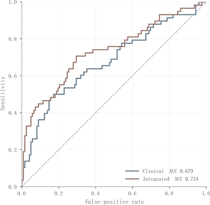
Figure 5: Cross-validated discrimination of the recurrence models. Receiver operating characteristic curves for the clinical-only and integrated clinical-plus-IHC models based on repeated stratified cross-validation. Abbreviations: AUC = area under the curve; IHC = immunohistochemistry. Please click here to view a larger version of this figure.
Supplementary Figure 1: Calibration and decision-curve analyses of clinical-only and integrated recurrence models. (A) Calibration plots comparing predicted probabilities with observed 24-month recurrence proportions for the clinical-only and integrated clinical-plus-IHC models; the dashed diagonal line represents perfect calibration. (B) Decision-curve analysis showing net benefit across threshold probabilities for the clinical-only and integrated models; treat-all and treat-none strategies are shown for reference. Abbreviations: IHC = immunohistochemistry. Please click here to download this file.
| Characteristic | Recurrence (n = 58) | No recurrence (n = 122) | p value |
| Age, years | 55.83 ± 4.40 | 57.02 ± 5.03 | 0.109 |
| BMI, kg/m² | 27.47 ± 3.14 | 26.66 ± 2.88 | 0.100 |
| Postmenopausal status | 31/58 (53.4%) | 74/122 (60.7%) | 0.450 |
| Previous hysteroscopic polypectomy | 14/58 (24.1%) | 23/122 (18.9%) | 0.533 |
| Systolic blood pressure, mmHg | 142.86 ± 11.96 | 142.20 ± 13.42 | 0.741 |
| Fasting plasma glucose, mg/dL | 123.05 ± 20.39 | 125.14 ± 20.78 | 0.524 |
| Triglycerides, mmol/L | 2.56 ± 0.97 | 2.32 ± 0.90 | 0.108 |
| HDL-cholesterol, mmol/L | 0.99 ± 0.21 | 1.00 ± 0.20 | 0.817 |
| Hemoglobin, g/L | 129.22 ± 13.74 | 122.50 ± 17.04 | 0.005 |
| Estradiol, pg/mL | 44.10 ± 24.67 | 45.91 ± 28.96 | 0.665 |
| FSH, IU/L | 46.2 (32.1–61.8) | 48.7 (33.4–64.9) | 0.685 |
| Endometrial thickness, mm | 9.18 ± 2.34 | 7.97 ± 2.30 | 0.001 |
| Maximum polyp diameter, cm | 1.71 ± 0.50 | 1.55 ± 0.53 | 0.043 |
| Multiple polyps | 26/58 (44.8%) | 29/122 (23.8%) | 0.007 |
| Abnormal uterine bleeding | 33/58 (56.9%) | 41/122 (33.6%) | 0.005 |
| Diffuse-strong p53 phenotype | 31/58 (53.4%) | 36/122 (29.5%) | 0.003 |
| High Ki-67 expression (≥10%) | 27/58 (46.6%) | 30/122 (24.6%) | 0.005 |
| Chronic endometritis | 15/58 (25.9%) | 23/122 (18.9%) | 0.378 |
Table 1: Baseline clinicopathological characteristics according to 24-month recurrence status. Baseline clinical, laboratory, imaging, operative, and pathological characteristics are compared between women with and without documented endometrial polyp recurrence within 24 months after hysteroscopic polypectomy. Data are presented as mean ± SD, median (IQR), or n/N (%) as appropriate. Two-sided p-values represent unadjusted between-group comparisons and were not used to determine inclusion in the multivariable model. Abbreviations: BMI = body mass index; FSH = follicle-stimulating hormone; HDL = high-density lipoprotein; IQR = interquartile range; SD = standard deviation.
| Predictor | Adjusted OR | 95% CI | p value |
| Diffuse-strong p53 phenotype | 2.74 | 1.32–5.68 | 0.007 |
| Ki-67 ≥10% | 1.85 | 0.88–3.90 | 0.107 |
| Multiple polyps | 2.74 | 1.25–6.02 | 0.012 |
| Abnormal uterine bleeding | 2.29 | 1.12–4.65 | 0.023 |
| Endometrial thickness, per mm | 1.25 | 1.05–1.48 | 0.010 |
| Age, per year | 0.97 | 0.88–1.07 | 0.537 |
| Postmenopausal status | 0.87 | 0.33–2.32 | 0.785 |
| Previous hysteroscopic polypectomy | 0.92 | 0.39–2.19 | 0.852 |
| Chronic endometritis | 1.02 | 0.41–2.55 | 0.966 |
| BMI, per kg/m² | 1.06 | 0.94–1.20 | 0.315 |
Table 2: Prespecified multivariable logistic regression for 24-month recurrence. Adjusted odds ratios and 95% confidence intervals are shown for the 10 prespecified predictors entered simultaneously into the multivariable logistic regression model. No univariable significance-based variable selection was applied. Diffuse-strong p53 was defined as strong 3+ nuclear staining involving ≥50% of lesional glandular epithelial nuclei, and Ki-67 ≥10% was the prespecified primary operational threshold. Abbreviations: BMI = body mass index; CI = confidence interval; OR = odds ratio.
| Predictor | Adjusted HR | 95% CI | p value |
| Diffuse-strong p53 phenotype | 2.27 | 1.32–3.89 | 0.003 |
| Ki-67 ≥10% | 1.69 | 0.99–2.91 | 0.056 |
| Multiple polyps | 2.27 | 1.28–4.03 | 0.005 |
| Abnormal uterine bleeding | 1.86 | 1.08–3.21 | 0.026 |
| Endometrial thickness, per mm | 1.20 | 1.07–1.35 | 0.002 |
| Age, per year | 0.97 | 0.91–1.04 | 0.413 |
| Postmenopausal status | 1.07 | 0.51–2.21 | 0.865 |
| Previous hysteroscopic polypectomy | 0.88 | 0.47–1.65 | 0.684 |
| Chronic endometritis | 0.82 | 0.43–1.58 | 0.562 |
| BMI, per kg/m² | 1.07 | 0.98–1.17 | 0.138 |
Table 3: Multivariable Cox proportional hazards model for time to recurrence. Adjusted hazard ratios and 95% confidence intervals are shown for the supportive Cox proportional hazards model using the same prespecified covariate set as the primary logistic regression model. Patients without documented recurrence were censored at the end of follow-up. The Cox analysis was supportive rather than the primary inferential analysis. Abbreviations: BMI = body mass index; CI = confidence interval; HR = hazard ratio.
Supplementary Table 1: Cohort accounting, follow-up, recurrence ascertainment, and missing-data profile. (A) Participant screening, exclusions, and final cohort accounting. (B) Follow-up and recurrence ascertainment. (C) Missing-data profile for the outcome and variables included in the prespecified primary multivariable model. Recurrence required documented imaging or hysteroscopy after complete removal of the index lesion; telephone contact alone was not sufficient to establish recurrence. The ≤3-month sensitivity analysis assessed the possibility that very early lesions represented residual rather than newly recurrent disease. Abbreviations: BMI = body mass index; HRT = hormone replacement therapy; MetS = metabolic syndrome. Please click here to download this file.
Supplementary Table 2: Operational definitions of pathology, prevalence, and interobserver reproducibility. The table summarizes the operational definitions and recurrence-group distributions of the prespecified p53 immunohistochemical phenotypes, Ki-67 expression, and chronic endometritis, together with interobserver agreement. Diffuse-strong p53 was the prespecified primary exposure and represents a study-defined operational phenotype rather than the WHO/ICCR p53-abnormal molecular category used for endometrial carcinoma. Null and cytoplasmic patterns were retained separately and grouped with diffuse-strong staining only in the prespecified expanded abnormal-pattern sensitivity analysis. None of these immunohistochemical categories was interpreted as molecular confirmation of TP53 mutation. Abbreviations: HPF = high-power field; ICCR = International Collaboration on Cancer Reporting; WHO = World Health Organization; κ = Cohen’s kappa. Please click here to download this file.
Supplementary Table 3: Prediction-model specifications, coefficients, and classification characteristics of the individual immunohistochemical markers. (A) Specifications, coefficients, and repeated cross-validated performance of the p53-only, Ki-67-only, clinical-only, and integrated clinical-plus-IHC models. Age, BMI, and endometrial thickness were centered for prediction-model specification; model composition was fixed before resampling. (B) Full-cohort 2 × 2 classification characteristics for diffuse-strong p53 and Ki-67 ≥10%. Panel B uses the full cohort of 180 participants and is presented descriptively to clarify the sensitivity and specificity estimates; p53 and Ki-67 are not presented as standalone diagnostic tests. Abbreviations: AUC = area under the curve; BMI = body mass index; CI = confidence interval; CV = cross-validation; FN = false negative; FP = false positive; IHC = immunohistochemistry; OR = odds ratio; TN = true negative; TP = true positive. Please click here to download this file.
Supplementary Table 4: Internal validation, calibration, decision analysis, and sensitivity analyses. The table summarizes repeated cross-validated discrimination and prediction error, calibration measures, exploratory decision-curve analysis, and prespecified sensitivity analyses using alternative Ki-67 thresholds, an expanded abnormal-pattern p53 definition, and exclusion of recurrences documented within 3 months after the index procedure. Repeated stratified cross-validation was used for internal validation. Decision-curve analysis was exploratory. Alternative Ki-67 thresholds were evaluated only in sensitivity analyses and did not redefine the prespecified primary cutoff. Abbreviations: AUC = area under the curve; CI = confidence interval; DCA = decision-curve analysis; OR = odds ratio; ΔAUC = difference in area under the curve. Please click here to download this file.
토론
This study found that 24-month recurrence of EPs in women with MetS was independently associated with diffuse-strong p53 phenotype and selected clinical features, whereas the adjusted association for Ki-67 was directionally positive but statistically uncertain. In the prespecified multivariable model, diffuse-strong p53 phenotype, multiple polyps, AUB, and greater endometrial thickness remained associated with recurrence after adjustment for age, menopausal status, BMI, previous polypectomy, chronic endometritis, and Ki-67. In exploratory internal validation, the integrated model showed modestly higher repeated cross-validated discrimination than the clinical-only model (AUC 0.734 vs. 0.679; ΔAUC 0.056, 95% CI 0.006–0.105). Diffuse-strong p53 staining showed a consistent association with recurrence in both the primary logistic model and the supportive Cox model. This finding should be interpreted in light of the study-specific p53 definition rather than as evidence that these benign polyps belonged to the WHO/ICCR p53-abnormal carcinoma category. The prognostic relevance of p53-abnormal molecular classification has been demonstrated in endometrial carcinoma, including high-risk disease13, but this carcinoma framework is distinct from the operational phenotype used in the present study. Strong p53-immunoreactive foci have previously been described in morphologically benign endometrial polyps11, providing a biologically plausible basis for investigating p53 immunophenotype in benign lesions. Nevertheless, the present data do not establish that diffuse-strong p53 staining represents a premalignant process or a causal mechanism of recurrence. However, because TP53 sequencing was not performed and all index lesions were histologically benign, the present data do not establish premalignant transformation or a causal TP53-mediated mechanism of recurrence.
Ki-67, by contrast, captures proliferative activity rather than genomic stress. In women with MetS, such proliferative drive might be sustained by the intersecting effects of chronic inflammation, insulin resistance, and a relatively estrogenic hormonal milieu. Ki-67 expression has been investigated as a proliferative biomarker in endometrial cancer, including studies evaluating imaging features associated with Ki-67 expression levels14. Symptom-related differences in Ki-67 expression have also been reported in endometrial polyps9, whereas evidence for an independent association between Ki-67 and benign polyp recurrence remains limited. The findings provide only exploratory support for Ki-67, because the direction of association was positive, but the adjusted confidence interval included the null value. The direction of association was similar when 5% and 15% thresholds were examined, but these sensitivity analyses do not establish 10% as an optimized or clinically validated cutoff.
Clinical characteristics also contributed independent information in the adjusted analyses. EPs may also be detected in asymptomatic postmenopausal women15, indicating that symptom status alone should not be regarded as a specific marker of lesion biology or recurrence. Atypical EPs represent a clinically distinct entity because of their reported association with concurrent endometrial cancer16; such lesions were excluded from the present cohort. Multiple EPs have been associated with CE in reproductive-aged women17; in the present cohort, multiplicity remained independently associated with recurrence, although our data do not establish an inflammatory or surgical mechanism. AUB is closely related to the clinical presentation and evaluation of endometrial polyps18; in the present cohort, AUB was independently associated with recurrence, although the data cannot determine whether this relationship reflects lesion biology, detection patterns, or other clinical factors. Endometrial thickness has been studied in relation to endometrial lesions in asymptomatic postmenopausal women19; in the present cohort, each additional millimeter was associated with a 25% increase in adjusted odds of recurrence, which should be interpreted as a clinical correlate rather than evidence of a specific hormonal mechanism. Previous observational studies have examined risk factors for EPs among women with AUB20 and for malignant pathology within endometrial polyps21. Other retrospective studies have addressed the management of endometrial lesions22 and predictors of malignancy in EPs among women with postmenopausal bleeding23. These outcomes are clinically relevant but distinct from postoperative recurrence after complete polypectomy.
In addition to multivariable logistic regression and exploratory predictive modeling, Kaplan–Meier survival analysis was incorporated to examine the time-dependent dynamics of recurrence. Kaplan–Meier curves were used to display the accumulation of recurrences over 24 months and complement point estimates from logistic regression. The unadjusted Kaplan–Meier patterns were directionally consistent with the time-to-event estimates but should not be interpreted as mechanistic evidence. Notably, p53 was consistently associated with recurrence across analyses. By contrast, the Ki-67 estimate was less precise after multivariable adjustment, and the unadjusted Kaplan–Meier pattern should not be interpreted as evidence of an independent effect. The time-to-event analyses provided supportive information on the timing of recurrence but were not considered independent confirmation of the primary logistic model.
The unadjusted Kaplan–Meier curves showed earlier recurrence accumulation among women with multiple polyps or AUB, whereas the corresponding adjusted associations were estimated in the Cox model. In the adjusted Cox model, diffuse-strong p53 phenotype, multiple polyps, AUB, and greater endometrial thickness were associated with time to recurrence, whereas the estimate for Ki-67 remained borderline.
The Kaplan–Meier and Cox analyses provided complementary information on the timing of recurrence. The exploratory prediction analysis showed that adding p53 and Ki-67 to the clinical model increased the repeated cross-validated AUC from 0.679 to 0.734, corresponding to a modest ΔAUC of 0.056. These findings suggest that IHC information may provide limited incremental discrimination beyond clinical characteristics, but they do not establish a clinically validated recurrence-stratification tool. However, given the retrospective single-center design and lack of external validation, the predictive implications should be regarded as exploratory rather than immediately practice-changing.
Several limitations warrant consideration. The retrospective nature of the study may have introduced selection bias. Requiring ascertainable 24-month recurrence status and excluding women with incomplete baseline or pathological information may have introduced additional selection bias, although patient flow and missingness are reported explicitly. Exclusion of women who received hormonal therapy, tamoxifen, or hormone-replacement therapy during follow-up also represents a post-baseline eligibility condition and may have introduced additional selection bias. Menstrual-cycle phase was not consistently documented in premenopausal women, leaving potential residual confounding of endometrial thickness and Ki-67 despite adjustment for menopausal status.
Because 13 of 58 recurrences (22.4%) were documented by ultrasonography without repeat hysteroscopic and histopathological confirmation, some outcome misclassification cannot be excluded; moreover, symptomatic women may have had greater opportunities for diagnostic reassessment. Although exclusion of events documented within 3 months produced directionally similar estimates, retrospective data cannot completely distinguish very early recurrence from residual disease. No participant in the analytic cohort received Zhuyang Huazheng Formula, and no protocolized pharmacologic treatment specifically intended to prevent polyp recurrence was administered as part of the study. Women receiving hormonal therapy, tamoxifen, or hormone-replacement therapy during follow-up were excluded by design; nevertheless, retrospective capture of non-protocolized postoperative care may still permit some exposure misclassification. Although procedures were undertaken by experienced hysteroscopists, small variations in operative technique are difficult to exclude in routine practice. All slides were independently reviewed by two pathologists, with separate interobserver agreement estimates of κ = 0.84 for p53, κ = 0.81 for Ki-67, and κ = 0.79 for chronic endometritis. Finally, the prediction analyses were internally validated using repeated cross-validation but have not undergone external validation, so their generalizability, calibration in other populations, and clinical utility remain uncertain. Further studies should validate these findings in larger cohorts with broader ethnic and clinical representation and should test whether emerging molecular classifiers provide incremental predictive value beyond p53 and Ki-67. Such evaluation should include external validation with calibration assessment and direct comparison with alternative modeling strategies, including time-to-event approaches, to establish reliability for clinical use.
In this retrospective cohort of women with MetS, diffuse-strong p53 phenotype, multiple polyps, AUB, and greater endometrial thickness were independently associated with 24-month recurrence after hysteroscopic polypectomy, whereas the adjusted association for Ki-67 remained uncertain. Adding p53 and Ki-67 to clinical variables was associated with a modest improvement in internally estimated discrimination, but the incremental value of these markers requires external validation before any clinical application is considered.
공개 사항
The authors declare that they have no competing interests. All authors have read and approved the final manuscript and agree to its submission for publication.
감사의 글
This work was supported by the Scientific Research Plan Project of Hebei Administration of Traditional Chinese Medicine (Grant No. 2023256; Study on the Effect of Zhuyang Huazheng Formula on the Expression of Ki-67 and P53 Related to Symptoms in Endometrial Polyps). No participant in the present analytic cohort received Zhuyang Huazheng Formula during postoperative follow-up.
재료
| 이름 | 회사 | 카탈로그 번호 | 댓글 |
|---|---|---|---|
| 0.9% Sodium Chloride Injection | Sichuan Kelun Pharmaceutical Co., Ltd. | H51021158 | Uterine distension and irrigation fluid used during hysteroscopic procedures. |
| 10% neutral buffered formalin | Servicebio Technology Co., Ltd. | G1101 | Fixative used for endometrial polyp tissue specimens before paraffin processing. |
| Adhesion microscope slides | Jiangsu Citotest Labware Manufacturing Co., Ltd. | 188105 | Positively charged slides used for 4-μm paraffin tissue sections. |
| Automatic immunohistochemistry staining system | Dako / Agilent Technologies | Autostainer Link 48 | Immunohistochemical staining platform used under the pathology laboratory standard operating procedure. |
| CD138 primary antibody | Dako / Agilent Technologies | M7228 | Used to identify stromal plasma cells for chronic endometritis assessment; chronic endometritis was defined as ≥5 CD138-positive stromal plasma cells across 10 non-overlapping high-power fields, with epithelial staining excluded from the count. |
| Citrate antigen retrieval buffer, pH 6.0 | ZSGB-BIO | ZLI-9064 | Antigen retrieval buffer used at 95 °C for 20 min during immunohistochemistry. |
| DAB chromogen kit | Dako / Agilent Technologies | K3468 | Chromogen used for brown immunohistochemical signal development. |
| Electrosurgical generator | ERBE Elektromedizin GmbH | VIO 300D | Electrosurgical unit used with the hysteroscopic resection system where applicable. |
| Electronic medical record system | Winning Health Technology Group Co., Ltd. | N/A | Source of baseline demographic, clinical, operative, treatment, and follow-up information. |
| EnVision detection system | Dako / Agilent Technologies | K5007 | Polymer-based immunohistochemical detection system used with DAB chromogen. |
| Hematoxylin staining solution | BASO Diagnostics Inc. | BA-4097 | Counterstaining reagent used after immunohistochemical signal development. |
| Hysteroscopic resectoscope set | KARL STORZ SE & Co. KG | 26033BA / 26163BN | Hysteroscopic resection instruments used for endometrial polypectomy and removal of the visible attachment or base. |
| Hysteroscopy system | KARL STORZ SE & Co. KG | IMAGE1 S / HOPKINS II hysteroscope | Used for hysteroscopic evaluation, polypectomy, and confirmation of complete removal of the index lesion by direct inspection of the uterine cavity. |
| Ki-67 primary antibody, clone MIB-1 | Dako / Agilent Technologies | M7240 | Primary antibody used for Ki-67 assessment; the labeling index was scored in glandular epithelial hotspot areas, with ≥10% used as the prespecified primary operational threshold. |
| Known p53-positive control tissue | Hospital pathology laboratory | N/A | Positive control included in p53 immunohistochemistry quality-control runs. |
| Laboratory information system | Kingstar Winning Medical Information Technology Co., Ltd. | N/A | Source of preoperative laboratory measurements, including fasting glucose, lipids, haemoglobin, estradiol, and FSH. |
| Light microscope | Olympus Corporation | BX53 | Used by pathologists for review and scoring of histopathological and immunohistochemical slides. |
| Negative control slide | Hospital pathology laboratory | N/A | Processed with omission of the primary antibody as a negative immunohistochemistry control. |
| p53 primary antibody, clone DO-7 | Dako / Agilent Technologies | M7001 | Primary antibody used for p53 immunophenotyping; the primary diffuse-strong phenotype was defined as strong 3+ nuclear staining in ≥50% of lesional glandular epithelial nuclei. |
| Paraffin embedding system | Leica Biosystems | HistoCore Arcadia | Embedding of formalin-fixed endometrial tissue in paraffin. |
| Paraffin tissue processor | Leica Biosystems | ASP300S | Automated processing of fixed tissue specimens before paraffin embedding. |
| Pathology information system | Leica Biosystems / local hospital pathology archive | N/A | Source of histopathology, chronic endometritis, and immunohistochemistry records. |
| Picture archiving and communication system | Neusoft Medical Systems Co., Ltd. | N/A | Used to retrieve and review transvaginal ultrasonography and other imaging records. |
| R software | R Foundation for Statistical Computing | Version 4.2.2 | Statistical analysis platform used for descriptive analyses, prespecified multivariable logistic regression, Kaplan–Meier analysis, multivariable Cox regression, repeated stratified cross-validation, ROC/AUC analysis, Brier score, calibration assessment, decision-curve analysis, and sensitivity analyses. |
| Rotary microtome | Leica Biosystems | RM2235 | Preparation of 4-μm paraffin sections for histopathology and immunohistochemistry. |
| Slide drying oven | Thermo Fisher Scientific | Heratherm OMS60 | Drying of paraffin tissue sections before staining. |
| Structured data-collection form | Study team | N/A | Prespecified case-report template used by trained investigators for retrospective data extraction and reconciliation. |
| Telephone follow-up form | Study team | N/A | Used only to supplement follow-up status when needed; telephone contact alone was not considered sufficient evidence of recurrence. |
| Tonsil tissue control | Hospital pathology laboratory | N/A | Proliferative control tissue used for Ki-67 immunohistochemistry quality control. |
| Transvaginal ultrasound probe | GE Healthcare | RIC5-9-D | High-frequency endovaginal probe used for pelvic ultrasonography. |
| Transvaginal ultrasound system | GE Healthcare | Voluson E8 | Used to measure endometrial thickness and maximum polyp diameter and, during follow-up, to document intracavitary recurrence when repeat hysteroscopy was not performed. |
| Uterine distension pump | KARL STORZ SE & Co. KG | Endomat SELECT | Used to maintain uterine cavity distension during hysteroscopic procedures. |
참고문헌
- Nijkang NP, Anderson L, Markham R, Manconi F. Endometrial polyps: pathogenesis, sequelae and treatment. SAGE Open Med. 2019;7:2050312119848247.
- Vitale SG, et al. Endometrial polyps: an evidence-based diagnosis and management guide. Eur J Obstet Gynecol Reprod Biol. 2021;260:70-7.
- Pavone ME. Predicting the recurrence of endometrial polyps: a commentary. Fertil Steril. 2018;109(3):445.
- Arthur RS, et al. Metabolic syndrome and risk of endometrial cancer in postmenopausal women: a prospective study. Cancer Causes Control. 2019;30(4):355-63.
- Sakaue T, et al. Obesity-induced extracellular vesicles proteins drive the endometrial cancer pathogenesis: therapeutic potential of HO-3867 and metformin. Oncogene. 2024;43(49):3586-97.
- Wang N, Zhang Y, Liu B. Demographic and clinical features of endometrial polyps in patients with endometriosis. Biomed Res Int. 2016;2016:1460793.
- Qu D, Liu Y, Zhou H, Wang Z. Chronic endometritis increases the recurrence of endometrial polyps in premenopausal women after hysteroscopic polypectomy. BMC Womens Health. 2023;23(1):88.
- Jia M, et al. The potential value of Ki-67 in prognostic classification in early low-risk endometrial cancer. Cancer Control. 2023;30:10732748231206929.
- Atak Z, Turhan EI, Rahimli Ocakoglu S, Uyaniklar OO. Symptom-related Ki-67 expression in endometrial polyps. Eur J Obstet Gynecol Reprod Biol. 2022;272:139-43.
- Huvila J, et al. Subclonal p53 immunostaining in the diagnosis of endometrial carcinoma molecular subtype. Histopathology. 2023;83(6):880-90.
- Sho T, et al. Expression of p53 in endometrial polyps with special reference to the p53 signature. Histol Histopathol. 2016;31(7):751-8.
- Third Report of the National Cholesterol Education Program (NCEP) Expert Panel on Detection, Evaluation, and Treatment of High Blood Cholesterol in Adults (Adult Treatment Panel III) final report. Circulation. 2002;106(25):3143-421.
- León-Castillo A, et al. Molecular classification of the PORTEC-3 trial for high-risk endometrial cancer: impact on prognosis and benefit from adjuvant therapy. J Clin Oncol. 2020;38(29):3388-97.
- Ding SX, et al. Radiomics model based on multi-sequence MRI for preoperative prediction of Ki-67 expression levels in early endometrial cancer. Sci Rep. 2023;13(1):22052.
- Weigel GM, Baison GN, Mihalov L, Mupombwa T. Prevalence of and risk factors for endometrial polyps among asymptomatic postmenopausal women with uterovaginal prolapse. Am J Obstet Gynecol. 2025;232(2):196.e1-.e6.
- de Rijk SR, Steenbergen ME, Nieboer TE, Coppus SF. Atypical endometrial polyps and concurrent endometrial cancer: a systematic review. Obstet Gynecol. 2016;128(3):519-25.
- Guo L, et al. Multiple endometrial polyps is associated with higher risk of chronic endometritis in reproductive-aged women. J Obstet Gynaecol Res. 2021;47(1):389-96.
- Clark TJ, Stevenson H. Endometrial polyps and abnormal uterine bleeding (AUB-P): what is the relationship, how are they diagnosed and how are they treated? Best Pract Res Clin Obstet Gynaecol. 2017;40:89-104.
- Liu W, Bai W. Association of endometrial thickness with lesions in postmenopausal asymptomatic women: risk factors and diagnostic thresholds. BMC Womens Health. 2025;25(1):105.
- Bakour SH, Gupta JK, Khan KS. Risk factors associated with endometrial polyps in abnormal uterine bleeding. Int J Gynaecol Obstet. 2002;76(2):165-8.
- Cetin F, Kayar İ, Birge Ö, Goc G. Malignancy risk factors based on endometrial polyp. BMC Womens Health. 2024;24(1):567.
- Jiang T, et al. Do endometrial lesions require removal? A retrospective study. BMC Womens Health. 2019;19(1):61.
- Ghoubara A, Sundar S, Ewies AAA. Predictors of malignancy in endometrial polyps: study of 421 women with postmenopausal bleeding. Climacteric. 2018;21(1):82-7.