전침 치료는 고속핵(nucleus tractus solitarius) 내의 신경 활동을 회복시킴으로써 만성 심부전과 병적 심장 리모델링을 완화합니다. 이러한 치료적 중추 신경 조절은 후보 생물학적 표적인 전사 인자 YY1의 상향 조절과 관련이 있습니다.
연구 논문
만성 심부전에서 전기침의 핵심 기전: 고립로핵의 정량적 단백질체학을 통한 통찰
* These authors contributed equally
이 논문에서
요약
초록
만성 심부전(CHF)은 자율신경 조절 장애와 교감신경 과활성화로 인해 발생하는 전 세계적인 심혈관 사망의 주요 원인입니다. 전기침(EA)은 중추 신경 조절을 통해 심장 보호 효과를 나타내지만, 주요 자율신경 통합 센터인 고립로핵(NTS) 내의 구체적인 분자 표적은 여전히 불분명합니다. 이를 조사하기 위해 좌전하행 관상동맥 결찰을 통해 CHF 쥐 모델을 구축하였습니다. 쥐들에게 양측 HT7 혈위에 7일 연속으로 EA를 시행하였습니다. 심장 기능과 심근 손상은 심초음파 검사, 효소결합면역흡착측정법(ELISA) 및 조직학적 염색을 사용하여 평가하였습니다. NTS 신경 활동은 c-Fos 면역형광법과 생체 내 전기생리학적 방법으로 측정하였습니다. 정량적 단백질체학 및 생물정보학을 이용하여 분자 기전을 탐구하였으며, 핵심 표적은 quantitative PCR 및 Western blotting을 통해 검증하였습니다. EA는 좌심실 구출률을 개선하고 심근 섬유화를 완화하며 혈청 내 심부전 마커를 감소시켰습니다. 동시에, EA는 NTS 내의 자발적 뉴런 발화율, 국소 전위 에너지 및 c-Fos 발현을 회복시켰습니다. 단백질체 프로파일링 결과, EA에 의해 조절되는 85개의 핵심 차별 발현 단백질이 확인되었습니다. 하향 조절된 58개 단백질과 상향 조절된 27개 단백질의 기능적 계층화 결과, 각각 세포외 기질 조직화와 ATP 의존적 염색질 리모델링에 관여함이 밝혀졌습니다. 위상 네트워크 매핑, 경로 풍부도 분석 및 조직 발현 프로파일링을 통합한 단계적 스크리닝 접근법을 통해 전사 인자인 YY1이 매우 반응성이 높은 후보 표적으로 식별되었습니다. 생체 내 검증을 통해 EA가 YY1 발현을 상향 조절함을 확인하였습니다. 종합적으로, EA는 병리적인 심장 리모델링을 완화하고 NTS 뉴런 활동을 회복시켰습니다. YY1이 이러한 중추 신경 조절과 관련된 후보 표적으로 확인되었으며, 이는 CHF에서 EA 효과의 기저에 있는 메커니즘에 대한 분자적 통찰을 제공합니다.
서론
만성 심부전(CHF)은 다양한 심혈관 질환의 말기 단계로 널리 인식되고 있으며, 전 세계적으로 심혈관 사망의 주요 원인으로 남아 글로벌 의료 체계에 막대한 부담을 주고 있습니다1. CHF의 병태생리학적 진행은 주로 지속적인 교감신경 과활성화와 부교감신경의 감소로 특징지어지는 자율신경계 조절 장애에 의해 추진됩니다2. 따라서 자율신경계의 항상성을 회복하는 것은 병리적인 심장 리모델링을 완화하고 전반적인 심장 기능을 개선하기 위한 핵심적인 치료 전략으로 간주됩니다3.
전침(Electroacupuncture, EA)은 전통 침술의 현대적 발전 형태이며, 심혈관 질환 관리에 있어 그 임상적 효능을 널리 인정받고 있습니다4. 본 연구팀의 이전 연구를 포함한 광범위한 연구들을 통해, 이 치료법이 주로 중추 자율신경계를 조절함으로써 유의미한 심장 보호 효과를 나타낸다는 점이 강조되었습니다. 구체적으로, 신문혈(HT7)에 시행하는 전침 치료가 표적 중추 신경 기전을 통해 과도한 교감신경 유출을 효과적으로 억제하고 교감-부교감 신경의 균형을 회복시킴으로써, CHF의 병리적 진행을 개선할 수 있음을 입증하였습니다5,6,7,8.
연수(medulla oblongata)에 위치한 고속핵(nucleus tractus solitarius, NTS)은 중추 자율 신경 균형을 유지하는 데 필수적인 통합 센터입니다. 이곳은 심혈관 반사와 말초 감각 입력의 주요 중계소 역할을 하며, 교감 신경과 미주 신경 톤 사이의 역동적인 평형을 조절합니다9. 이러한 해부학적 및 기능적 중요성에도 불구하고, CHF 치료를 위한 EA 처치 중 이 중요한 뇌줄기 센터 내에서 발생하는 구체적인 분자적 변화는 여전히 대부분 알려지지 않은 상태입니다. 생물정보학 분석과 결합된 첨단 고처리량 단백질체학(high-throughput proteomics)은 이러한 중추 신경생물학적 과정에 관여하는 복잡한 단백질 네트워크와 신호 전달 폭포(signaling cascades)를 편향 없이 매핑할 수 있는 강력한 도구를 제공합니다10.
따라서 본 연구의 목적은 CHF 상태에서 HT7 혈위의 EA가 심장 기능과 NTS 신경세포 활동에 미치는 영향을 조사하고, EA 치료와 관련된 NTS 내의 분자적 변화를 규명하는 것입니다. 본 연구에서는 in vivo 전기생리학과 정량적 단백질체학, 생물정보학 분석 및 분자적 검증을 통합함으로써, CHF에서 EA 매개 중심 신경 조절과 관련된 후보 분자 표적을 식별하고자 하였습니다.
프로토콜
All methods involving vertebrate animals were performed in compliance with the Animal Care and Use Committee of Anhui University of Chinese Medicine, with approval number AHUCM-rats-2024145. The reagents and the equipment used are listed in the Table of Materials.
Animal preparation and grouping
Clean-grade male Sprague-Dawley rats, aged 8 weeks and weighing 200 to 250 g, were used for all experiments. The animals were housed in a controlled environment maintained at 24 ± 2 °C and 50%–60% relative humidity, under a standard 12 h light-dark cycle. All rats were provided ad libitum access to food and water and underwent a 1-week acclimatization period prior to any experimental procedures.
For Experiment I, which evaluated functional and central neural activity, 24 rats were randomly divided into four groups of six animals each: Sham, CHF, EA, and Sham EA groups (Figure 1A). For Experiment II, which evaluated molecular targets and high-throughput proteomics, 36 rats were randomly assigned to three groups of 12 each: Sham, CHF, and EA. The sample sizes were determined based on the Resource Equation Method to adhere to animal welfare ethics and the reduction principle while ensuring statistical validity (E value = 20 for Experiment I; E value = 33 for Experiment II)11. This sample size strategy was consistent with protocols used in previous studies on EA for cardiovascular diseases8,12. Post hoc power analyses indicated that statistical power exceeding 0.90 supported the adequacy of this allocation. The Sham EA group was excluded from Experiment II because preliminary functional evaluations indicated a lack of therapeutic efficacy.
Establishment of the chronic heart failure model
The rats were fasted for 12 h prior to surgery and had free access to water. Anesthesia was induced with 3% isoflurane and maintained at 1.5%–2% isoflurane during the procedure. As isoflurane is a volatile anesthetic, a proper scavenging system was used, and the procedure was performed in a well-ventilated area to prevent occupational exposure.
Standard limb electrocardiograms (ECG) were continuously recorded. A left thoracotomy was performed to expose the heart, and the left anterior descending coronary (LAD) artery was permanently ligated 2 to 3 mm below the left atrial appendage using a 7-0 non-absorbable suture13. Successful ischemia was confirmed by identifying an ST-segment elevation of at least 0.2 mV on the ECG (Figure 1B). Intraperitoneal injections of penicillin (200,000 U/mL) and subcutaneous injections of carprofen (5 mg/kg) were administered daily for three consecutive days postoperatively to prevent infection and alleviate pain.
For the sham control cohort, an identical thoracotomy procedure was performed, followed by superficial needle insertion at the matching anatomical location without ligation of the LAD coronary artery. Identical postoperative care and monitoring were provided. Echocardiographic assessment was performed at 4 weeks post-surgery. The model was considered successfully established when the left ventricular ejection fraction (LVEF) was ≤45%8 (Figure 1C). Animals that failed to meet these criteria, presented preoperative baseline ECG abnormalities, or experienced premature mortality were systematically excluded. Supplementary rats were subjected to identical surgical protocols to immediately replace excluded animals and maintain a uniform sample size.
Electroacupuncture intervention
In accordance with established comparative anatomical protocols14, the bilateral HT7 acupoints on the Hand-Shaoyin Heart Meridian were located at the palmar aspect of the transverse wrist crease near the ulnar border, directly within the radial depression of the flexor carpi ulnaris tendon. The local area was routinely disinfected. Two sterile disposable acupuncture needles (0.25 x 25 mm) were advanced perpendicularly into each HT7 acupoint area to a target depth of 2 to 3 mm and positioned approximately 1 mm apart to remain strictly within the identical acupoint region. For video documentation, the insertion depth was visually verified by pre-marking the needle shaft at the 3 mm position prior to insertion.
One needle was attached to the cathodic terminal of the EA apparatus, and the adjacent needle was attached to the anodic terminal to establish a localized electrical circuit (Figure 1D). A continuous electrical waveform was delivered at a constant frequency of 2 Hz and an intensity of 1 mA, calibrated to produce observable yet mild muscle twitches in the corresponding limbs, for 30 min daily over seven consecutive days under isoflurane anesthesia.
A minimal needling protocol was applied to the sham intervention cohort to account for nonspecific somatosensory responses15,16. Sterile needles were inserted superficially to a depth of 0.5–1 mm at identical HT7 locations without attaching electrical leads or administering current, matching the 30 min anesthetic duration. The baseline Sham and CHF control groups were subjected to identical daily isoflurane exposure (1.5%–2% isoflurane for 30 min daily over seven consecutive days) without needle insertion or electrical stimulation. This procedure standardized the anesthetic baseline across all experimental cohorts and controlled for potential neurosuppressive or cardioprotective artifacts induced by repetitive volatile anesthesia.
Echocardiography assessment and animal euthanasia
Following the intervention, the rats were anesthetized with 3% isoflurane for induction and 1% for maintenance, and secured in a supine position on the animal platform. Chest hair was carefully removed using a depilatory agent, and an acoustic coupling agent was uniformly applied to the exposed skin. Cardiac function was evaluated using a digital ultrasound system equipped with an 18 MHz echo transducer. M-mode echocardiography was recorded to measure LVEF and left ventricular fractional shortening (LVFS), and the mean value from 3 technical replicates was calculated for final analysis.
Blood was drawn from the abdominal aorta while the animals remained under anesthesia to prevent subsequent blood loss and coagulation and to ensure sample quality for enzyme-linked immunosorbent assay (ELISA). The rats were euthanized immediately by continuous induction with 5% isoflurane until cessation of heartbeat and respiration.
Tissue samples were subsequently allocated according to the experimental design. For Experiment I, 3 rats per group were used to harvest the heart for hematoxylin and eosin (HE)/Masson staining and the brain for c-Fos immunofluorescence. The remaining three rats were allocated for in vivo electrophysiology. For Experiment II, bilateral NTS tissues were extracted from six rats per group for proteomics. Tissues from the remaining six rats were harvested for molecular verification: three were used for Western blotting and qPCR, and three were used exclusively for qPCR.
Enzyme-linked immunosorbent assay
The collected blood samples were allowed to coagulate naturally at 4 °C for 10–20 min. The samples were centrifuged at 1,000 x g for 15 min at 4 °C to separate the serum, and the supernatants were collected. When analysis was not performed immediately, serum samples were stored at −80 °C until required. Serum concentrations of N-terminal pro-brain natriuretic peptide (NT-proBNP) and cardiac troponin T (cTnT) were quantified using specific ELISA kits according to the standard instructions. Absorbance values were measured, and biomarker levels were calculated based on standard curves.
Histological and immunofluorescence staining
Harvested heart tissues were rinsed in pre-cooled 0.9% sodium chloride solution, sectioned 5 mm above the apex, and embedded in paraffin. The tissue sections were deparaffinized and rehydrated. HE staining was performed by applying hematoxylin for 10 min, followed by eosin counterstaining for 2.5 min. Masson staining was performed by applying hematoxylin for 60 s and Masson's trichrome for 30–60 s. The sections were differentiated in 6%–8% phosphoric acid and counterstained with light green for 5 min. The collagen volume fraction (CVF) was quantified to evaluate myocardial fibrosis.
Harvested brain tissues were fixed in 4% paraformaldehyde, dehydrated in graded sucrose solutions, and sectioned at a thickness of 30 µm. As paraformaldehyde is toxic and a suspected carcinogen, it was handled inside a chemical fume hood while appropriate personal protective equipment was worn. The sections were blocked with a buffer containing 0.5% Triton X-100 and 3% bovine serum albumin, then incubated overnight at 4 °C with a primary antibody against c-Fos. The sections were then washed with phosphate-buffered saline and incubated with corresponding secondary antibodies for 2 h at room temperature in the dark. The slices were mounted with a fluorescence quenching solution containing 4',6-diamidino-2-phenylindole (DAPI), and the c-Fos-positive neurons were imaged.
In vivo electrophysiology
Rats were anesthetized with isoflurane at 3% for induction and 1% for maintenance and secured in a brain stereotaxic apparatus. The scalp was shaved and disinfected. A midline incision was made to expose the bregma, and the dura mater was removed. A cranial hole was precisely drilled above the NTS region based on the following stereotaxic coordinates relative to Bregma: anteroposterior (AP) ±12.6 mm, mediolateral (ML) ±0.9–2.1 mm, and dorsoventral (DV) ±7.6–8.1 mm.
Following completion of the electrophysiological recordings, the exact stereotaxic localization of the electrode tip was verified. A direct current of 1 mA was briefly applied to the recording electrode for 10 s to create a small electrolytic lesion. Post-experiment, the brainstem was sectioned, and the lesion tract was histologically confirmed to be anatomically localized within the NTS.
An eight-channel (2 x 4) microelectrode array was implanted and slowly advanced to the target brain region at a rate of 5 µm/s using a motorized micromanipulator. Once stable neural activity was established, recordings were acquired continuously for 400 s. A multichannel acquisition system was used to capture neuronal spike discharges (filtered at 150–8,000 Hz, sampling rate 40 kHz) and local field potentials (LFP; filtered at 0.7–400 Hz, sampling rate 1 kHz).
Spike-sorting software was used to eliminate typical high-amplitude interference signals and artifacts via waveform cross-correlation. Subsequent neuronal signal analysis was performed using neural-analysis software. Neurons were classified as active only when they exhibited a spontaneous mean firing rate >2 Hz and maintained a stable signal-to-noise ratio >3:1. The 2 Hz threshold was selected based on the established physiological characteristics of autonomic-related NTS neurons; it effectively excluded predominantly silent cells and low-frequency respiratory-rhythmic bursts, thereby ensuring that the analysis focused on tonically active neurons mediating basal cardiovascular regulation17,18,19,20. Mean neuronal firing frequency and spike discharge raster plots were generated based on these criteria. Energy spectrograms of the 2D LFP and 3D power spectral density (PSD) topographies were constructed to assess neural oscillatory energy characteristics.
Following the recording, the incision was sutured, and the rats were monitored until they fully recovered from anesthesia. Penicillin was administered via intraperitoneal injection, and carprofen (5 mg/kg, s.c.) was administered daily for three consecutive days.
Data-independent acquisition quantitative proteomics analysis
Total protein was extracted with a lysis buffer and quantified by the bicinchoninic acid method. The proteins were denatured, subjected to reductive alkylation, and digested with trypsin at 37 °C for 2 h. The resulting peptides were desalted using a C18 column, concentrated at 45 °C, and resuspended for subsequent mass spectrometry analysis.
Peptides were loaded onto a trap column (5 µm C18, 300 µm x 5 mm) equilibrated with 96% buffer A (0.1% formic acid in water). The peptides were separated along a high-throughput ultra-high-performance liquid chromatography analytical column using a rigorously optimized gradient of buffer A and buffer B (0.1% formic acid in 80% acetonitrile).
Mass spectrometry was performed in positive ion data-independent acquisition (DIA) mode with an electrospray voltage of 1.9 kV. Full mass spectrometer scans were acquired over a mass range of 380 to 980 m/z at a resolution of 240,000, with an automatic gain control (AGC) target of 500% and a maximum injection time of 3 ms. MS/MS spectra were acquired at a resolution of 80,000 (AGC target: 500%; maximum injection time: 3 ms; RF lens amplitude: 40%). Precursor ions were fragmented using higher-energy collisional dissociation with an isolation window of 2 Th and a normalized collision energy of 25% under a defined cycle time of 0.6 s.
Raw mass spectrometry data were processed using DIA-NN software. A spectral library was generated using the internal deep learning algorithm, and the match-between-runs function was applied for quantification against the Rattus norvegicus reference proteome database, with trypsin digestion and up to 2 missed cleavages. The final quantitative protein matrix was filtered using a strict 1% false discovery rate threshold at both the precursor-ion and protein levels.
Bioinformatics analysis and hub gene mining
Bioinformatics analyses and data visualizations were performed primarily using the R programming environment alongside specialized network visualization software. To ensure methodological transparency and reproducibility, all custom R scripts used throughout these analytical processes were provided in the Supplementary File 1. Initial data quality control and overall distribution assessments were performed using the ropls package. Protein identification/quantification overview statistics were compiled; partial least squares discriminant analysis (PLS-DA) was performed across the three groups; orthogonal partial least squares discriminant analysis (OPLS-DA) was performed for pairwise comparisons; and sample correlation heatmaps were generated using the pheatmap package.
Core differentially expressed proteins were screened using thresholds comprising a p-value <0.05, fold change >1.2, and variable importance in projection score >1.0. Double volcano plots were generated using the ggplot2 package, intersections were identified using the ggVennDiagram package, and hierarchical clustering heatmaps were plotted using the pheatmap package to visualize the intersecting proteins. Gene Ontology (GO) and Kyoto Encyclopedia of Genes and Genomes (KEGG) enrichment analyses were performed separately for the upregulated and downregulated proteins using the enrichR package based on the Enrichr database.
All differentially expressed proteins were mapped to the STRING database with a confidence threshold of 0.4 to construct the protein-protein interaction network. The interaction data were imported into network visualization software. The cytoHubba plugin was applied to calculate topological scores, and the top 18 high-scoring genes were extracted based on node color depth. The 18 hub candidates underwent pathway enrichment analysis, and the top 10 most prominent proteins were selected to assess their expression levels across human tissues using the Genotype-Tissue Expression (GTEx) database. The most critical hub gene was identified based on human brain expression levels and network topological scores. The Cistrome Data Browser was used to predict downstream transcription factors and histone modifications associated with the specific hub gene.
Western blot and quantitative polymerase chain reaction
Total protein was extracted from NTS tissues using a lysate mixture containing phenylmethanesulfonyl fluoride lysis buffer, PMSF, and phosphatase inhibitors. The samples were homogenized using an ultrasonic cell disruptor and centrifuged at 13,800 x g for 20 min at 4 °C to collect the supernatants. Protein concentrations were determined using the bicinchoninic acid protein assay kit. A total of 20 µg of protein from each sample was separated by 10% sodium dodecyl sulfate-polyacrylamide gel electrophoresis and subsequently transferred onto polyvinylidene fluoride membranes.
The membranes were blocked with 5% non-fat milk in Tris-buffered saline Tween (TBST) for 1 h at room temperature and incubated overnight at 4 °C with specific primary antibodies targeting Yin Yang 1 (YY1) and β-actin as the housekeeping protein. The membranes were washed with TBST for 5 min three times and incubated with a horseradish peroxidase-conjugated secondary antibody for 1.5 h at room temperature. Target bands were visualized using an enhanced chemiluminescence kit, captured using an imaging system, and quantified relative to β-actin.
Total RNA was extracted from NTS tissues using an RNA extraction reagent. RNA concentration and purity were measured using a spectrophotometer, and first-strand cDNA was synthesized using a reverse transcription kit. Quantitative real-time polymerase chain reaction (PCR) was performed using a SYBR Green PCR kit on a real-time PCR system. The specific primer sequences used for amplification, along with their respective NCBI Reference Sequence accession numbers, were as follows: YY1 (NM_001002271.1), forward 5'-AGCGTTCGTTGAGAGCTCAA-3' and reverse 5'-AGCCTTCGAATGTGCACTGA-3'; DPF2 (NM_001014264.1), forward 5'-TGTGATGACTGTGACCGTGG-3' and reverse 5'-TCCAAACACAGGTGGCAACT-3'; DMAP1 (NM_001007705.1), forward 5'-GGTGTGACATTACGGAGCCA-3' and reverse 5'-TCCAGCAGCATCTGTTCCAG-3'; and glyceraldehyde-3-phosphate dehydrogenase (GAPDH) (NM_017008.4), forward 5'-GGCACAGTCAAGGCTGAGAATG-3' and reverse 5'-ATGGTGGTGAAGACGCCAGTA-3'. Relative mRNA expression levels were calculated using the 2-ΔΔCt method and normalized against the internal reference gene GAPDH.
Statistical analysis
All experimental data were analyzed using statistical software and presented as the mean ± standard deviation (SD). Normality was assessed using the Shapiro-Wilk test, and homogeneity of variance was evaluated using the Brown-Forsythe test.
For comparisons between two independent groups, the Student's t-test or the paired t-test was used for data that met the normality criteria. Data that failed to satisfy normality assumptions were analyzed using the Mann-Whitney U test or the Wilcoxon signed-rank test. For comparisons among multiple groups, one-way analysis of variance, followed by the Holm-Sidak post hoc test, was used when the assumptions of normality and homogeneity were met. The Kruskal-Wallis H test, followed by the Dunn post hoc test, was used when these assumptions were not met.
Correlations were assessed using the Pearson correlation coefficient calculated using the Hmisc package in R. Corresponding p-values were adjusted using the Benjamini-Hochberg false discovery rate method via the stats package to control for the inflated type I error rate due to multiple testing. General statistical significance was set at p <0.05.
결과
전침 치료가 만성 심부전 쥐의 심장 기능을 개선하고 심근 손상을 완화한다
EA가 CHF에 미치는 영향을 평가하기 위해, LAD 관상동맥 결찰을 통해 쥐 모델을 구축한 후 특정 EA 또는 Sham EA 중재를 실시하였습니다. 심초음파 검사, 생화학적 분석 및 조직학적 염색을 포함한 체계적인 평가가 수행되었습니다(그림 2A). 기능적으로 CHF 군은 심실 확장 및 수축력 손상을 나타냈습니다. EA 처리는 CHF 쥐에서 관찰된 LVEF 및 LVFS의 감소를 개선시킨 반면, Sham EA 군은 CHF 군과 유사한 심장 기능 수준을 보였습니다(그림 2B 및 그림 2C).
또한, EA는 혈청 NT-proBNP 및 cTnT 수치를 낮추었으며, 이는 심장 부담의 감소와 심근 손상의 완화를 나타냅니다. 반면, Sham EA 그룹은 CHF 그룹과 유사한 생화학적 파라미터를 보였습니다(그림 2E,F). 조직학적 분석 결과, CHF 그룹에서는 심근 구조 손상과 섬유화가 나타났습니다. EA 처리는 심근 구조를 개선하고 CVF를 감소시킨 반면, Sham EA 그룹은 CHF 그룹과 유사한 병리적 변화와 CVF 수치를 유지했습니다(그림 2D). 종합적으로, EA는 CHF에 대해 심장 보호 효과를 나타낸 반면, Sham EA 중재는 유사한 치료 결과를 생성하지 않았습니다.
전침 치료는 만성 심부전 쥐의 고속핵 뉴런을 활성화한다
NTS가 EA 치료에 대한 중추적 반응을 매개하는지 확인하기 위해, 신경 흥분 마커인 c-Fos 염색을 통해 신경세포 활성화를 평가하였습니다. 면역형광 이미징(그림 3A)과 정량적 세포 수 측정(그림 3B) 결과, CHF 군의 NTS 내 c-Fos 양성 신경세포 수는 Sham 군에 비해 더 적었습니다. 그러나 EA 치료는 c-Fos 발현을 Sham 군의 기저 수준과 유사한 수준으로 증가시켰습니다. 말초 기능 결과와 마찬가지로, Sham EA 군은 CHF 군과 유사한 c-Fos 발현 수준을 보였습니다.
이러한 NTS 뉴런의 기능적 상태를 평가하기 위해 생체 내(in vivo) 전기생리학적 신호를 기록하였습니다. 스파이크 방전 추적도(그림 3C)와 정량적 평균 발화율(그림 3D)은 조직학적 결과와 일치하였습니다. CHF 조건에서 NTS 뉴런의 자발적 발화 빈도가 감소하였습니다. 이와 대조적으로, EA 중재는 뉴런의 발화율을 높인 반면, Sham EA 절차에서는 이러한 전기생리학적 변화가 나타나지 않았습니다.
이러한 신경 활성화 패턴은 국소 전위(local field potential) 분석을 통해 뒷받침되었습니다. 2D LFP 컬러 스케일 스펙트로그램과 3D 전력 스펙트럼 밀도 맵(그림 3E)에 나타난 바와 같이, CHF 상태 동안 NTS 내의 신경 진동 에너지는 감쇠되었습니다. EA 처치 후에는 신경 주파수 스펙트럼 전반에 걸쳐 전력 강도가 증가하여 네트워크 활성화를 나타냈으며, 이는 Sham EA 군에서는 나타나지 않았습니다.
이러한 중추 활성화의 생리학적 관련성을 조사하기 위해, 말초 심장 지표와 중추 NTS 신경 활성을 통합하여 피어슨 상관관계 분석을 수행하였습니다(그림 3F). 분석 결과, NTS 내의 평균 발화율과 c-Fos 발현은 LVEF와 양의 상관관계를 보였습니다. 반대로, 이러한 중추 신경 활성 지표들은 혈청 심부전 마커인 NT-proBNP와 음의 상관관계를 나타냈습니다. 이러한 상관관계는 EA에 의해 조절되는 NTS의 신경 활성화가 말초 심장 보호 효능과 연결되어 있음을 시사합니다.
전침 치료는 고속핵의 전반적인 단백질체 프로파일을 변화시킨다
이전의 기능 평가 과정에서 Sham EA 그룹의 치료 효능이 관찰되지 않았으므로, 단백질체학 단계에는 Sham, CHF 및 EA 그룹만 포함되었습니다. 조직 수집 전, 그룹당 12마리의 쥐를 대상으로 EA의 치료 효과를 평가하기 위해 심초음파 검사를 실시하였으며, 해당 결과는 부록 그림 1A에 제시되어 있습니다. 이 평가 이후, 각 그룹에서 6개의 생물학적 반복 시료를 채취하여 질량 분석 기반의 정량 단백질체 분석을 수행하였습니다.
단백질체 데이터의 신뢰성과 전반적인 분포를 평가하기 위해 품질 관리 및 다변량 분석을 수행하였습니다. 식별된 단백질의 총 수는 Sham 그룹에서 5,914개, CHF 그룹에서 5,980개, EA 그룹에서 5,836개였으며, 이는 세 실험 그룹 간에 단백질 검출 수준이 유사함을 나타냅니다(그림 4A 및 보충 표 1). 부분 최소 제곱 판별 분석 결과 Sham, CHF, EA 그룹 간의 명확한 구분이 확인되었으며, 이는 질환 상태 및 EA 중재와 관련된 뚜렷한 전역적 단백질체 프로파일이 존재함을 시사합니다(그림 4B).
쌍별 직교 부분 최소 제곱 판별 분석(Pairwise orthogonal partial least squares discriminant analysis)을 통해 Sham 그룹과 CHF 그룹 간(그림 4C), 그리고 CHF 그룹과 EA 그룹 간(그림 4D)의 분리가 더욱 명확히 입증되었습니다. 이에 상응하는 순열 검정(permutation tests) 결과는 각 모델의 신뢰성을 뒷받침하였습니다. 또한, Pearson 상관관계 히트맵은 단백질체 샘플들 사이에서 그룹 내 높은 상관관계와 뚜렷한 그룹 간 패턴을 보여주었으며, 이는 생물학적 반복 실험의 재현성을 뒷받침합니다(그림 4E). 종합적으로, 이러한 품질 관리 및 다변량 분석 결과는 단백질체 데이터셋이 이후의 차등 발현 단백질 식별 및 생정보학 분석을 수행하기에 충분히 견고함을 나타냅니다.
전침 치료는 고속핵 내의 핵심 차등 발현 단백질 및 신호 전달 경로를 조절한다
EA의 분자적 타겟을 탐색하기 위해 단백질 변화를 분석하였다. 화산 플롯(volcano plot)을 통해 Sham 군 대비 CHF 군, 그리고 CHF 군 대비 EA 군의 상향 조절 및 하향 조절된 단백질의 분포를 나타내었다(그림 5A, 보충 표 2 및 보충 표 3). 치료 타겟을 식별하기 위해 교집합 분석을 수행하였다. 벤 다이어그램에 나타낸 바와 같이, CHF 군에서 상향 조절되었으나 EA 치료 후 하향 조절된 58개의 단백질과, CHF 군에서 하향 조절되었으나 EA 중재로 인해 상향 조절된 27개의 단백질이 확인되었다(그림 5B 및 보충 표 4). 이 스크리닝을 통해 총 85개의 핵심 차별 발현 단백질을 도출하였다. 계층적 군집 분석 히트맵을 통해 EA 중재가 이 85개 단백질의 비정상적인 발현 패턴을 조절하여, 그 프로파일을 Sham 기저 상태로 이동시켰음을 확인하였다(그림 5C).
이들 핵심 타겟의 생물학적 기능을 결정하기 위해, 두 개의 서로 다른 단백질 서브셋에 대해 GO 및 KEGG 농축 분석을 각각 수행하였습니다. EA에 의해 발현이 감소된 58개 단백질 서브셋의 경우, GO 버블 플롯에서 세포외 기질 조직화 및 세포외 구조 조직화를 포함한 생물학적 과정, 외부 캡슐화 구조 및 콜라겐 삼량체와 같은 세포 구성 성분, 그리고 콜라겐 결합 및 글루타메이트 수용체 결합을 포함한 분자 기능이 유의하게 농축된 것으로 나타났습니다(그림 5D 및 보충 표 5). 이후 코드 다이어그램과 산키 다이어그램을 통해 시각화한 KEGG 경로 분석 결과, 이 58개 단백질이 단백질 소화 및 흡수, 근육 세포의 세포골격, 그리고 TGF-β 신호 전달 경로를 포함한 캐스케이드에 참여하고 있음이 밝혀졌습니다(그림 5E 및 보충 표 6). 이러한 위상학적 시각화를 통해 COL3A1, COL14A1, MYH1, DCN 및 DPF2를 포함한 특정 단백질들이 여러 병리 경로를 연결하는 주요 노드임이 강조되었습니다.
반대로, EA 중재에 의해 상향 조절된 27개 단백질 서브셋에 대해 기능적 주석 작업이 수행되었습니다. GO 농축 분석 결과, 세포 성장 조절 및 DNA 템플릿 전사 신장으로의 양성 조절과 같은 생물학적 과정뿐만 아니라 칼슘 의존적 단백질 결합 및 테트라피롤 결합을 포함하는 분자 기능과의 관련성이 나타났습니다(그림 5F 및 부록 표 7). 이에 대응하는 KEGG 코드 및 Sankey 다이어그램을 통해 이들 단백질을 ATP 의존적 염색질 리모델링 및 polycomb 억제 복합체를 포함하는 핵심 경로에 매핑하였습니다(그림 5G 및 부록 표 8). 특히, 이러한 네트워크 매핑을 통해 이들 중심 신호 전달 네트워크를 연결하는 YY1, E2F2, ENTPD5와 같은 핵심 단백질들을 분리해 낼 수 있었습니다. 이러한 종합적인 기능적 계층화는 이후의 핵심 표적 검증을 위한 구조적 토대가 되었습니다.
네트워크 토폴로지와 발현 프로파일링을 통해 전침 중재의 효과와 관련된 후보 타겟으로 YY1을 식별함
85개의 차등 발현 단백질 중 허브 유전자를 결정하기 위해 단백질-단백질 상호작용 네트워크를 구축하였습니다. 위상 분석을 통해 높은 연결성 점수를 가진 18개의 노드를 식별하였습니다(그림 6A 및 부록 표 9). 이어서, 이 18개의 허브 단백질만을 대상으로 KEGG 경로 분석을 수행하였습니다. 해당 코드 다이어그램과 산키 다이어그램은 이러한 허브들과 ATP 의존성 염색질 리모델링, 폴리콤 억제 복합체, TGF-β 신호 전달 경로를 포함하여 농축된 신호 전달 폭포 사이의 복잡한 관계를 매핑하였습니다(그림 6A 및 부록 표 10). 이러한 기능적 프로파일링을 바탕으로, 상위 10개의 유의하게 농축된 단백질을 추가 스크리닝을 위해 선정하였습니다. EA의 치료 표적이 중추 신경계에 위치하므로, 이 10개의 후보군을 GTEx 인간 조직 발현 데이터베이스를 이용하여 평가하였습니다(그림 6B 및 부록 그림 1B–J). 분석 결과, YY1(그림 6B), DMAP1(부록 그림 1C) 및 DPF2(부록 그림 1D)만이 뇌 조직에서 발현되는 것으로 나타났습니다.
EA에 매우 민감하게 반응하는 후보 표적을 분리하기 위해, NTS 조직을 이용한 in vivo 분자 실험을 수행하였습니다. 3가지 뇌 풍부 후보군의 전사 반응성을 평가하기 위해 정량적 실시간 PCR을 이용하였습니다. CHF 조건에서 YY1의 mRNA 발현은 감소하였으며 EA 처리 후에는 증가한 반면, DPF2와 DMAP1의 mRNA 발현 수준은 실험군 간에 통계적으로 유의미한 변화를 보이지 않았습니다(그림 6C). 이러한 전사 스크리닝을 통해 DPF2와 DMAP1은 추가 검증 대상에서 제외되었으며, YY1이 주요 반응 후보임을 확인하였습니다. 이어진 Western blot 분석 결과, Sham 기저치와 비교하여 CHF 군에서 YY1 단백질 수준이 감소한 반면, EA 중재는 이러한 경향을 상쇄하여 YY1 단백질 발현을 증가시켰습니다(그림 6D 및 그림 6E). 이러한 실험 결과는 EA가 NTS 내에서 YY1 발현을 상향 조절한다는 것을 나타냅니다.
YY1 발현을 조절하는 상위 조절 기전을 조사하기 위해, 이와 관련된 히스톤 수정 및 전사 인자를 분석하였습니다. 이러한 결과는 Cistrome Data Browser에서 도출된 컴퓨터 생물정보학 예측에만 기반한 것이며, 직접적인 실험적 ChIP-seq 검증을 나타내는 것이 아님을 유의해야 합니다. 후성유전학적 예측 결과, YY1 유전자 자리에 대해 H3K27ac, H3K27me3, H3K4me3를 포함한 히스톤 수정이 높은 조절 잠재력 점수를 보였습니다(그림 6F). 동시에, 조절 잠재력 점수 분석을 통해 CDK9, PRDM1, ELL2와 같은 전사 인자들이 YY1의 상위 조절 인자인 것으로 확인되었습니다(그림 6G). 이러한 생물정보학적 예측은 NTS에서 YY1 발현을 제어하는 잠재적인 후성유전학적 및 전사 네트워크를 보여줍니다.
요약하자면, 제시된 데이터는 EA 중재가 CHF 쥐의 전신 심장 기능을 효과적으로 개선하고 심근 손상을 완화한다는 것을 입증합니다. 이러한 말초 심장 보호 효과는 중추적으로 NTS 내의 뉴런 발화 활동 및 신경 진동 에너지의 회복을 동반합니다. 또한, 정량적 단백질체 스크리닝을 통해 85개의 핵심 차등 발현 단백질을 식별하였으며, 전사 인자인 YY1을 매우 반응성이 높은 후보 표적으로 강조하였습니다. 종합적으로, 이러한 결과는 CHF에 대한 EA의 치료 효능이 NTS 신경 기능의 회복과 밀접하게 연관되어 있으며, YY1의 상향 조절이 이러한 중추 신경생물학적 과정에 관여할 수 있음을 시사합니다.
데이터 가용성
본 연구의 결과를 뒷받침하기 위해 생성 및 분석된 모든 데이터는 원고와 부록 파일 1(Supplementary File 1)에 포함되어 있습니다. 공공 데이터 접근성 요구 사항을 준수하기 위해, 본 연구의 근거가 되는 모든 이용 가능한 로우 데이터는 Zenodo 공공 저장소에 기탁되었으며 다음 링크를 통해 자유롭게 접근할 수 있습니다: https://doi.org/10.5281/zenodo.2030720. 추가적인 지원 데이터는 합리적인 요청이 있을 경우 교신 저자로부터 제공받을 수 있습니다.
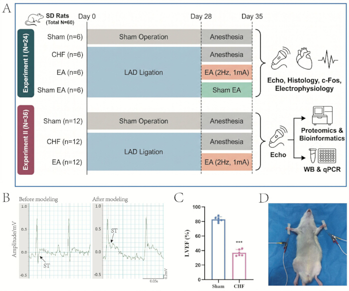
그림 1: 실험 설계, 만성 심부전 쥐 모델 구축 및 전침 중재. (A) 두 개의 독립적인 실험 시리즈에 대한 타임라인과 그룹 할당을 포함한 전체 실험 흐름도. (B) LAD 관상동맥 결찰 전후의 대표적인 심전도 기록으로, 화살표는 심근 허혈을 확인하는 ST분절 상승을 나타냄. (C) 성공적인 모델 구축을 확인하기 위한 수술 4주 후 LVEF의 정량적 분석 (생물학적 반복 n = 6, 평균 ± SD). ***p < 0.001 vs. Sham 그룹. (D) 양측 HT7 혈위에서의 EA 중재. 이 그림의 더 큰 버전을 보시려면 여기를 클릭하십시오.
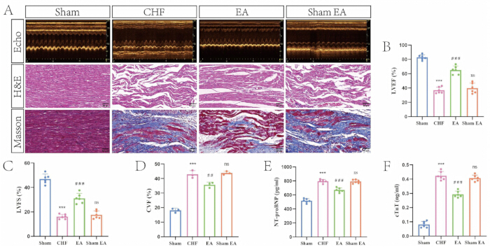
그림 2: 전기침 치료가 만성 심부전 쥐의 심장 기능을 개선하고 심근 손상을 완화한다. (A) 4가지 실험군에 따른 대표적인 M-모드 심초음파 영상과 심근 조직의 hematoxylin and eosin 및 Masson trichrome 염색 이미지(스케일 바 = 50 µm). (B) 그룹별 LVEF의 정량 분석(생물학적 반복 n = 6, 평균 ± SD). ***p < 0.01 vs. Sham군; ###p < 0.01 vs. CHF군; ns, CHF군 대비 유의미하지 않음. (C) 그룹별 LVFS의 정량 분석(생물학적 반복 n = 6, 평균 ± SD). ***p < 0.01 vs. Sham군; ###p < 0.01 vs. CHF군; ns, CHF군 대비 유의미하지 않음. (D) 그룹별 CVF의 정량 분석(생물학적 반복 n = 3, 평균 ± SD). ***p < 0.01 vs. Sham군; ##p < 0.01 vs. CHF군; ns, CHF군 대비 유의미하지 않음. (E) 그룹별 혈청 NT-proBNP 수치의 정량 분석(생물학적 반복 n = 6, 평균 ± SD). ***p < 0.01 vs. Sham군; ###p < 0.01 vs. CHF군; ns, CHF군 대비 유의미하지 않음. (F) 그룹별 혈청 cTnT 수치의 정량 분석(생물학적 반복 n = 6, 평균 ± SD). ***p < 0.01 vs. Sham군; ###p < 0.01 vs. CHF군; ns, CHF군 대비 유의미하지 않음. 여기를 클릭하여 이 그림의 확대 버전을 확인하십시오.
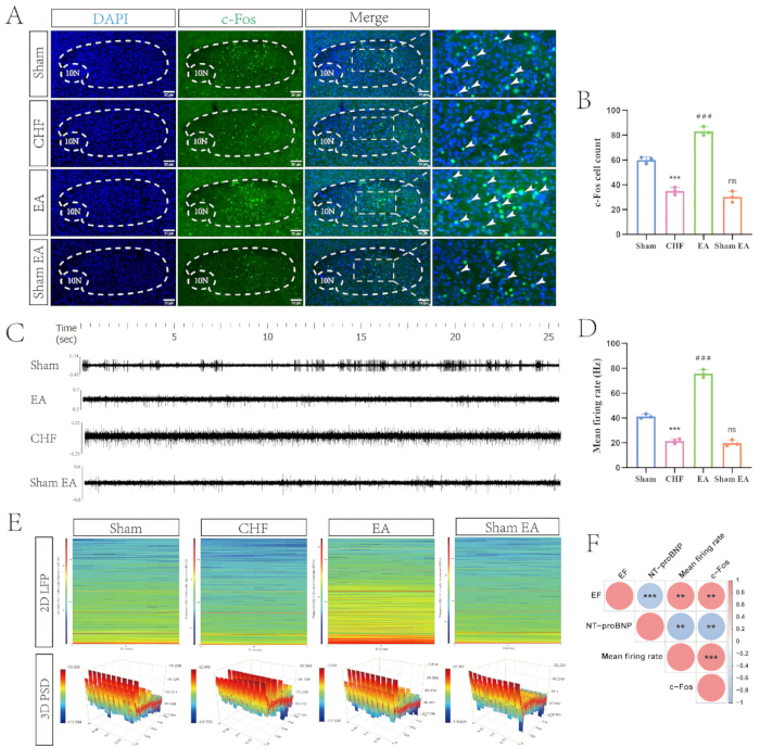
그림 3: 전기침 치료가 만성 심부전 쥐의 고립로핵(nucleus tractus solitarius) 내 신경 세포 활동을 회복시킨다. (A) 그룹별 NTS의 c-Fos 발현을 나타내는 대표적인 면역형광 이미지. 핵은 DAPI로 대조 염색되었으며, c-Fos 양성 세포는 녹색으로 염색되었다. 점선은 해부학적 영역을 나타내며, 흰색 화살표 머리는 대표적인 c-Fos 양성 뉴런을 가리킨다 (스케일 바 = 30 µm). (B) 그룹별 c-Fos 양성 세포 수의 정량적 분석 (생물학적 반복 n = 3, 평균 ± SD). ***p < 0.01 vs. Sham 그룹; ###p < 0.01 vs. CHF 그룹; ns, CHF 그룹 대비 유의미하지 않음. (C) 그룹별 NTS의 대표적인 생체 내(in vivo) 전기생리학적 스파이크 방전 추적선. (D) 그룹별 평균 뉴런 발화율의 정량적 분석 (생물학적 반복 n = 3, 평균 ± SD). ***p < 0.01 vs. Sham 그룹; ###p < 0.01 vs. CHF 그룹; ns, CHF 그룹 대비 유의미하지 않음. (E) NTS 내의 신경 진동 에너지를 보여주는 대표적인 2D 국소 전위 스펙트로그램 및 3D 전력 스펙트럼 밀도 지형도. (F) 말초 심혈관 표현형과 중추 신경 활동 파라미터 간의 관계를 평가한 상관관계 행렬. 빨간색과 파란색 원은 각각 양의 상관관계와 음의 상관관계를 나타낸다. **p < 0.01; ***p < 0.01. 이 그림의 확대 버전을 보려면 여기를 클릭하십시오.
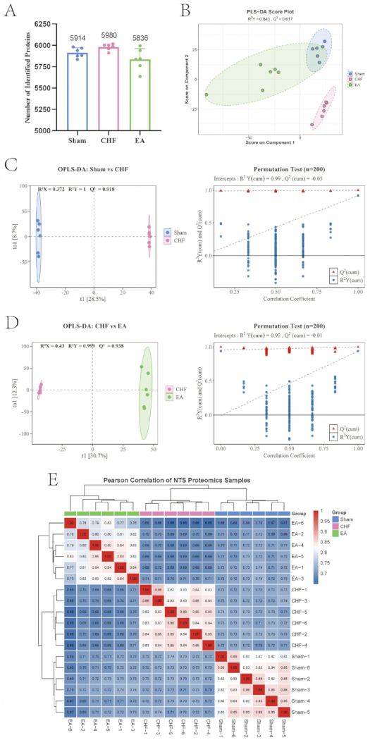
그림 4: 고속핵(nucleus tractus solitarius) 내 단백질체 프로파일의 품질 관리 및 다변량 통계 평가. (A) 3가지 실험군 전체에서 확인된 단백질의 정량적 개요(생물학적 반복 n = 6). (B) 군 간의 전반적인 공간적 분포와 분리를 보여주는 부분 최소 제곱 판별 분석(Partial least squares discriminant analysis) 스코어 플롯. (C) Sham 군과 CHF 군 비교에 대한 모델 신뢰도를 검증하는 직교 부분 최소 제곱 판별 분석(Orthogonal partial least squares discriminant analysis) 스코어 플롯 및 해당 순열 검사(20회 순열). (D) CHF 군과 EA 군 비교에 대한 모델 신뢰도를 검증하는 직교 부분 최소 제곱 판별 분석 스코어 플롯 및 해당 순열 검사(20회 순열). (E) 모든 단백질체 샘플에 걸쳐 군 내 재현성과 군 간 분산을 평가한 피어슨 상관계수의 계층적 군집 분석 히트맵. 여기를 클릭하여 이 그림의 더 큰 버전을 확인하십시오.
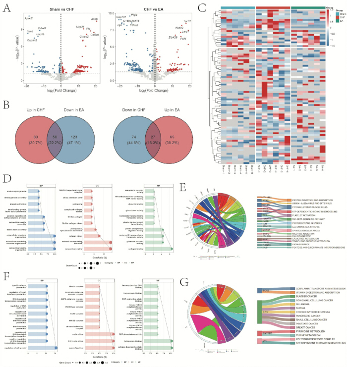
그림 5: 고립로핵에서 전기침 치료에 의해 조절되는 핵심 차별 발현 단백질의 식별 및 기능 농축 분석. (A) Sham 대 CHF 및 CHF 대 EA 비교에서 상향 조절 및 하향 조절된 단백질의 분포를 보여주는 화산 도표(Volcano plots). (B) EA 중재 후 하향 조절 및 상향 조절된 핵심 단백질을 식별하기 위한 교집합 분석 벤 다이어그램. (C) 핵심 차별 발현 단백질의 발현 프로파일에 대한 계층적 군집 분석 히트맵. (D) 하향 조절된 단백질 서브셋에 대해 농축된 생물학적 과정, 세포 구성 성분 및 분자 기능을 상세히 나타낸 GO 버블 도표. (E) 하향 조절된 단백질에 의해 농축된 신호 전달 경로를 매핑한 KEGG 코드 및 산키 다이어그램(Sankey diagrams). (F) 상향 조절된 단백질 서브셋에 대해 농축된 생물학적 과정, 세포 구성 성분 및 분자 기능을 상세히 나타낸 GO 버블 도표. (G) 상향 조절된 단백질에 의해 농축된 신호 전달 경로를 매핑한 KEGG 코드 및 산키 다이어그램. 이 그림의 확대 버전을 보려면 여기를 클릭하십시오.
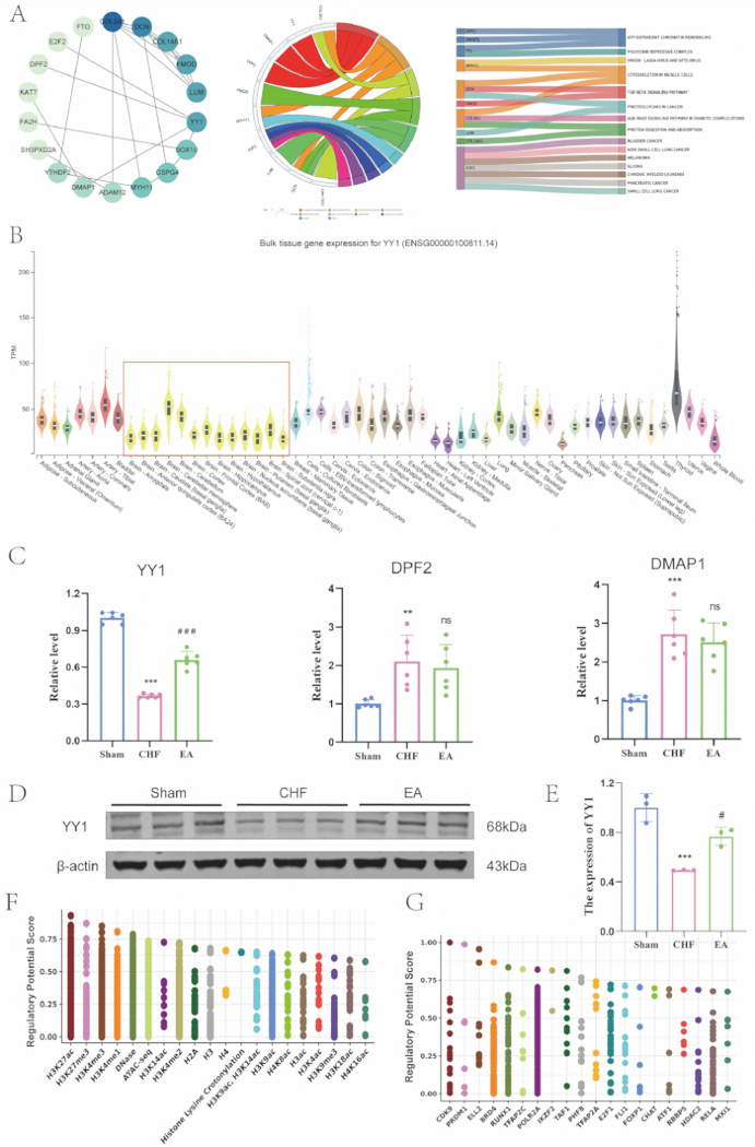
그림 6: 네트워크 토폴로지, 조직 발현 프로파일링 및 분자적 검증을 통해 전침과 관련된 후보 타겟으로 YY1을 식별함. (A) 식별된 허브 단백질의 단백질-단백질 상호작용 네트워크, 코드 다이어그램 및 생키 다이어그램. (B) GTEx 데이터베이스에서 도출된 YY1의 인간 조직 발현 프로파일이며, 빨간색 상자는 뇌 조직을 강조함. (C) 각 군의 NTS에서 YY1, DPF2 및 DMAP1 mRNA 발현의 정량적 분석 (생물학적 반복 n = 6, 평균 ± SD). **p < 0.01, ***p < 0.01 vs. Sham 군; ###p < 0.01 vs. CHF 군; ns, CHF 군 대비 유의미하지 않음. (D) 각 군의 NTS에서 YY1 및 β-actin의 대표적인 웨스턴 블롯 밴드. (E) 각 군의 YY1 단백질 발현의 정량적 분석 (생물학적 반복 n = 3, 평균 ± SD). ***p < 0.01 vs. Sham 군; #p < 0.05 vs. CHF 군. (F) 조절 잠재력 점수를 기반으로 YY1 유전자좌를 조절하는 후성유전적 히스톤 수정의 생물정보학적 예측. (G) 조절 잠재력 점수를 기반으로 YY1 유전자좌를 조절하는 상위 전사 인자의 생물정보학적 예측. 이 그림의 더 큰 버전을 보시려면 여기를 클릭하십시오.
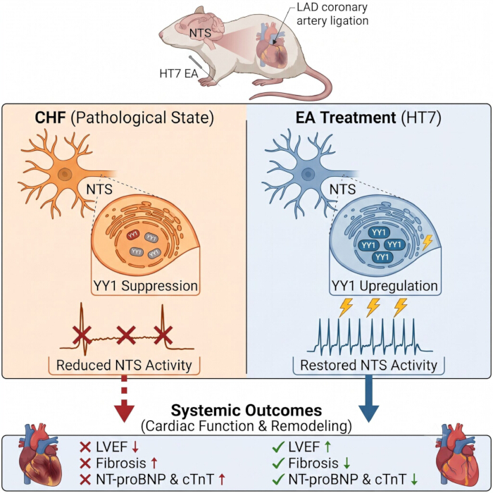
그림 7: HT7 전침 자극, YY1 상향 조절, 고속핵(nucleus tractus solitarius)의 신경 활동 회복 및 만성 심부전 개선 사이의 연관성에 대한 제안 모델. (왼쪽) 병리적 상태: LAD 관상동맥 결찰로 유도된 CHF 모델에서는 NTS 내의 신경 활동이 감소하며, 전사 인자인 YY1의 억제가 동반됩니다. 이러한 중추 신경 억제는 LVEF 감소, 심근 섬유화 증가, 혈청 심부전 마커 상승으로 나타나는 병리적 심장 리모델링과 연관되어 있습니다. (오른쪽) EA 치료: HT7 혈위의 EA 중재는 NTS 내 YY1 발현을 상향 조절하고 신경 발화를 회복시킵니다. 이러한 중추 신경 조절은 심장 보호 효과를 발휘하여 병리적 리모델링을 완화하고 전신 심장 기능을 개선합니다. 이 그림의 확대 버전을 보려면 여기를 클릭하십시오.
보충 그림 1: 프로테오믹 코호트의 심장 기능 평가 및 후보 허브 유전자의 인간 조직 발현 프로필. (A) 그룹 간 LVEF 및 LVFS의 정량 분석 (생물학적 반복 횟수 n = 12, 평균 ± SD). ***p < 0.01 vs. Sham 그룹; ###p < 0.01 vs. CHF 그룹. (B-J) GTEx 데이터베이스에서 도출된 COL3A1 (B), DMAP1 (C), DPF2 (D), FMOD (E), MYH1 (F), E2F2 (G), LUM (H), DCN (I) 및 COL14A1 (J)을 포함한 후보 허브 유전자의 인간 조직 발현 프로필.이 파일을 다운로드하려면 여기를 클릭하십시오.
보충 표 1: 고속핵(nucleus tractus solitarius) 내 글로벌 프로테옴의 종합적인 식별 및 정량 행렬.이 표는 모든 실험 코호트에 걸친 전체 정량 프로테오믹스 데이터셋을 나타냅니다. 주요 열에는 Protein ID(UniProt accession number), Gene(gene symbol), First.Protein.Description(기능적 주석) 및 Sham, CHF, EA 그룹(그룹당 n = 6)의 각 생물학적 반복 실험에 대한 정규화된 단백질 강도 값이 포함됩니다.이 파일을 다운로드하려면 여기를 클릭하십시오.
보충 표 2: Sham 그룹 대 CHF 그룹 비교에서 단백질의 OPLS-DA variable importance in projection 점수.이 표는 OPLS-DA 모델에 따라 Sham 그룹과 CHF 그룹을 구분하는 특징들을 나열합니다. 주요 열에는 feature(특정 단백질/유전자의 식별자)와 VIP_value(Variable Importance in Projection)가 포함되며, VIP_value는 각 특징이 그룹 분리에 기여하는 정도를 나타냅니다(임계값 VIP > 1.0).이 파일을 다운로드하려면 여기를 클릭하십시오.
보충 표 3: CHF 대 EA 비교 시 단백질의 OPLS-DA 변수 중요도 투영(variable importance in projection) 점수. 이 표는 OPLS-DA 모델을 기반으로 CHF 그룹과 EA 그룹을 구분하는 특징들을 나열합니다. 표 S2와 마찬가지로, 특징 열과 그에 해당하는 VIP_value가 포함되어 있으며, EA 중재에 가장 민감하게 반응하는 특징들을 강조합니다.여기를 클릭하여 이 파일을 다운로드하십시오.
보충 표 4: 전기침에 의해 조절된 85개의 핵심 차별 발현 단백질의 상세 목록.이 표는 CHF에 의해 유의하게 변했고 이후 EA 중재에 의해 역전된 단백질들의 교집합을 제공합니다. 주요 열로는 Protein_ID, Gene, log2FC (발현 변화를 나타내는 log2 Fold Change), 통계적 유의성을 위한 P_value (임계값 p < 0.05), 그리고 모델 기여도를 평가하는 VIP_value가 포함됩니다.이 파일을 다운로드하려면 여기를 클릭하십시오.
보조 표 5: 하향 조절된 핵심 단백질 서브셋의 유전자 온톨로지(Gene Ontology) 풍부성 분석.이 표는 EA에 의해 하향 조절된 58개 단백질의 유전자 온톨로지(GO) 기능적 주석을 보여줍니다. 주요 열로는 ONTOLOGY(생물학적 과정 [BP], 세포 구성 성분 [CC], 분자 기능 [MF]로 분류됨), 기능적 Description, p.adjust(다중 테스트에 대해 보정된 p-값), 그리고 각 용어에 풍부하게 포함된 특정 유전자에 매핑되는 geneID가 있습니다.이 파일을 다운로드하려면 여기를 클릭하십시오.
보충 표 6: 발현이 감소된 핵심 단백질 서브셋의 Kyoto Encyclopedia of Genes and Genomes 경로 풍부도 분석.이 표는 발현이 감소된 58개 단백질에 대해 풍부하게 나타난 신호 전달 폭포를 보여줍니다. 주요 열에는 경로 명칭(Term), 풍부도의 통계적 유의성을 나타내는 보정 p-값, 그리고 각 경로에 관여하는 구체적인 교차 유전자(Genes)가 포함되어 있습니다.이 파일을 다운로드하려면 여기를 클릭하십시오.
보충 표 7: 상향 조절된 핵심 단백질 서브셋의 유전자 온톨로지(Gene Ontology) 풍부도 분석.이 표는 EA에 의해 상향 조절된 27개 단백질의 유전자 온톨로지(GO) 기능 주석을 보여줍니다. 형식 및 열 정의(ONTOLOGY [BP, CC, MF], Description, p.adjust, geneID)는 표 S5에 상세히 설명된 내용과 동일합니다.이 파일을 다운로드하려면 여기를 클릭하십시오.
보충 표 8: 상향 조절된 핵심 단백질 서브셋의 Kyoto Encyclopedia of Genes and Genomes 경로 농축 분석.이 표는 상향 조절된 27개 단백질의 농축된 신호 전달 폭포를 보여줍니다. 형식 및 열 정의(용어, 보정된 p-값, 유전자)는 표 S6에 상세히 설명된 내용과 동일합니다.이 파일을 다운로드하려면 여기를 클릭하십시오.
보충 표 9: 단백질-단백질 상호작용 네트워크에서 도출된 18개 허브 단백질의 위상적 연결성 점수.이 표는 핵심 허브 유전자를 식별하는 데 사용된 네트워크 위상 매개변수를 요약한 것입니다. 주요 열에는 유전자 이름과 Degree, Betweenness Centrality, Closeness Centrality와 같은 중심 네트워크 지표가 포함되며, 이는 생물학적 네트워크 내에서 각 노드의 상대적 중요도와 연결성을 정량화합니다.이 파일을 다운로드하려면 여기를 클릭하십시오.
보충 표 10: 18개 네트워크 허브 단백질의 Kyoto Encyclopedia of Genes and Genomes 경로 농축 분석.이 표는 식별된 18개 허브 단백질에 의해 독점적으로 농축된 특정 신호 전달 경로를 나타냅니다. 주요 열에는 경로 용어(Term), 유의성을 나타내는 보정 p-값, 그리고 기능적 농축을 유도하는 각각의 유전자(Genes)가 포함됩니다.이 파일을 다운로드하려면 여기를 클릭하십시오.
보충 파일 1: 데이터 분석 및 시각화에 사용된 R 스크립트 및 자르지 않은 웨스턴 블롯 이미지.보충 파일에는 Pearson 상관관계 분석, 이중 화산 플롯 생성 및 EA-역전 단백질 식별, 유전자 온톨로지(GO) 풍부도 분석, 계층적 클러스터링 히트맵 생성, Kyoto Encyclopedia of Genes and Genomes (KEGG) 풍부도 시각화, 직교 부분 최소 제곱 판별 분석(OPLS-DA) 및 순열 검정, 부분 최소 제곱 판별 분석(PLS-DA), 단백질체 샘플 상관관계 히트맵 생성, 그리고 벤 다이어그램 분석에 사용된 R 스크립트가 포함되어 있습니다.이 파일을 다운로드하려면 여기를 클릭하십시오.
토론
본 연구에서는 CHF 쥐 모델에서 HT7 혈위의 EA에 의한 중추 기전을 조사하였다. 생리학적 및 조직학적 데이터는 EA가 LVEF를 개선하고 심근 섬유화를 완화했음을 나타냈다. 이러한 말초 변화와 병행하여, in vivo 전기생리학적 기록 및 신경 매핑을 통해 EA가 NTS 내의 신경 세포 발화율을 회복시켰음이 확인되었다. 질량 분석 기반 단백질체학을 통해 NTS의 분자 경관을 매핑하였으며, 중재에 의해 비정상적인 발현 패턴이 조절된 85개의 차별 발현 단백질을 식별하였다. 이후 위상 분석 및 조직 발현 프로파일링을 통해 전사 인자인 YY1을 후보 생물학적 표적으로 분리하였다. 분자 검증 결과, EA가 YY1 발현을 상향 조절하는 것으로 나타났다. 이러한 발견은 EA에 대한 중추 분자 네트워크를 구축하며, YY1이 중추 자율 신경 조절과 CHF의 완화를 연결하는 잠재적인 신경생물학적 표적임을 시사한다. 종합적인 중추 기전은 그림 7에 요약되어 있다.
NTS는 중추 심혈관 반사와 자율신경 조절의 통합 센터 역할을 합니다21,2. 심혈관 질환의 맥락에서, CHF는 교감신경 과활성화와 미주신경 감소로 특징지어지는 지속적인 자율신경 불균형을 수반합니다23. 이 질환의 병태생리학적 진행 과정에서 말초 압수용기의 민감도가 감소하며, 이는 NTS로 전달되는 흥분성 구심성 입력의 감소를 초래합니다24. 결과적으로, 이러한 신경 둔화는 미주외측연수의 미측 활성화를 감소시키고, 이어 전측 미주외측연수의 탈억제를 유발하여 과도한 중추 교감신경 유출로 이어집니다25. 이러한 기전과 병행하여, 전기침(EA)의 전기생리학적 및 조직학적 데이터는 CHF 상태에서 NTS 뉴런의 자발적 발화 빈도와 c-Fos 발현이 감소했음을 나타냈습니다. 표적 신경 조절 접근법으로서, EA로 생성된 체성감각 구심성 신호는 이론적으로 말초 신경을 통해 척수 후각으로 전달되어 NTS를 포함한 뇌간 구조로 상행하는 것으로 제안됩니다26. 연구 결과, HT7 혈위에서의 EA 중재가 이 뇌 영역의 신경 진동 에너지를 증가시키고 뉴런 발화율을 회복시켰음을 보여주었습니다. 생리학적으로, 회복된 NTS 뉴런 활성은 연수의 억제성 중간 뉴런의 활성화를 촉진하며, 이를 통해 과활성화된 교감신경 전운동 뉴런을 억제하는 동시에 부교감신경 원심성 신호를 강화합니다27. 이러한 중추 신경 기전을 말초 심혈관 표현형과 연결했을 때, 상관관계 분석 결과 이러한 중추 신경 활성화가 LVEF와는 양의 상관관계를, 말초 심부전 마커인 NT-proBNP와는 음의 상관관계를 가짐을 뒷받침했습니다. 이러한 발견은 EA가 NTS 뉴런의 병리적 억제를 완화하고 중추 자율신경 항상성을 촉진함으로써 말초 심장 보호 효과를 나타낼 수 있음을 시사합니다.
이러한 중추 신경 조절의 기저에 있는 분자 경로를 규명하기 위해 NTS의 단백질체 지형을 분석하였다. 질량 분석 기반의 단백질체 분석을 통해 중재에 의해 발현 패턴이 조절되는 85종의 단백질을 식별하였다. 기능적 층화 분석 결과, EA에 의해 하향 조절된 58종의 단백질 하위 집합이 세포외 기질 구성 및 transforming growth factor β-신호 전달 경로에 현저하게 관여하는 것으로 나타났다. 중추신경계의 세포외 기질은 시냅스 말단을 물리적으로 감싸고 국소 이온 항상성을 조절하는 perineuronal net을 포함하여 조직화된 격자 구조를 형성한다28. 심부전과 같은 만성 병리적 스트레스 상황에서, 비정상적인 세포외 기질 침착은 뇌간 내에 제한적인 미세환경을 조성한다29. 이러한 구조적 리모델링은 시냅스의 구조적 가소성을 물리적으로 저해하며 신경 회로를 교감신경 과활동 상태로 유지시킨다30. EA는 DCN과 더불어 COL3A1 및 COL14A1과 같은 구조 단백질의 발현을 조절함으로써 이러한 병리적 세포외 장벽을 리모델링한다. 동시에, 관련 신호 전달 폭포의 농축—이는 NTS 내에 실제 근세포가 존재함을 의미하기보다 일반적인 구조적 및 integrin 매개 변화를 나타내는 "cytoskeleton in muscle cells" KEGG 경로로 분류됨—은 세포외 변화가 세포 내 구획으로 전달됨을 시사한다31. 이러한 기계적 자극 전달 신호는 수지상 돌기 가시의 역동적인 회전을 회복시키고 자율신경 반사궁의 시냅스 전달 및 효율을 조절한다32.
세포 외 구조적 재편성을 넘어, 중재에 의해 상향 조절된 27개 단백질의 기능적 주석 분석 결과 ATP 의존적 염색질 리모델링의 관여가 나타났습니다. 지속적인 심혈관 자율신경 불균형은 후성유전학적 제한에 의해 유지되며, 여기서 응축된 염색질은 신경 억제를 조절하는 유전자의 프로모터 영역에 대한 접근성을 제한합니다3. ATP 의존적 염색질 리모델링 복합체는 ATP 가수분해 에너지를 사용하여 뉴클레오솜을 슬라이딩시키거나 제거함으로써 활성 전사를 위한 게놈 유전자좌를 개방합니다34. DNA 템플릿 전사 연장의 양성 조절과 더불어 이 경로의 농축은 EA가 후성유전학적 수준에서 작용하여 질병 관련 전사 기억을 리모델링함을 시사합니다35. 세포 외 미세환경의 구조적 이완은 핵 내 후성유전학적 변형으로 이어져, YY1 및 E2F2와 같은 핵심 단백질에 의해 연결되는 신경 보호 유전자 네트워크의 조화로운 전사를 촉진합니다36,37. EA의 치료 효능을 매개하는 생물학적 표적을 체계적으로 분리하기 위해 단계적 스크리닝 전략을 적용했습니다. 85개의 차별 발현 단백질을 위상 네트워크 매핑 및 경로 농축 분석을 통해 필터링하여 10개의 주요 허브 후보를 식별했습니다. 이후 조직 발현 프로파일링을 통해 YY1과 DPF2, 그리고 DMAP1만이 인간 뇌 조직에서 발현됨을 확인했습니다. 인비보(In vivo) 분자 검증 결과, 전사 반응성의 부족으로 인해 DPF2와 DMAP1이 제외되었으며, 이를 통해 YY1이 매우 반응성이 높은 후보 표적으로 확인되었습니다. YY1은 중추신경계 발달과 신경 가소성을 제어하는 아연 집게 전사 인자입니다38. 성숙한 신경 회로에서 YY1은 미토콘드리아 항상성을 유지하고 산화 스트레스를 완화함으로써 신경 보호 조절자로 기능합니다39. CHF의 병리적 진행 과정에서 지속적인 혈역학적 스트레스와 말초 염증은 뇌줄기 뉴런의 대사 기능 장애와 구조적 손상을 유발합니다40. 실험 데이터는 CHF 조건 하에서 NTS의 YY1 발현이 억제되는 반면, EA 중재가 이러한 병리적 억제를 상쇄했음을 보여주었습니다. 경험적 검증보다는 순수하게 컴퓨터 생물정보학 예측에 기반하여, YY1은 H3K27ac 및 H3K4me3와 같은 활성 히스톤 변형과 상호작용하여 하위 신경 보호 유전자 네트워크의 전사를 조율하는 것으로 가설이 세워졌습니다41. YY1 활성의 회복은 뉴런 대사 경로의 복구를 촉진하고 시냅스 연결의 구조적 무결성을 증진할 수 있습니다. 결과적으로, 조절된 NTS 뉴런은 말초 압수용기 반사 입력을 처리하는 능력이 향상되고 하행 교감신경 전운동 경로에 대해 억제 제어력을 행사하여, CHF와 관련된 심혈관 기능 저하를 개선합니다42.
이러한 발견에도 불구하고, 몇 가지 방법론적 한계에 대해 엄격한 고려가 필요합니다. 첫째, 현재의 실험 설계는 직접적인 기능적 증거 없이 상관관계 기반의 오믹스 및 발현 프로파일링에 의존하였습니다. 향후 연구에서는 NTS 내 바이러스 벡터 매개 넉다운 또는 과발현과 같은 표적 유전자 조작을 경험적 ChIP-seq 또는 CUT&Run과 병행하여 포함해야 합니다.&첫째, YY1의 인과적 역할을 확고히 규명하고 그 하위 후성유전학적 네트워크를 검증하기 위한 태그 분석(Tag assays)이 수행되지 않았습니다. 둘째, 단백질체학 단계에서 가짜 전침(Sham EA) 코호트를 제외함으로써 비특이적 체성감각 자극에 의해 유도된 분자적 시그니처를 확정적으로 배제하는 능력에 제한이 있었습니다. 그러나 현재의 설계에서는 가짜 전침 기저치와 비교하여 CHF 군과 EA 군을 비교하는 것이 EA 특이적 분자 변화를 분리해내는 데 충분한 것으로 간주됩니다. 이는 가짜 전침 및 CHF 군을 포함한 모든 비개입 코호트가 매일 가해지는 이소플루란 마취 노출에 대해 엄격한 기저 표준화를 거쳤으며, 이를 통해 가장 심각한 비특이적 신경학적 스트레스 교란 변수를 효과적으로 최소화했기 때문입니다. 또한, 초기 기능 데이터에서 가짜 전침은 CHF 군과 비교하여 유의미한 심혈관 또는 신경 활성화를 유발하지 않았으며, 이는 전류가 없는 표면적 체성감각 자극이 실질적인 중추 분자 재프로그래밍을 유도하는 데 필요한 임계치에 도달하지 않음을 시사합니다. 셋째, 본 연구에는 표준 비혈위 대조군이 부족하여 HT7 혈위의 특이성을 확정적으로 확인하는 데 한계가 있습니다. 더욱이, 제안된 HT7-NTS 구심성 신경 경로는 직접적인 신경 추적이나 신경 차단 검증 없이는 추측 단계에 머물러 있습니다. 마지막으로, 생체 내 전기생리학적 평가는 특정 흥분성 및 억제성 신경세포 하위 집단을 구분하지 않고 단일 시점에서 전반적인 NTS 활성을 평가했으며, 장기적인 신경 가소성을 측정하지 않았습니다. 후속 연구에서는 이러한 핵심적인 공백을 해결하여 중추 신경생물학적 메커니즘을 정교화해야 합니다.
최종적으로, 이러한 결과들이 상관관계적 성격임을 인정하면서 이를 통합함으로써, 본 연구는 EA가 NTS 뉴런 기능을 회복시켜 병리적인 심장 리모델링을 완화한다는 가치 있는 분자적 프레임워크를 제공합니다. 이러한 데이터는 YY1이 중추 심혈관 신경 조절을 위한 반응성이 높은 후보 타겟임을 강조하며, 심부전 관리에서 침술 치료의 임상적 적용을 위한 새로운 신경생물학적 통찰을 제시합니다.
공개 사항
모든 저자는 이해상충이 없음을 선언합니다. 또한 저자들은 원고 작성 과정의 어느 단계에서도 인공지능 도구가 사용되지 않았음을 선언합니다.
감사의 글
본 연구에 기여한 모든 연구자분께 진심으로 감사드리며, 실험 동물들의 희생에 깊은 경의를 표합니다. 본 연구는 중국 국가중점연구개발프로그램(grant no. 202YFC35050 및 202YFC350502)과 Xu Nenggui 대학 수준 인재 지원 프로그램(grant no. DT24002)의 지원을 받아 수행되었습니다. 지원 기관은 연구 설계, 데이터 수집 및 분석, 출판 결정 또는 원고 작성에 관여하지 않았습니다.
재료
| 이름 | 회사 | 카탈로그 번호 | 댓글 |
|---|---|---|---|
| 성체 수컷 Sprague-Dawley 랫드 | Liaoning Changsheng Biotechnology Co., Ltd. | 번호: SCXK (LIAO) 2025-01 | RGD_70508 |
| 침 acupuncture needles | Suzhou Tianxie Acupuncture Instrument Co., Ltd. | — | — |
| DAPI 포함 항퇴색 봉입제 | Aibixin Biotechnology Co., Ltd. | abs9235-25ml | — |
| Anti-YY1 1차 항체 | Proteintech Group, Inc | 6281-1-Ig | AB_288164 |
| Anti-Beta Actin 1차 항체 | 609-1-Ig | AB_2687938 | |
| 소혈청알부민 (BSA) | Sigma Odellie Trading Co., Ltd. | V9093 | — |
| BCA 단백질 정량 분석 키트 | Shanghai Biyuntian Biotechnology Co., Ltd. | P012S | — |
| c-Fos (9F6) 토끼 mAb | Sai Xin Tong Biological Reagents Co., Ltd. | 250S | AB_24721 |
| Cytoscape | Cytoscape 컨소시엄 | 버전 3.2.1 | SCR_03032 |
| ChamQ Universal SYBR qPCR Master Mix | Vazyme Biotech Co., Ltd. | Q71-02 | — |
| 전침 장치 | Nanjing Jisheng Medical Technology Co., Ltd. | HANS-20A | — |
| 8채널 (2 × 4) 미세전극 어레이 | Kewa Suzhou Medical Technology Co., Ltd. | - | — |
| ELISA 키트 (NT-proBNP) | Wuhan Enzyme Immuno-Biotechnology Co., Ltd. | MM-0329R1 | — |
| ELISA 키트 (cTnT) | MM-0795R2 | — | |
| 강화 화학발광 키트 | Proteintech Group, Inc | PK1001 | — |
| 염소 항-토끼 IgG | Aimee Technology Co., Ltd. | 111-545-003 | AB_2338046 |
| HRP-접합 염소 항-마우스 IgG 2차 항체 | BeiJing Cowin Biotech Co.,Ltd. | CW0102 | 입력된 텍스트가 없습니다. 번역할 내용을 제공해 주십시오. |
| HiScript III RT SuperMix | Vazyme Biotech Co., Ltd. | R323-01 | — |
| ImageJ 소프트웨어 | 미국 국립보건원 | 버전 4.0 | SCR_03070 |
| 이미징 시스템 | Bio-Rad Laboratories, Inc | ChemiDoc MP | |
| 다채널 소동물 마취기 | RWD Life Science Co., Ltd | R510-2-10 | — |
| 현미경 | Olympus Optical Co., Ltd. | DP72 | — |
| NeuroExplorer 소프트웨어 | Beijing Plexon Technology Co., Ltd. | 버전 5.0 | SCR_01818 |
| Offline Sorter 소프트웨어 | Beijing Plexon Technology Co., Ltd. | 버전 4.7.2 | SCR_0012 |
| Orbitrap Astral | Thermo Fisher Scientific | — | — |
| Powerlab 표준 사지 II 리드 시스템 | AD Instruments International Trading Co., Ltd. | ML18 | — |
| PepMap Neo 트랩 컬럼 | Thermo Fisher Scientific | — | — |
| 포스파타아제 억제 | Shanghai Biyuntian Biotechnology Co., Ltd. | P1081 | — |
| R 소프트웨어 | — | 버전 4.5.2 | SCR_01905 |
| RIPA 용해 완충액 | Yeasen Biotechnology Co., Ltd. | 2015ES60 | — |
| RNAiso Plus | TaKaRa Biotechnology Co., Ltd. | 9108 | — |
| 소동물 디지털 초음파 시스템 | Feiyino Technology Co., Ltd. | VINNO6 연구실 | — |
| 통계 소프트웨어 | GraphPad 소프트웨어 | 버전 8.0 | SCR_02798 |
| OmniPlex 다채널 획득 시스템 | Beijing Plexon Technology Co., Ltd. | 버전 1.20.0 | — |
| Triton X-10 | Beijing Solab Technology Co., Ltd. | 22298142 | — |
| Vanquish Neo UHPLC 시스템 | Thermo Fisher Scientific | — | — |
| µPAC Neo 고처리량 컬럼 | Thermo Fisher Scientific | — | — |
참고문헌
- Valente V, et al. The global epidemiology of heart failure: a comprehensive and contemporary review. Eur J Heart Fail. 2026.
- Triposkiadis F, et al. The sympathetic nervous system in heart failure physiology, pathophysiology, and clinical implications. J Am Coll Cardiol. 2009;54(19):1747-62.
- Arshad MS, et al. Sympathetic nervous system in heart failure: targets for treatments. Curr Hypertens Rep. 2025;27(1):20.
- Fan H, et al. The hypotensive role of acupuncture in hypertension: clinical study and mechanistic study. Front Aging Neurosci. 2020;12:138.
- Zuo H, et al. Electroacupuncture alleviates acute myocardial ischemic injury in mice by regulating the β1 adrenergic receptor and post-receptor protein kinase A signaling pathway. Acupunct Med. 2024;42(6):342-55.
- Wu HS, et al. Neural mechanism of HT7 electroacupuncture in myocardial ischemia: critical role of the paraventricular nucleus oxytocin system. Front Neurosci. 2025;19:1678938.
- Kun W, et al. Electroacupuncture ameliorates cardiac dysfunction in myocardial ischemia model rats: a potential role of the hypothalamic-pituitary-adrenal axis. J Tradit Chin Med. 2023;43(5):944-54.
- Xu W, et al. Electroacupuncture ameliorates chronic heart failure: the role of CRH neurons in the paraventricular nucleus of the hypothalamus. Front Neurosci. 2026;20:1741523.
- Andresen MC, Kunze DL. Nucleus tractus solitarius--gateway to neural circulatory control. Annu Rev Physiol. 1994;56:93-116.
- Craft GE, et al. Recent advances in quantitative neuroproteomics. Methods. 2013;61(3):186-218.
- Charan J, Kantharia ND. How to calculate sample size in animal studies? J Pharmacol Pharmacother. 2013;4(4):303-6.
- Zhou J, et al. Electroacupuncture pretreatment mediates sympathetic nerves to alleviate myocardial ischemia-reperfusion injury via CRH neurons in the paraventricular nucleus of the hypothalamus. Chin Med. 2024;19(1):43.
- Litwin SE, et al. Serial echocardiographic assessment of left ventricular geometry and function after large myocardial infarction in the rat. Circulation. 1994;89(1):345-54.
- Shu Q, et al. Electroacupuncture alleviates myocardial ischemia-reperfusion injury by inhibiting hypothalamic paraventricular nucleus neurons projecting to the rostral ventrolateral medulla. Eur J Neurosci. 2024;60(5):4861-76.
- Wan F, et al. Electroacupuncture improves cerebral blood flow in vascular cognitive impairment mice by activating the locus coeruleus-prefrontal cortex circuit. Neuroscience. 2025;583:33-42.
- Xu R, et al. Electroacupuncture ameliorates incisional pain via suppressing IL-33 signaling-related macrophage infiltration and ROS overproduction in incised skin. Chin Med. 2026;21(1):26.
- Quiroga RQ, et al. Unsupervised spike detection and sorting with wavelets and superparamagnetic clustering. Neural Comput. 2004;16(8):1661-87.
- Goldwyn JH, et al. Gain control with A-type potassium current: IA as a switch between divisive and subtractive inhibition. PLoS Comput Biol. 2018;14(7):e1006292.
- Buzsáki G. Large-scale recording of neuronal ensembles. Nat Neurosci. 2004;7(5):446-51.
- Zhang J, Mifflin SW. Responses of aortic depressor nerve-evoked neurones in rat nucleus of the solitary tract to changes in blood pressure. J Physiol. 2000;529(Pt 2):431-43.
- Benarroch EE. The central autonomic network: functional organization, dysfunction, and perspective. Mayo Clin Proc. 1993;68(10):988-1001.
- Scheitz JF, et al. Bidirectional brain-heart interactions in health and disease. Nat Rev Neurol. 2026;22(4):209-25.
- Floras JS. Sympathetic nervous system activation in human heart failure: clinical implications of an updated model. J Am Coll Cardiol. 2009;54(5):375-85.
- Iannetta D, et al. Dissecting the exercise pressor reflex in heart failure: a multi-step failure. Auton Neurosci. 2025;259:103269.
- Sved AF, et al. Baroreflex dependent and independent roles of the caudal ventrolateral medulla in cardiovascular regulation. Brain Res Bull. 2000;51(2):129-33.
- Liu Y, et al. Neurophysiological basis of electroacupuncture stimulation in the treatment of cardiovascular-related diseases: vagal interoceptive loops. Brain Behav. 2024;14(10):e70076.
- Zhou W, Benharash P. Effects and mechanisms of acupuncture based on the principle of meridians. J Acupunct Meridian Stud. 2014;7(4):190-3.
- Fawcett JW, et al. The roles of perineuronal nets and the perinodal extracellular matrix in neuronal function. Nat Rev Neurosci. 2019;20(8):451-65.
- Moon S, Ito Y. Vasculature cells control neuroglial co-localization and synaptic connection in a central nervous system tissue mimic system. Hum Cell. 2023;36(6):1938-47.
- Ferrer-Ferrer M, Dityatev A. Shaping synapses by the neural extracellular matrix. Front Neuroanat. 2018;12:40.
- Rabinowitch I, et al. Understanding neural circuit function through synaptic engineering. Nat Rev Neurosci. 2024;25(2):131-9.
- McGeachie AB, et al. Stabilising influence: integrins in regulation of synaptic plasticity. Neurosci Res. 2011;70(1):24-9.
- Qiu L, et al. RNA modification: mechanisms and therapeutic targets. Mol Biomed. 2023;4(1):25.
- Clapier CR, et al. Mechanisms of action and regulation of ATP-dependent chromatin-remodelling complexes. Nat Rev Mol Cell Biol. 2017;18(7):407-22.
- Du W, et al. Mechanisms of chromatin-based epigenetic inheritance. Sci China Life Sci. 2022;65(11):2162-90.
- Rashid F, et al. Mechanomemory of nucleoplasm and RNA polymerase II after chromatin stretching by a microinjected magnetic nanoparticle force. Cell Rep. 2024;43(7):114462.
- Weintraub AS, et al. YY1 is a structural regulator of enhancer-promoter loops. Cell. 2017;171(7):1573-88.e28.
- Zurkirchen L, et al. Yin Yang 1 sustains biosynthetic demands during brain development in a stage-specific manner. Nat Commun. 2019;10(1):2192.
- Cunningham JT, et al. mTOR controls mitochondrial oxidative function through a YY1-PGC-1alpha transcriptional complex. Nature. 2007;450(7170):736-40.
- van Weperen VYH, Vaseghi M. The brain-heart axis: effects of cardiovascular disease on the CNS and opportunities for central neuromodulation. Nat Rev Neurosci. 2026;27(3):159-77.
- Lam JC, et al. YY1-controlled regulatory connectivity and transcription are influenced by the cell cycle. Nat Genet. 2024;56(9):1938-52.
- Montuoro S, et al. Neuroimmune cross-talk in heart failure. Cardiovasc Res. 2025;121(4):550-67.