All procedures involving animal models have been reviewed by the local institutional animal care committee and the JoVE veterinary review board.
1. Local Tumor Induction via Surgical Adeno-cre Infection
- Preparation of animals for surgery
NOTE: Virtually any conditional ("floxed") mutation can be induced via the here described method. The use of mutations in genes relevant to colorectal cancer, such as Apc, Kras, or Tp53, is recommended. The efficiency of Cre recombination is dependent on the size of the construct to be excised. Large floxed sequences are excised less efficiently. The recombination of all alleles should be confirmed in the tumors by PCR.
- For the development of colorectal tumors, use a cross of the following conditional alleles for a basic model of CRC (MGI database number in brackets):
Apctm2Rak (MGI: 3688435)
Krastm4Tyj (MGI: 2429948)
Tp53tm2Tyj (MGI: 3039263)
- If a fluorescence reporter allele is required (e.g., to detect micrometastases), use the following allele:
Gt(ROSA)26Sortm6(CAG-ZsGreen1)Hze (MGI: 3809522)
NOTE: All above strains are available via the NCI Mouse Repository or the Jackson Laboratory. No fasting is required as all remaining fecal matter can be flushed out before the adenoviral infection. Preoperative fasting leads to higher perioperative mortality and constitutes tremendous stress for small rodents.
- Use sevoflurane at 3 - 3.5 vol% for general anesthesia. A loss of the toe pinch reflex indicates sufficient anesthesia.
- Before the first incision, inject 0.05 mg/kg of buprenorphine subcutaneously.
- Cover the eyes of the anesthetized mouse with ophthalmic ointment to avoid desiccation of the cornea.
- Place the mouse in a supine position on a small table. Use non-traumatic adhesive tapes to restrain the mouse.
- Shave the abdomen with an electric shaver (depilatory cream can be used alternatively) and disinfect with alcohol swabs or iodine. Use the contact time recommended by the manufacturer.
- Cover the surgical field with sterile drapes.
NOTE: The use of perioperative antibiotics is optional and subject to institutional guidelines.
- Use sterile single-use or sterilized instruments for all surgical procedures.
- Midline laparotomy and exposure of the colon
- Use scissors (scalpels can be used alternatively) to make a midline incision (~ 15 mm) on the skin of the lower abdomen.
- Pick up the abdominal wall musculature with forceps and carefully incise it with scissors, opening the abdominal cavity.
- Identify the distal colon, only touch it with atraumatic forceps. Clamp the colon with a delicate clamp (e.g., a Micro Serrefine vascular clamp) approximately 15 mm proximally of the anus.
NOTE: Give special attention to the vulnerability of the colon at all times. Perforation inevitably leads to peritonitis and sepsis and requires euthanasia of the animal.
- Segmental colon infection with Adeno-cre virus
- Insert a flexible Teflon tube transanally and carefully advance it until it reaches the lumen occlusion achieved by the clamp previously placed 15 mm from the anal verge. Do not use excessive force as this may lead to perforation.
- Cannulate the tube with a 30G cannula, connect a standard 1 mL syringe, and flush the colon with normal saline to evacuate remaining fecal matter. This may require several mL of saline.
- Once the distal colon is empty, remove the tube and replace it with a fresh Teflon tube and again position it directly distal to the clamp as described above.
- Occlude the colon with a second clamp ~ 3 mm distal to the proximal clamp (i.e., over the inserted tube, ~ 12 mm from the anal verge), resulting in a 3 mm isolated segment to be infected.
NOTE: For distal occlusion of the segment, Fogarty coronary artery clips have proven to be most suitable as they are rubberized, leading to tight occlusion of the colon despite the intraluminal tube between the clamp's branches.
- Use a second syringe (standard 1 mL with a 30G cannula) to carefully inject 50 - 80 µL of 0.25% Trypsin-EDTA into the clamped colon segment and incubate for 10 min. Leave the cannula and the syringe attached to the Teflon tube to prevent the fluid from leaking back.
NOTE: The colon must be inflated to break up the mucosal barrier and reach the crypt stem cells, yet not too inflated to avoid perforation of the clamped segment.
- First, remove the distal clamp and then the trypsin tube.
- Flush the distal colon with ~ 500 µL of normal saline to remove the remaining trypsin.
- Insert a new Teflon tube, put the distal clamp back in place, and inflate the colon segment with 50 - 80 µL of adenoviral solution (1011 plaque-forming units (PFU)/mL in phosphate-buffered saline) and incubate for 30 min (Figure 2A).
NOTE: Do not spill viral solution as contact with adeno-cre may lead to tumor development in any tissue of conditionally mutant mice.
- Remove the clamps and the tube.
- Closure of the abdomen and postoperative recovery
- Close the abdominal wall with 6-0 rapidly absorbable running sutures (e.g., polydioxanone (PDS)).
- Close the skin with surgical wound clips.
- Place the mouse on a heating pad set to 38 °C until fully recovered from the anesthesia.
- Administer another bolus of 0.05 mg/kg buprenorphine i.p. 12 h after surgery, followed by additional buprenorphine boluses every 12 h if needed.
- Monitor the mice at least once daily for signs of distress due to tumor growth.
2. Colonoscopy
NOTE: Depending on the conditional mutations used, adenoviral infection leads to endoscopically visible tumors within 2 - 4 weeks. Therefore, perform the first postoperative colonoscopy 2 weeks after the adenoviral induction and repeat every 2 weeks. A commercially available system is recommended for murine colonoscopy.
- Preparation of animals for colonoscopy
NOTE: No fasting is required. The remaining fecal matter is usually well-formed in the distal colon and can be pushed beyond the tumor during colonoscopy, making the stressful process of repeated fasting unnecessary.
- Use sevoflurane at 3 - 3.5 vol% for general anesthesia. A loss of the toe pinch reflex indicates sufficient anesthesia.
- Cover the eyes of the anesthetized mice with ophthalmic ointment to avoid desiccation of the cornea.
- Restrain the mice in a supine position on a small table.
- Colonoscopy
- Insert the scope (diameter 1.9 mm; length 10 cm) into the intestinal tract through the anus and carefully insufflate air under visual control to distend the colon. Do not insufflate more air than required for the examination.
NOTE: For air insufflation, the anti-fog air pump of the colonoscopy system may be used. If no anti-fog air pump is available, any other air pump with very low-pressure settings can be used or pressurized air with a delicate pressure-reducing valve. Carbon dioxide (CO2) easily leads to acidosis in small rodents and must therefore be avoided.
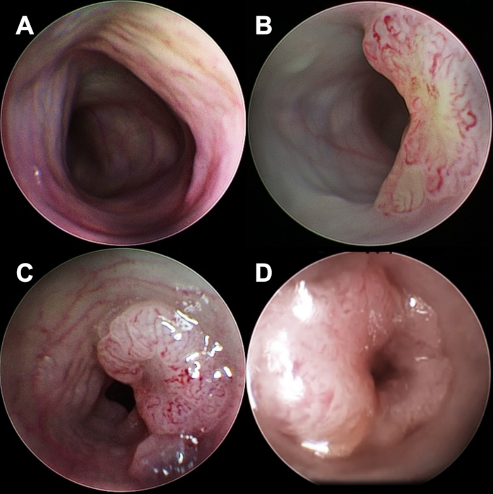
- Carefully push the scope forward until a mucosal lesion in the distal colon can be identified (Figure 1A - 1D).
- Save endoscopic images for later evaluation. An endoscopic scoring system for intraluminal tumors has been described before.
- Carefully remove the scope and place the mouse on a heating pad set to 38 °C until it has fully recovered from the anesthesia.