This video demonstrates the use of transcranial magnetic stimulation (TMS) to modulate motor neuron activity and measure muscle responses via electromyography (EMG). It outlines the steps for locating the motor hotspot, determining stimulation thresholds, and assessing the effects of subthreshold TMS (subTMS) on cortical inhibition.
Een abonnement op JoVE is vereist om deze inhoud te bekijken. Log in of start vandaag met uw gratis proefperiode.
Methodenartikel
Transcranial Magnetic Stimulation for Modulating Muscle Response in Humans
905 weergaven
⸱
29 augustus 2025
In dit artikel
Samenvatting
Protocol
All procedures involving human participants have been performed in compliance with the institutional, national, and international guidelines for human welfare and have been reviewed by the local institutional review board.
- Transcranial magnetic stimulation (TMS)
- Fix the reflective markers on the participant's forehead with double-sided adhesive tape.
NOTE: Reflective markers allow for the constant delivery of TMS to the target area over M1 by using a neuronavigation system (see Figure 1). The advantage of the neuronavigation system is that the coil position can be recorded relative to the skull position in space and can be checked at any time throughout the entire experiment. - Use a 95-mm focal figure-of-eight coil attached to a TMS stimulator to deliver stimuli to the contralateral motor cortical hand area.
NOTE: The induced current must be directed from posterior to anterior and must be delivered in reverse mode. The waveform should be monophasic. - Find the optimal position (hot spot) of the coil relative to the skull for eliciting motor evoked potentials (MEPs) in the first dorsal interosseous (FDI) muscle by performing a classical mapping procedure.
- Start by placing the coil approximately 0.5 cm anterior to the vertex and over the midline, with the coil handle pointing at 45° towards the contralateral forehead.
NOTE: This will ensure that the induced current flow is approximately perpendicular to the central sulcus. - To get the participants used to the TMS stimuli, start at intensities below 25% of maximum stimulator output (MSO). Then, start to increase the stimulation intensity and move the coil in the medio-lateral and rostro-frontal direction to discover the hot spot.
- Start by placing the coil approximately 0.5 cm anterior to the vertex and over the midline, with the coil handle pointing at 45° towards the contralateral forehead.
- Once the hot spot is found, record the optimal position with the neuronavigation system. Determine the active motor threshold (aMT) by adjusting the intensity of the stimulator output. Define the aMT as the minimum intensity required to evoke MEP peak-to-peak amplitudes in the electromyography (EMG) of the FDI larger than 0.1 mV in three out of five consecutive trials.
- Fix the reflective markers on the participant's forehead with double-sided adhesive tape.
- SubTMS-induced EMG suppression (see Figure 2B).
- Prepare the weight representing 10% of maximal force (Fmax) by filling the bottle of water.
NOTE: The 10% of Fmax is selected based on the Fmax (the best of 3 trials) performed. In the subthreshold TMS protocol, only 10% of the Fmax has to be selected, as it has previously been shown that fatigue has an influence on subTMS-induced EMG suppression. For the same reason, the subTMS session must be carried out in a separate session. The volume of water used here is between 0.3 L (smallest 30% of Fmax) and 1.2 L (biggest 30% of Fmax). - Instruct participants about the task; the motor task consists of holding the index finger in the target position by counteracting the light weight of 10%.
- As the participants remain relaxed in a comfortable position, they find the optimal intensity for eliciting subTMS-EMG suppression, without giving any instruction about the focus of attention. To do this, successively diminish in steps of 2% MSO from the aMT determined previously.
- While they are still seated in a relaxed and comfortable position, have the participants perform two separate isometric index finger abductions at 10% of Fmax and record the EMG signal of the FDI. During this isometric index finger abduction, record (by pressing the "record" button on the recording software) 20 trials with and 20 trials without TMS, with a randomized interstimulus interval (ISIs) ranging from 0.8 to 1.1 s in a 100-ms time window.
NOTE: This interval ensures that participants do not have to perform the motor task for too long and therefore minimizes fatiguing effects. After each series, check the subTMS-induced EMG suppression.- Apply a full-wave rectification by converting all negative amplitudes to positive amplitudes in the EMG signals. Average the EMG signals using time-normalized averaging.
NOTE: The onset of subTMS-EMG suppression is defined as the moment when the difference between the trials with and those without TMS is negative for at least 4 ms in a time window from 20 to 50 ms after the TMS: EMGDiff = EMGWithout- EMGWith.
- Apply a full-wave rectification by converting all negative amplitudes to positive amplitudes in the EMG signals. Average the EMG signals using time-normalized averaging.
- Repeat step 2.3 until the optimal stimulation intensity is found, indicated by the largest EMG suppression.
NOTE: The optimal intensity is found at around 80% of aMT. - While they remain seated in a relaxed and comfortable position, have the participants perform two separate isometric index finger abductions.
- During this isometric index finger abduction, record (by pressing the "record" button on the recording software) 40 trials with and 40 trials without TMS, with randomized ISIs.
- Between each series, allow a break of a minimum of 5 min to minimize any bias that may be induced by fatigue.
- Prepare the weight representing 10% of maximal force (Fmax) by filling the bottle of water.
Toegang beperkt. Log in of start een proefperiode om deze inhoud te bekijken.
Resultaten
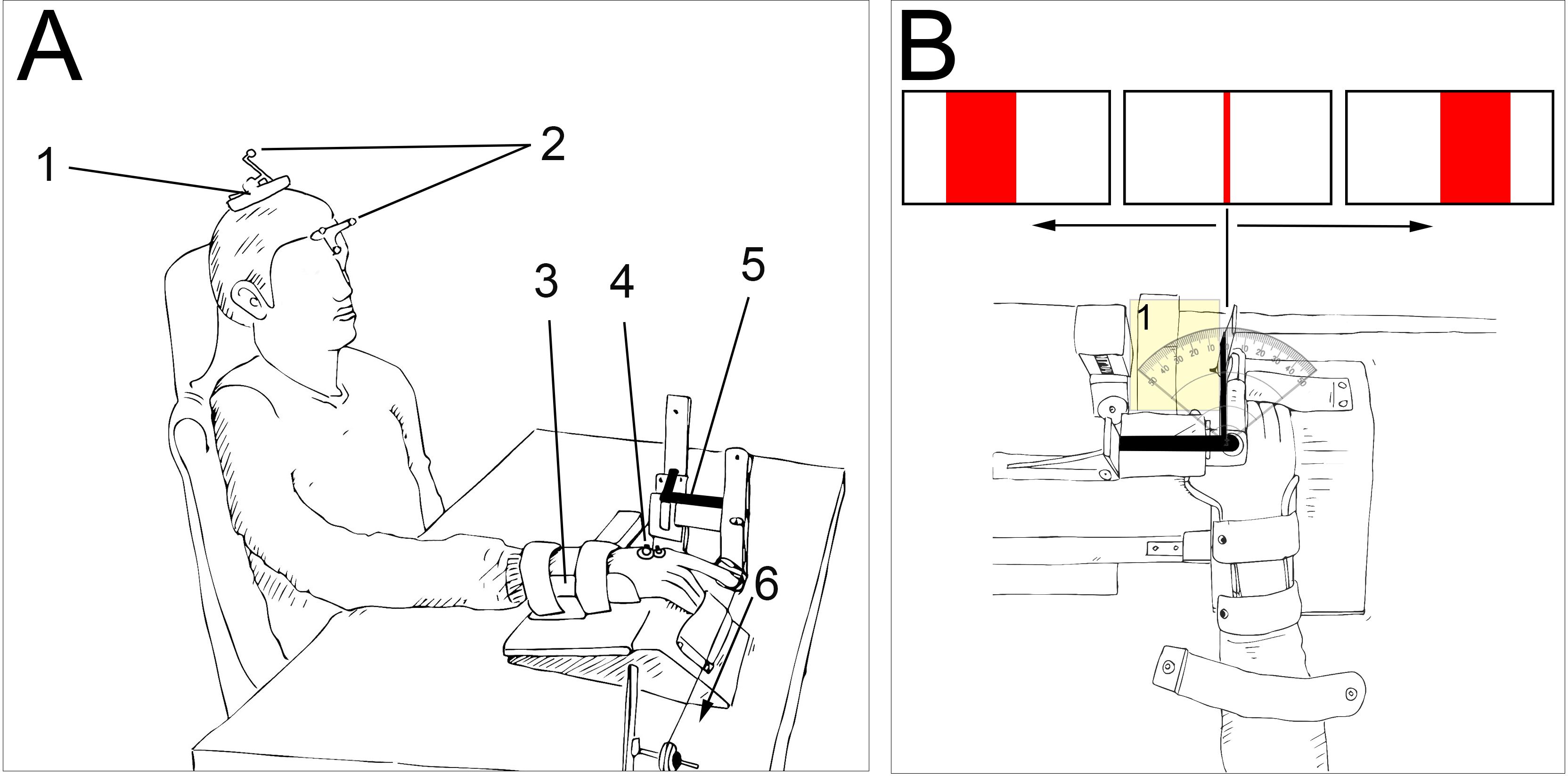
Figure 1. Experimental setup. A. 1. The TMS ...
Toegang beperkt. Log in of start een proefperiode om deze inhoud te bekijken.
Materialen
| Naam | Bedrijf | Catalogusnummer | Opmerkingen |
|---|---|---|---|
| BlueSensor P | Ambu A/S, Bellerup, Denmark | - | Ag/AgCl surface electrodes for EMG |
| Polaris Spectra | Northern Digital, Waterloo, ON, Canada | - | neuronavigation system, active or passive markers tracker |
| Localite TMS Navigator Version 2.0.5 | LOCALITE GmbH, Sankt Augustin, Germany | - | navigation system for transcranial magnetic stimulation (TMS) |
| MagVenture MagPro X100 | MagVenture A/S, Farum, Denmark | 9016E0711 | Transcranial magnetic stimulator |
| MagVenture D-B80 | MagVenture A/S, Farum, Denmark | 9016E0431 | TMS coil (figure-of-eight) |
| Goniometer | N/A | - | Custom-made goniometer |
| Othopedic splint | N/A | - | Custom-made splint |
| Recording software | LabView based | - | Custom-made script |
