This video demonstrates the procedure for performing electroconvulsive therapy in a human patient, highlighting the stimulation process and assessing seizure activity through pupillary response.
Een abonnement op JoVE is vereist om deze inhoud te bekijken. Log in of start vandaag met uw gratis proefperiode.
Methodenartikel
Performing Electroconvulsive Therapy and Assessing Seizure Efficacy via Pupillary Response
1.7K weergaven
⸱
29 augustus 2025
In dit artikel
Samenvatting
Protocol
All procedures involving human participants have been performed in compliance with the institutional, national, and international guidelines for human welfare and have been reviewed by the local institutional review board.
1. Electroconvulsive Therapy Procedure
1. Conduct electroconvulsive therapy (ECT) via bitemporal electrode stimulation using an ECT instrument. Set the initial electrical stimulus dose (%) at half the value of each individual’s age.
NOTE: Maximum stimulus dose is 100%. The electrical stimulation time is about 7-8 s.
2. Immediately after electrical stimulation, hold the automated infrared pupillometer over one of the patient’s eyes. At this time, if the patient closes their eyes, keep the patient’s eyelid open. Press the device button, and measure maximum (initial) resting pupil size or minimum pupil size after stimulation.
3. Perform controlled ventilation using a face mask with 100% oxygen until the patient begins breathing spontaneously.
4. Using an electroencephalogram (EEG), measure ictal regularity, seizure time, and greater postictal suppression by the psychiatrist (Figure 1 and Figure 2).
Toegang beperkt. Log in of start een proefperiode om deze inhoud te bekijken.
Resultaten
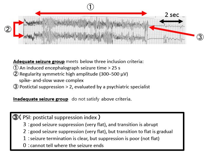
Figure 1. Electroencephalogram (EEG) results during a typical seizure.
Toegang beperkt. Log in of start een proefperiode om deze inhoud te bekijken.
Materialen
| Naam | Bedrijf | Catalogusnummer | Opmerkingen |
|---|---|---|---|
| Npi-100/automated infrared pupillometer | NeurOptics | ||
| Electroencephalogram (EEG) | Somatics Inc. | ECEF-4 | |
| Npi-200/automated infrared pupillometer | NeurOptics |
