This video demonstrates bacterial transinfection through antigen-specific T cell–dendritic cell interactions. Bacteria-infected dendritic cells presenting ovalbumin peptide are incubated with T cells, either directly or separated by a membrane. Direct contact allows immunological synapse formation, enabling bacterial transfer, while the membrane prevents interaction. Gentamicin treatment is then done to eliminate extracellular bacteria.
Een abonnement op JoVE is vereist om deze inhoud te bekijken. Log in of start vandaag met uw gratis proefperiode.
Methodenartikel
In Vitro Bacterial Transinfection of T Cells via Immunological Synapse Formation
495 weergaven
⸱
26 september 2025
In dit artikel
Samenvatting
Protocol
Note: Mice were bred in specific pathogen free (SPF) housing and they were euthanized by trained and qualified personnel using carbon dioxide (CO2) inhalation method.
1. Mouse Bone Marrow-derived Dendritic Cells (DCs) Differentiation and InfectionNote: Figure 1 summarizes this first step. All procedures should be carried out in the hood from this point on, using only sterile media, instruments, pipette tips and culture dishes.
- Mouse Bone Marrow-derived DCs Differentiation
- Dissect tibias and femurs from one C57/BL6 mouse and transfer them into a plastic dish with 10 ml of Roswell Park Memorial Institute (RPMI) medium.
- Cut each epiphysis off, with sterile scissors and expose bone marrow, which has a characteristic bright red color.
- Infuse the inside of the bone with 10 ml of phosphate-buffered saline (PBS) containing 0.5% bovine serum albumin (BSA) and 5 mM ethylenediaminetetraacetic acid (EDTA) (PBS/BSA/EDTA buffer) using a sterile syringe on a sterile Petri dish.
- Collect the cell suspension and centrifuge in a tube at 600 x g in a refrigerated centrifuge (4 °C).
- Resuspend the cells in 1 ml of Ammonium-Chloride-Potassium (ACK) lysing buffer (0.15 M Ammonium Chloride (NH4Cl), 10 mM Potassium Bicarbonate (KHCO3), and 0.1 mM EDTA) and incubate for 30 sec at room temperature (RT) in order to eliminate red blood cells.
- Add 10 ml of RPMI 1640 medium with 10% fetal bovine serum (FBS) and filter through a cell strainer (70 μm) to remove bone fragments and cell clumps before centrifuging at the same speed (600 xg) because clumps were eliminated by filtering.
- Count the cells and adjust to 5 x 10⁵ cells/ml with RPMI 10% FBS (containing 50 μM B-mercaptoethanol, 1 mM sodium pyruvate) and add 20 ng/ml recombinant murine granulocyte-macrophage colony-stimulating factor (rm-GM-CSF) as a final concentration.
- Add this suspension to a sterile, microbiological quality, 15 cm Petri dish, and culture in a CO2 incubator (37 °C, 5% CO2).
- Every three days, spin down both non-adherent cells and detached cells with 5 mM EDTA in PBS and resuspend at 5 x 10⁵ cells/ml of cell density, with fresh medium containing 20 ng/ml rm-GM-CSF.
- Maturation of DCs and Antigen Loading
- At day 9, resuspend the cells at a density of 5 x 10⁵ cells/ml in a medium containing 20 ng/ml rm-GM-CSF. Then, add 20 ng/ml of lipopolysaccharide (LPS) to allow high Major Histocompatibility Complex or MHC-II expression on cells and incubate in a non-tissue culture-treated sterile 15 cm Petri dish for 24 hr.
- After one day of LPS-stimulation, stain some DC cells with antibodies against mouse CD11c, MHC-II, and Granulocyte receptor-1 or Gr-1 to check DC differentiation by flow cytometry. An example of flow cytometry analysis of DCs is shown in Figures 3A and 3B.
- Incubate DCs (5 x 10⁵ cells/sample) with an anti-mouse-CD16/CD32 monoclonal antibody at 2.5 μg/ml of final concentration in 50 μl PBS/BSA/EDTA buffer per sample. Afterward, add 50 μl of the antibodies against MHC-II (I-A/I-E), CD11c, and Gr-1 conjugated with different fluorochromes at 1:100, 1:200, and 1:500 in PBS/BSA/EDTA buffer, respectively.
- Finally, wash DCs with PBS/BSA/EDTA buffer and analyze by flow cytometry according to the manufacturer's protocol.
Note: If the cells are well differentiated, they are ready for antigen loading.
- Wash DCs with RPMI 10% FBS (without antibiotics) and incubate them with 10 μg/ml OTII peptide of ovoalbumin (OVAp) (OVA 323-339; I S Q A V H A A H A E I N E A G R) on a plastic tube in 1 ml of RPMI 10% FBS medium per 5 x 10⁶ DCs for a minimum of 1 hr at 37 ºC. As a negative control, leave DCs without incubating with OVAp.
- Infection of DCs
- Infect DCs at a multiplicity of infection (MOI) of 10 bacteria (10 bacteria per DC) for 1 hr in a CO2 incubator (37 °C, 5% CO2).
- Wash DC cells 3x in PBS and 1x in RMPI 10% FBS and centrifuge at 450 x g in order to wash most of the extracellular bacteria. Finally, resuspend cells in RMPI 10% FBS medium (without antibiotics) at 20 x 106 cells/ml.
2. Isolation of CD4+ T Lymphocytes from OTII Transgenic MiceNote: Figure 1 summarizes this second step. Lymph nodes should be used instead of spleen to isolate CD4+ T cells, because the proportion of CD4+ lymphocytes in lymph nodes (~50%) is larger than in spleen (~25%) and therefore the purification would be more effective.
- Make a Single-Cell Suspension of Lymph Nodes
- Remove inguinal, axillary, brachial, cervical, and mesenteric lymph nodes from OTII transgenic mice and transfer them into a plastic dish with 10 ml of RPMI 10% FBS medium.
- Grind lymph nodes under a sterile hood using two frosted microscope slides. Place lymph nodes on a frosted side of one microscope slide, and rub with frosted side of the second slide until organs have been ground.
- Wash single cell suspension in PBS/BSA/EDTA buffer.
- Filter the cells through a cell strainer (70 μm) to remove connective tissue and wash with PBS/BSA/EDTA buffer.
- Resuspend the cells in 1 ml of ACK lysis buffer and incubate for 1 min at RT in order to eliminate red blood cells.
- Isolation of CD4+ T Cells
- Wash again as in step 2.1.3 and count cells to resuspend them at 100 x 106 cells /ml in PBS/BSA/EDTA buffer. Add biotinylated antibodies against CD8, IgM, B220, CD19, MHC class II (I-Ab), CD11b, CD11c, and DX5 at 1:250 for 30 min on ice for later negative selection of CD4+ T cells.
- Wash cells in PBS/BSA/EDTA buffer and incubate the cells (100 x 106 cells/ml) with streptavidin microbeads at the concentration recommended in manufacturer's protocol for 15 min on ice.
- Wash cells in PBS/BSA/EDTA buffer and filter them through a cell strainer (30 μm) before CD4+ T cell isolation.
- Isolate CD4+ T cells by negative selection using a magnetic cell separation machine according to the manufacturer's protocol.
- Count the cells and adjust to 4 x 10⁶ cells/ml with RPMI 10% FBS medium (without antibiotics).
- Fix and keep on ice isolated CD4+ T cells (3 x 10⁵ cells) to check the purity by flow cytometer. An example of CD4+ T cells purification is shown in Figure 3C.
3. T Cell Transinfection Measurement by Gentamicin Protection AssayNote: Figure 2 summarizes this third step of the protocol.
- T cell Transinfection
1. Incubate isolated CD4+ T cells with infected DC cells (1:1) (the DCs were infected for 1 hr and washed to eliminate free bacteria) for 30 min to allow immune synapse formation in a CO2 incubator (37 °C, 5% CO2).
2. Add 0.5 ml of isolated CD4+ T cells (2 x 10⁶ cells) and 0.1 ml of infected DCs (2 x 10⁶ cells) on a 24-well culture plate. As a negative control, infect directly 0.5 ml of CD4+ T cells at an MOI of 10 bacteria for 30 min.
3. Include additional controls separating infected DCs from T cells using a polycarbonate transwell barrier (with 3 μm of pore size), impeding DC-T physical contact. Add 0.1 ml of DCs inside the transwell and 0.5 ml of CD4+ T cells into the lower compartment of the well (24-well plate).
4. Finally, complete with RMPI medium until there is 0.6 ml in each well to make equal volumes in all samples.
5. After 30 min of immune synapse formation, add 100 μg/ ml of gentamicin and incubate for 1 hr before collecting cells in a CO2 incubator (37°C, 5% CO2).
Toegang beperkt. Log in of start een proefperiode om deze inhoud te bekijken.
Resultaten
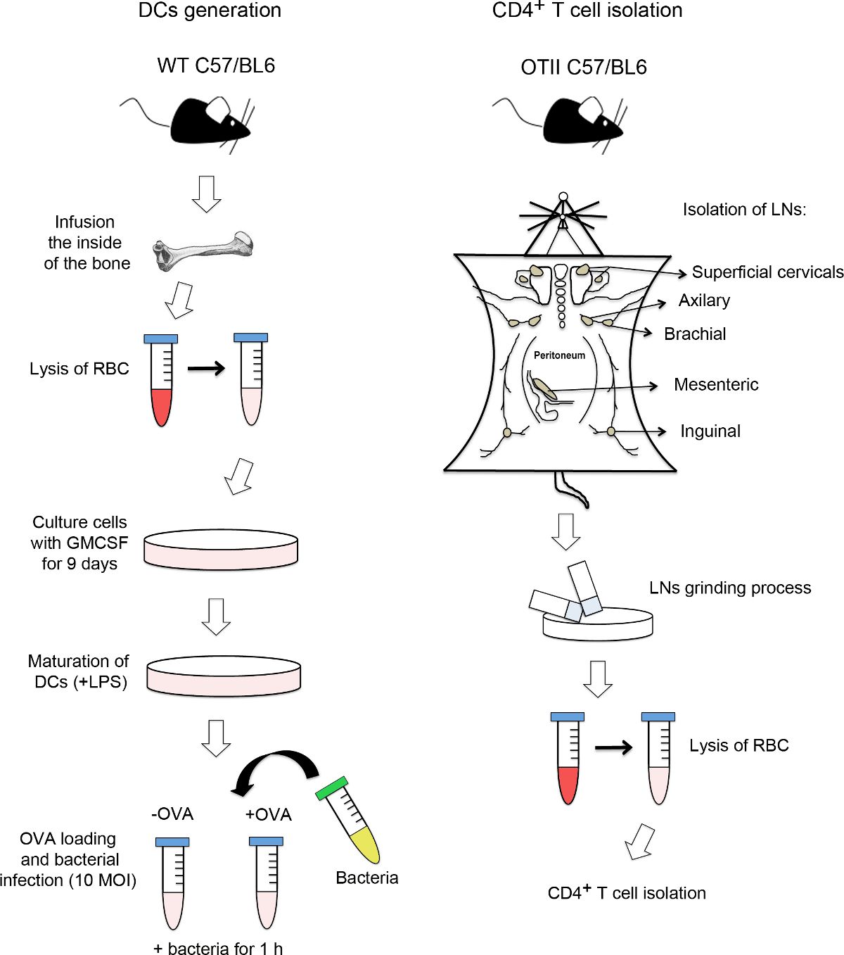
Figure 1. Flow chart of DC generation and CD4+ T cells isolation. On the left-hand side...
Toegang beperkt. Log in of start een proefperiode om deze inhoud te bekijken.
Materialen
| Naam | Bedrijf | Catalogusnummer | Opmerkingen |
|---|---|---|---|
| RPMI | Fisher Scientific | SH3025501 | |
| r-GMCSF | Peprotech | 315-03 | |
| LPS | SIGMA | L2630-10mg | |
| Na Pyruvate | Thermo Scientific | SH3023901 | |
| 2-ME | Gibco | 31350-010 | |
| OVAp OTII (323–339) | GenScript | --- | |
| Cell Strainer 70uM | BD | 352350 | |
| 30uM Syringe Filcons Sterile | BD | 340598 | |
| AutoMacs Classic | Miltenyi Biotec | 130-088-887 | |
| Gentamicin | Normon | 624601.6 | |
| Transwell | Costar | 3415 | |
| LB | Pronadisa | 1231 | |
| Agar | Pronadisa | 1800 | |
| Paraformaldehyde 16% | Electron Microscopy Sciences | 15710 | |
| Triton X-100 | |||
| CD8 biot | BD Biosciences | 553029 | |
| IgM Biot | ImmunoStep | Clone RMM-1 | |
| B220 Biot | BD Biosciences | 553086 | |
| CD19 biot | BD Biosciences | 553784 | |
| MHC-II Biot (I-A/I-E) | BD Biosciences | 553622 | |
| CD11b biot | Immunostep | 11BB-01mg | |
| CD11c biot | Immunostep | 11CB3-01mg | |
| DX5 biot | BD Biosciences | 553856 | |
| Gr-1 biot | BD Biosciences | 553125 | |
| CD16/CD32 | ImmunoStep | M16PU-05MG | |
| anti Salmonella | ABD Serotec | 8209-4006 | |
| CD11cPE | BD Biosciences | 553802 | |
| CD4-APC | Tonbo Biosciences | 20-0041-U100 | |
| Gr-1 APC | BD Biosciences | 553129 | |
| MHC-II (I-A/I-E) FITC | BD Biosciences | 553623 | |
| Alexa-Fluor 647 Goat Anti-Rabbit IgG (H+L) Antibody, highly cross-adsorbed | Invitrogen | A-21245 | |
| CMAC (7-amino-4-chloromethylcoumarin) | Life technologies | C2110 | |
| BSA | SIGMA | A7030-100G | |
| Streptavidin MicroBeads | Miltenyi Biotec | 130-048-101 | |
| BD FACSCanto II | BD Biosciences | --- |
