This video demonstrates a zebrafish embryo model for studying infections linked to implanted biomaterials. Injecting Staphylococcus aureus with polystyrene microspheres enhances bacterial survival, confirming the infection-promoting role of implanted materials.
Een abonnement op JoVE is vereist om deze inhoud te bekijken. Log in of start vandaag met uw gratis proefperiode.
Methodenartikel
In Vivo Analysis of Implant-Associated Bacterial Infections Using Zebrafish Embryos
436 weergaven
⸱
26 september 2025
In dit artikel
Samenvatting
Protocol
1. Preparation of Bacteria-only and Bacteria-Microspheres Suspensions
NOTE: The Staphylococcus aureus RN4220 strain expressing mCherry fluorescent protein (S. aureus-mCherry) is used. The S. aureus RN4220 strain is mutated in the virulence regulator gene agrA (accessory gene regulator A), and, therefore, may have relatively low virulence in the zebrafish embryo model. Other S. aureus strains or other bacterial species for biomaterial-associated infection (BAI) can be used.
- Take 4 to 5 colonies of S. aureus RN4220 bacteria from tryptic soya agar culture plates supplemented with 10 µg/mL chloramphenicol and culture the bacteria to mid-logarithmic growth phase in 10 mL of tryptic soy broth supplemented with 10 µg/mL chloramphenicol at 37 °C under shaking.
- During culture, dilute 100 µL of the bacterial suspension in 900 µL of sterile phosphate-buffered saline (PBS) in a cuvette (width of 1 cm) for an optical density (OD) measurement at 620 nm (OD620). Culture the bacteria until the OD620 reaches 0.4–0.8.
NOTE: An OD620 of 0.1 generally corresponds to 3.0 x 10⁷ colony-forming unit (CFU)/mL S. aureus. The OD620 of an inoculum of bacteria in the mid-logarithmic growth phase is between 0.4-0.8. Different time periods for culturing may be needed for other species and strains of bacteria.
- During culture, dilute 100 µL of the bacterial suspension in 900 µL of sterile phosphate-buffered saline (PBS) in a cuvette (width of 1 cm) for an optical density (OD) measurement at 620 nm (OD620). Culture the bacteria until the OD620 reaches 0.4–0.8.
- Centrifuge bacteria at 3,500 x g for 10 min and resuspend the pelleted bacteria in 1 mL of sterile PBS. Subsequently, wash the bacteria with sterile PBS 2 times, and finally re-suspend the bacteria in 1.1 mL of 4% (w/v) polyvinylpyrrolidone40 (PVP40) solution in PBS.
- Vortex this bacterial suspension and dilute 100 µL of the suspension in 900 µL of sterile PBS in a cuvette for the OD620 measurement. Adjust the concentration of the bacterial suspension with PVP40 solution. This is the "Bacteria-only" suspension.
- Biomaterials can be freely chosen to mix with the bacterial suspension. In the present study, commercial polystyrene (PS) microspheres (blue fluorescent, 10 µm) are used. To generate a bacteria-Microspheres suspension, centrifuge the microspheres for 1 min at 1,000 x g, room temperature, discard the supernatant and re-suspend the microspheres in the “Bacteria-only” suspension (in PVP40 solution).
- In order to inject approximately equal doses of bacteria in the presence and in the absence of microspheres, the concentration of Bacteria-Microspheres suspension needs to be two-thirds of the concentration of the “Bacteria-only” suspension (without microspheres). This is achieved by diluting the Bacteria-Microspheres suspension with 0.5 volume of PVP40 solution. Mix the suspensions by vortexing. Of note, this ratio (two-thirds) has been assessed for S. aureus. It is also appropriate for S. epidermidis, but it is advised to check whether this ratio also applies to other bacterial species and other sizes (than 10 µm) or shapes of biomaterials (than microspheres) to be injected.
- Check the concentration of the “Bacteria-only” and Bacteria-Microspheres suspension by quantitative culture of 10-fold serial dilutions as below: transfer 100 µL of the suspensions to a 96 well-plate and serially dilute by transferring 10 µL aliquots of the suspension into 90 µL of sterile PBS. Plate duplicate 10 µL aliquots of the undiluted and diluted suspensions on agarose plates, incubate the plates at 37 °C overnight, count the colonies and calculate the number of bacteria (colony-forming units, CFU).
2. Breeding, Harvesting, and Maintenance of Zebrafish Embryos
- Follow the general procedures for breeding, harvesting, and maintenance of zebrafish embryos, with modifications described below. Cross a family of wild-type Tupfel long fin (TL) zebrafish or zebrafish of the selected transgenic line (here, Mpeg1: Kaede) in a tank with a net added to induce the adult females to produce eggs after the light turns on, then separate adults from the produced eggs.
- The next day, collect the embryos and discard the non-transparent ones, which are not viable. Keep approximately 60 embryos per Petri dish (100 mm in diameter) in E3 medium and incubate at 28 °C. Remove dead and abnormal embryos and refresh the E3 medium daily.
3. Preparation of Injection Needles
- Prepare the glass microcapillary needles for injection using a micropipette puller instrument. Use the following settings: heat: 772, pull: 100, vel: 200, time: 40, gas: 75.
- Break the needle tip with forceps at the position where the needle has an outer diameter of approximately 20 µm (for 10 µm microspheres), using a light microscope with a scale bar in the ocular. Avoid needles with a very large opening size, as they will compromise the survival of the embryos.
NOTE: The opening may be chosen to be smaller or larger, depending on the size and shape of biomaterials to be injected. In the literature, injections using needles with an opening of approximately 50 µm have been reported to cause a significant decrease in embryo survival.
4. Injection of Bacteria-Only or Bacteria-Microspheres Suspension into Zebrafish Embryos
- Heat agarose solution (1–1.5% (wt) in demi-water) using a microwave oven and pour into a 100-mm Petri dish. Place a plastic mold template on top of the agarose solution in the Petri dish to create indentations in the agarose for placing embryos in proper positions and facilitating injections. Incubate at room temperature and remove the mold when the agarose solution has solidified.
- At 3 d post-fertilization, place the embryos in a 100 mm Petri dish containing 0.02% (w/v) 3-aminobenzoic acid (Tricaine) to anesthetize them. After 5 min, transfer the embryos to the agarose plate overlaid with E3 medium containing 0.02% (w/v) Tricaine and align them in one orientation for injection. For the Mpeg1: Kaede transgenic line first anesthetize embryos in E3 medium containing 0.02% (w/v) tricaine. Then, select embryos expressing green fluorescent proteins using a stereo fluorescence microscope.
- Load the needle with approximately 10 µL of the "Bacteria-only" or Bacteria-Microspheres suspension using a microloader pipette tip. Mount the needle onto a micromanipulator connected to the micro-injector. For the injector used here (see Table of Materials), use the following settings for injections of 2–3 nL: pressure: 300–350, back pressure: 0, time: 2 ms.NOTE: The settings for the micro-injector depend on the injector used. Injector settings may need to be adjusted for injections of bacteria mixed with biomaterials with other shapes or sizes.
- Use needles with the same opening for the injection of the "Bacteria-only" suspension and the Bacteria-Microspheres suspension. If the needle is broken or clogged, always change for a new needle for further injections.
- Insert the needle into the muscle tissue of embryos under a light microscope (Figure 1), at an angle of 45–60° between the needle and the body of embryos. Adjust the position of the needle in the tissue by gently moving it back and forth. Inject the embryos using a foot pedal connected to the micro-injector.
- After injection of fluorescent bacteria, score the embryos for successful infection under a stereo fluorescence microscope. Discard the embryos scored negative (no visible fluorescent bacteria or no visible fluorescent microspheres). Maintain embryos individually in E3 medium in 48-well plates. Refresh the medium daily.
5. Crushing of Embryos, Microscopic Scoring, and Quantitative Culture of Bacteria
- Score all embryos microscopically for the presence of fluorescent bacteria using a stereo fluorescence microscope, starting immediately after the injections, and on each subsequent day until the embryos are randomly selected for quantitative culture.
- Randomly select a few live infected embryos (5 to 6 in the present study) shortly after injection and transfer them individually to separate 2 mL microtubes using sterile tips. Remove the medium, wash the embryos gently with sterile PBS once and add 100 µL of sterile PBS.
- Add 2–3 sterile zirconia beads (2 mm in diameter) to each vial and crush the embryos using the homogenizer (see Table of Materials) at 3,500 rpm for 30 s. Culture the homogenate quantitatively as described in step 1.3.
NOTE: Other homogenizers may require different settings. - Randomly select multiple embryos on subsequent days after injection for quantitative culture according to step 5.3.
Toegang beperkt. Log in of start een proefperiode om deze inhoud te bekijken.
Resultaten
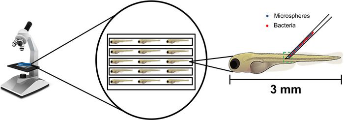
Figure 1: Schematic procedure of co-injection of Bacteria-Microspheres suspension into the tail muscle of zebrafish embryo...
Toegang beperkt. Log in of start een proefperiode om deze inhoud te bekijken.
Materialen
| Naam | Bedrijf | Catalogusnummer | Opmerkingen |
|---|---|---|---|
| Tryptic soya agar | BD Difco | 236950 | Media preparation unit at AMC |
| Tryptic soya broth | BD Difco | 211825 | |
| Polyvinylpyrrolidone40 | Applichem | A2259.0250 | |
| 10 µm diameter polystyrene microspheres (blue fluorescent) | Life technology/ThemoFisher | F8829 | |
| Glass microcapilary (1 mm O.D. x 0.78 mm I.D.) | Harvard Apparatus | 30-0038 | |
| Micropipette puller instrument | Sutter Instrument Inc | Flaming p-97 | |
| Light microscope LM 20 | Leica | MDG33 10450123 | |
| 3-aminobenzoic acid (Tricaine) | Sigma-Aldrich | E10521-50G | |
| Agarose MP | Roche | 11388991001 | |
| Stereo fluorescent microscope LM80 | Leica | MDG3610450126 | |
| Microloader pipette tips | Eppendorf | 5242956.003 | |
| Micromanipulator M3301 with M10 stand | World Precision Instruments | 00-42-101-0000 | |
| FemtoJet express micro-injector | Eppendorf | 5248ZO100329 | |
| Microtrube 2ml pp | Sarstedt | 72.693.005 | |
| Zirconia beads | Bio-connect | 11079124ZX | |
| MagNA lyser | Roche | 41416401 | |
| Chloramphenicol | Sigma-Aldrich | C0378 | |
| Gyrotory shaker (for bacterial growth) | New Brunswick Scientific | G10 | |
| Zebrafish incubator | VWR | Incu-line | |
| Cuvettes | BRAND | 759015 | |
| Centrifuge | Hettich-Zentrifugen | ROTANTA 460R | |
| Spectrometer | Pharmacia biotech | Ultrospec®2000 | |
| Forceps | Sigma-Aldrich | F6521-1EA | |
| 48 well-plates | Greiner bio-one | 677180 | |
| 96 well-plates | Greiner bio-one | 655161 | |
| Petri-dish | Falcon | 353003 | |
| Petri-dish | Biomerieux | NL-132 |
