This video demonstrates the initiator-dependent amplification of a suicide plasmid in E. coli. The plasmid, which carries an antibiotic resistance gene, replicates only in the presence of an initiator protein encoded by the host. After electroporation and incubation in recovery medium, the mixture is plated on antibiotic-containing agar. Bacteria with a plasmid survive, as its initiator protein allows expression of the plasmid's antibiotic-resistant gene.
Een abonnement op JoVE is vereist om deze inhoud te bekijken. Log in of start vandaag met uw gratis proefperiode.
Methodenartikel
Amplification of a Suicide Plasmid in a Bacterial Host
817 weergaven
⸱
26 september 2025
In dit artikel
Samenvatting
Protocol
1. Generation of null mutants in the flagella glycosylation island genes
NOTE: This method of mutagenesis is based on the allelic exchange of polymerase chain reaction (PCR) in-frame deletion products using the suicide vector pDM428 (GenBank: KC795686.1). Replication of pDM4 vector is lambda pir dependent, and the complete allelic exchange is coerced by utilizing the sacB gene located on the vector.
- Construction of PCR in-frame deletion products.
- Design two pairs of primers that amplify DNA regions upstream (A and B primers) and downstream (C and D primers) of the selected gene or region to be deleted (Figure 1B).
- Ensure that Primers A and D have more than 600 bp upstream from the start and downstream from the stop, respectively, of the gene or genes to be deleted. These two primers need to include a restriction site at the 5' end for an endonuclease that allows cloning in pDM4.
NOTE: The restriction site included at the 5' end of A and D primers should not be the same as sites within AB or CD amplicons. - Ensure that Primers B and C are within the gene to be deleted or inside the first and last gene, respectively, of the region to be deleted. Ensure that both primers (B and C) are in-frame and located between 5-6 codons inside the gene. Furthermore, ensure that both primers include at the 5' end a 21 bp complementary sequence (Table 1) that allows joining the DNA amplicons AB and CD.
- Grow the selected Aeromonas strain in 5 mL of tryptic soy broth (TSB) overnight (O/N) at 30 °C. Use a commercially available kit for genomic DNA purification and follow the manufacturer's instructions (Table of Materials). Quantify 2 µL of the purified DNA using a spectrophotometer.
NOTE: Use double-distilled water as a blank, with the instrument set to record absorbance at 260 nm. Record absorbance with 2 µL of purified DNA 3-5 times and calculate an average absorbance reading. Use the Beer-Lambert law to calculate the DNA concentration, where A = Ɛ.c.l. Wherein "A" is absorbance, "Ɛ" is the molar average extinction coefficient for DNA, "c" is DNA concentration, and "l" is the path length (refer to the manufacturer's instructions for the path length of each instrument). - Use 100 ng of purified chromosomal DNA as the template in two sets of asymmetric PCRs with A-B and C-D primers (Table of Materials). Use a 10:1 molar ratio of A:B and D:C primer pairs.
NOTE: Asymmetric PCRs allow the preferential amplification of one strand of the template over the other. - Analyze the PCR products by electrophoresis in a 1% agarose gel. Use TAE (40 mM Tris-acetate, 1 mM ethylenediaminetetraacetic acid (EDTA)) as gel running buffer and a power setting of 8 V/cm. To visualize DNA, add ethidium bromide (0.5 µg/mL) to the gel and visualize the PCR products using a bio-imaging station (Table of Materials).
- Excise the amplicons from the gel using a scalpel, purify them following the manufacturer's protocol (Table of Materials), and quantify them.
- Join AB and CD amplicons by their 21 bp overlapping sequences in the 3' end of PCR products (Figure 2). Mix 100 ng of each amplicon (AB and CD) in a PCR tube with PCR reagents (pre-AD PCR), but without primers, and extend using the thermocycler program. Then, add 100 µM of A and D primers to the reaction (AD PCR) and amplify as a single fragment.
- Analyze the PCR product (AD amplicon) by electrophoresis in a 1% agarose gel using the conditions described in step 1.1.6, excise the amplicon from the gel, purify it following the manufacturer's protocol, and quantify the amplicon (Table of Materials).
- Construction of pDM4 recombinant plasmid with the in-frame deletion.
- Grow an O/N culture of Escherichia coli cc18λ pir containing pDM4 in Luria-Bertani (LB) Miller broth (10 mL) with chloramphenicol (25 µg/mL) at 30 °C.
- Spin down the culture at 5,000 x g at 4 °C and suspend the pellet in 600 µL of sterile distilled water. Perform extraction and purification of plasmid pDM4 using a commercially available plasmid purification kit, following the provider's instructions (Table of Materials).
- Digest 1 µg of pDM4 and 400 ng of AD amplicon for 2 h with an endonuclease able to digest both ends of the AD amplicon and the cloning site of pDM4 (Figure 2). Follow the enzyme manufacturer's protocol for reaction conditions (Table of Materials).
- Clean up the digested plasmid and amplicon using a DNA purification kit and quantify them following the manufacturer's instructions (Table of Materials).
- To prevent religation of the linearized pDM4, remove the phosphate group from both 5' ends using the alkaline phosphatase enzyme following the manufacturer's instructions (Table of Materials). Then, clean up the treated plasmid and quantify it using a spectrophotometer as described in step 1.1.4 (Table of Materials).
- Combine 150 ng of the digested and phosphatase alkaline treated pDM4 with 95-100 ng of digested AD amplicon at a molar vector:insert ratio of 1:4. Prepare the ligation reaction in a volume of 15 µL, following the manufacturer's instructions (Table of Materials).
- Incubate O/N at 20 °C or over the weekend at 4 °C. After incubation, inactivate the T4 ligase at 70 °C for 20 min.
- Use ligation to introduce the plasmid into Escherichia coli MC1061λpir strain by electroporation. Use electrocompetent bacteria and 2 mm-gap electroporation cuvettes.
- Preparation of electrocompetent E. coli and electroporation of pDM4 recombinant plasmid
- Inoculate a single colony of E. coli MC1061λpir strain into Luria-Bertani (LB) Miller broth and grow O/N at 30 °C with 200 rpm shaking.
- Dilute 2 mL of the bacterial culture in 18 mL of LB broth and incubate at 30 °C with shaking (200 rpm) until an optical density (OD600) between 0.4-0.6 is attained.
- Pellet the bacterial culture by centrifugation at 5,000 x g and 4 °C for 15 min. Discard the supernatant and suspend the pellet in 40 mL of chilled distilled H2O.
- Repeat this cleaning step two more times to remove all culture salts. Finally, suspend the pellet in 4 mL of chilled distilled H2O and transfer 1 mL of the suspension to each of four conical 1.5 mL microfuge tubes.
- Pellet the bacterial culture by centrifugation at 14,000 x g and 4 °C for 5 min. Remove the supernatant without disturbing the pellet and suspend each pellet in 100 µL of chilled distilled H2O.
- Add 3-3.5 µL of the ligation mixture to each tube and incubate on ice for 5 min. Then, transfer the contents of each tube to a chilled 2 mm-gap electroporation cuvette and apply 2 kV, 129 Ω, and a time constant of around 5 ms.
NOTE: Pre-cool the electroporation cuvettes on ice. Maintain the bacteria on ice during the entire procedure. - After electroporation, add 250 µL of SOC (Super Optimal broth with Catabolite repression) medium to each of the four cuvettes to recover their contents, transfer them to a culture tube (about 1 mL), and incubate for 1 h at 30 °C with shaking (200 rpm).
- Plate the transformed cells on Luria-Bertani (LB) agar plates supplemented with chloramphenicol (25 µg/mL) to ensure all colonies growing on the plate have the plasmid backbone.
Table 1: Primers used for the construction of null mutants. Overlapping regions in primers B and C are underlined. The BamHI site is bolded in primers A and D.
| Primer name | Sequence in 5’ to 3’ direction | Used for |
| A. piscicola AH-3 | ||
| A-Flgi1 | CGCGGATCCGACTGTACCCGTTTCAATCA | fgi mutant |
| B-Flgi1 | CCCATCCACTAAACTTAAACAGATCACCTCGAACTCGAAA | |
| C-Flgi12 | TGTTTAAGTTTAGTGGATGGGGGAACCTTAAATGCCATGA | |
| D-Flgi12 | CGCGGATCCCAGTCTTCAGCTTCCATCC | |
| E-Flgi1 | ACCCGCTTCATTCGCTAT | |
| F-Flgi12 | TCCGATTTTCTGACTCAGGG | |
| A-Fgi4 | CGCGGATCCGATGCGTACGCTAATATGAA | fgi-4 mutant |
| B-Fgi4 | CCCATCCACTAAACTTAAACACATATTATCTTGCCCCTGAT | |
| C-Fgi4 | TGTTTAAGTTTAGTGGATGGGATGGAGCTAATCACTCGTTT | |
| D-Fgi4 | CGCGGATCCACATATCAACCCCCAAC | |
| E-Fgi4 | ATTTCCCTGCCAAATACG | |
| F-Fgi4 | CCTGCCAACAGGATGTAAG | |
| pDM4 vector | ||
| pDM4for | AGTGATCTTCCGTCACAGG | Insertions into the pDM4 vector |
| pDM4rev | AAGGTTTAACGG TTGTGGA | |
Toegang beperkt. Log in of start een proefperiode om deze inhoud te bekijken.
Resultaten
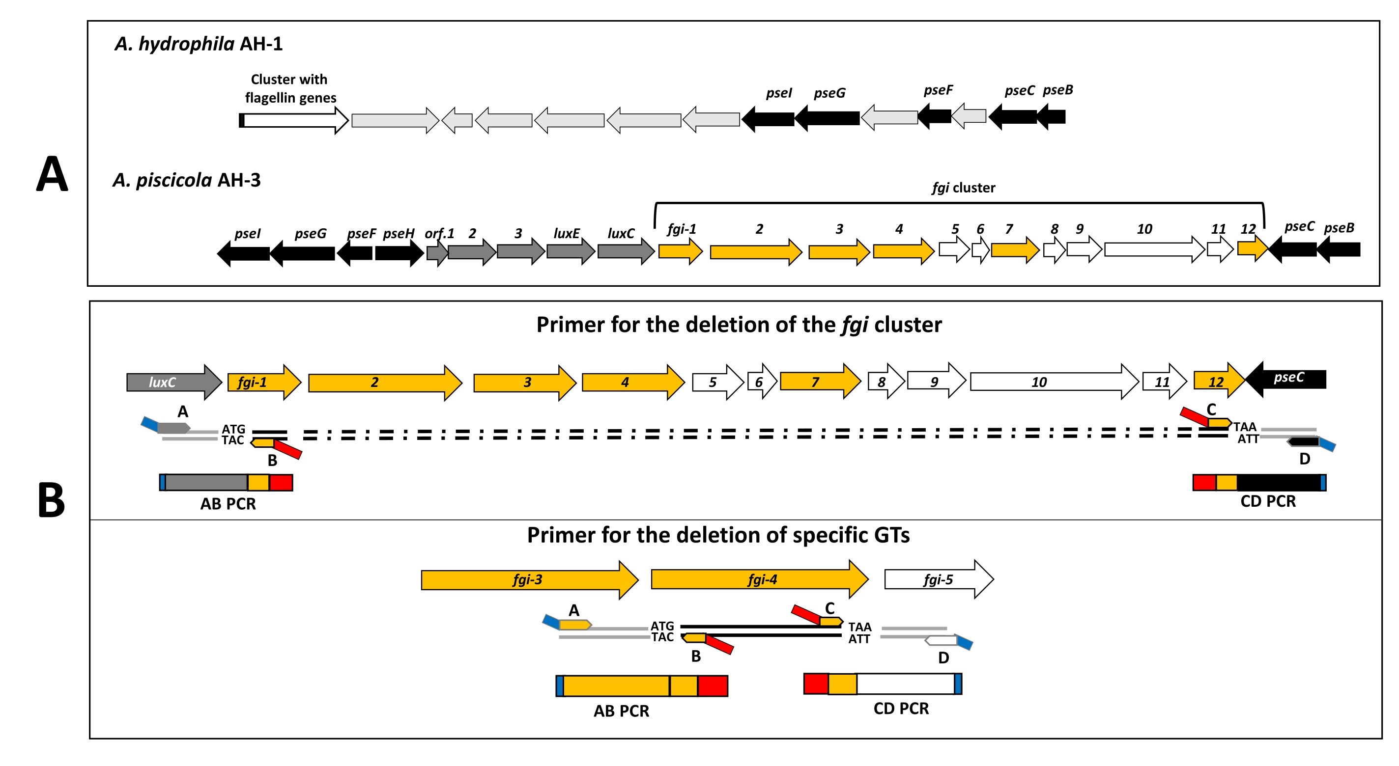
Figure 1: Bioinformatic detection of chromosomal regions and primer design. (A) Scheme o...
Toegang beperkt. Log in of start een proefperiode om deze inhoud te bekijken.
Materialen
| Naam | Bedrijf | Catalogusnummer | Opmerkingen |
|---|---|---|---|
| ABI PRISM Big Dye Terminator v. 3.1 Cycle Sequencing Ready Reaction Kit | Applied Biosystems | 4337455 | Used for sequencing |
| AccuPrime Taq DNA Polymerase, high fidelity | Invitrogen | 12346-086 | Used for amplification of AB, CD and AD fragments |
| Agarose | Conda-Pronadise | 8008 | Used for DNA electrophoresis |
| Alkaline phosphatase, calf intestinal (CIAP) | Promega | M1821 | Used to remove phosphate at the 5’ end |
| Bacto agar | Becton Dickinson | 214010 | Use for motility analysis |
| BamHI | Promega | R6021 | Used for endonuclease restriction |
| BglII | Promega | R6081 | Used for endonuclease restriction |
| BioDoc-It Imagin System | UVP | Bio-imaging station used for DNA visualization | |
| Biotaq polymerase | Bioline | BIO-21040 | Used for colony screening |
| Cytiva illustra GFX PCR DNA and Gel Band Purification Kit | Cytivia | 28-9034-71 | Used for purification of PCR amplicons and DNA fragments. |
| EDTA | Applichem | 131026.1211 | Used for DNA electrophoresis |
| Electroporation cuvettes 2 mm gap | VWR | 732-1133 | Used for transformation |
| Luria-Bertani (LB) Miller agar | Condalab | 996 | Used for Escherichia coli culture |
| Luria-Bertani (LB) Miller broth | Condalab | 1551 | Used for Escherichia coli culture |
| Nanodrop ND-1000 | NanoDrop Techonologies Inc | Spectrophotometer used for DNA quantification | |
| Rifampicin | Applichem | A2220,0005 | Used for triparental mating |
| SOC Medium | Invitrogen | 15544034 | Used for electroporation recovery |
| T4 DNA ligase | Invitrogen | 15224017 | Used for ligation reaction |
| Veriti 96 well Thermal Cycler | Applied Biosystems | Used for PCR reactions |
