This video demonstrates the method of capturing single fluorescently tagged Escherichia coli (E. coli) cells in microfluidic traps using a carbon-free suspension and controlled flow, followed by nutrient addition to trigger growth and fluorescence imaging of microcolonies.
Een abonnement op JoVE is vereist om deze inhoud te bekijken. Log in of start vandaag met uw gratis proefperiode.
Methodenartikel
Microfluidic Patterning and Fluorescence-Based Tracking of Single-Cell Bacterial Growth
542 weergaven
⸱
30 oktober 2025
In dit artikel
Samenvatting
Protocol
1. Bacterial patterning
- One day prior to the experiment, grow a population of Escherichia coli (strain MG1655 prpsM-GFP). Inoculate the culture directly from the frozen stock and grow overnight for 20 h in lysogeny broth (LB) medium in a shaker incubator at 37 °C. Add 50 µg/mL of kanamycin for the cells to retain the prpsM-GFP (green fluorescent protein) plasmid.
- On the day of the experiment, set the box incubator (Figure 1A) at 37 °C several hours before the experiment to ensure a uniform and stable temperature before starting. Then, set up the syringe pump and the heated glass plate on the microscope stage (Figure 1B,C), setting the temperature at the same temperature as the box incubator.
NOTE: The box incubator in which the entire system, including the microscope (Figure 1E), is enclosed ensures that a uniform and constant temperature is maintained throughout the entire experiment when the channel is flushed with medium. - Ninety minutes before the experiment, put the microfluidic chip in a vessel filled with 100% ethanol (EtOH) and flush the channel with 100% EtOH for at least 10 min. Place the microfluidic chip in a vacuum desiccator and degas for at least 30 min. Exchange the EtOH with distilled water and vacuum-treat the chip for at least 30 min. Put the microfluidic chip in the oven at 70 °C for 10 min to remove any traces of liquid left in the channel.
NOTE: This step is necessary to prevent bubble formation in the channel when flushed with culture medium. - Pipette 1 mL of 3-(N-morpholino)propanesulfonic acid (MOPS) medium (1x) into a centrifuge vial and add 10 µL of 0.132 M potassium phosphate (K2HPO4). Take the overnight culture out of the 37 °C incubator and aliquot 100 µL into the centrifuge vial and centrifuge the culture at 2,300 × g for 2 min. Gently pipette the supernatant out of the centrifuge vials, and resuspend the pellet in 1 mL of fresh MOPS medium with 0.015% v/v of Tween 20 and 0.01% v/v of potassium phosphate.
NOTE: The typical concentration of the bacterial suspension is in the range of 0.015-0.15 vol%, which ensures the formation of an extended and mobile accumulation zone and prevents the accumulation of particles on the substrate. Replacing the overnight medium with fresh medium minimizes the risk of releasing films of bacteria on the template. The lack of a carbon source in the fresh medium prevents cells from growing in the suspension during template patterning. A concentration of 0.015% v/v of Tween 20 in the suspension is necessary for the contact angle of the receding liquid on the PDMS template to fall within the optimal range, between 30 and 60°. - Load the bacterial suspension in a 1 mL syringe and connect the syringe to the chip through microfluidic tubing.
- To secure the connection between the syringe and the tubing, directly insert a needle with an outer diameter of 0.6 mm into the tubing. To avoid scattering any suspension remaining in the inlet vicinity across the channel, flush fresh medium through the hole used as an outlet during the patterning process (Figure 2A-III), rather than the hole used as the inlet.
- Mount the syringe on the syringe pump and inject the suspension into the microfluidic chip through the inlet located at the upstream part of the channel until the suspension covers the template region with traps.NOTE: During the liquid injection process, air can escape through an outlet located at the downstream end of the channel.
- Set the syringe pump to withdraw the bacterial suspension (Figure 2A-I) at a flow rate of 0.07-0.2 µL/min, which in the described geometry corresponds to a meniscus receding speed of 80-100 µm/min.
- Monitor the patterning process via microscope software.
NOTE: Here, a 10x magnification was used to monitor the receding meniscus on the template, and a 20x magnification was used to monitor the deposition of individual bacteria into the microfabricated traps.
- Once the template has been patterned with cells (Figure 2A-II), increase the withdrawal flow rate to quickly empty the microfluidic channel and flush it with fresh LB that was previously degassed for at least 30 min and prewarmed at 30 °C.
- Set the syringe pump at a flow rate of 2 µL/min to gently flush the channel. Once the channel has been filled, increase the flow rate (15 µL/min) according to the specific experimental needs.
- Acquire images of growing bacteria at the desired magnification and time interval.
Toegang beperkt. Log in of start een proefperiode om deze inhoud te bekijken.
Resultaten
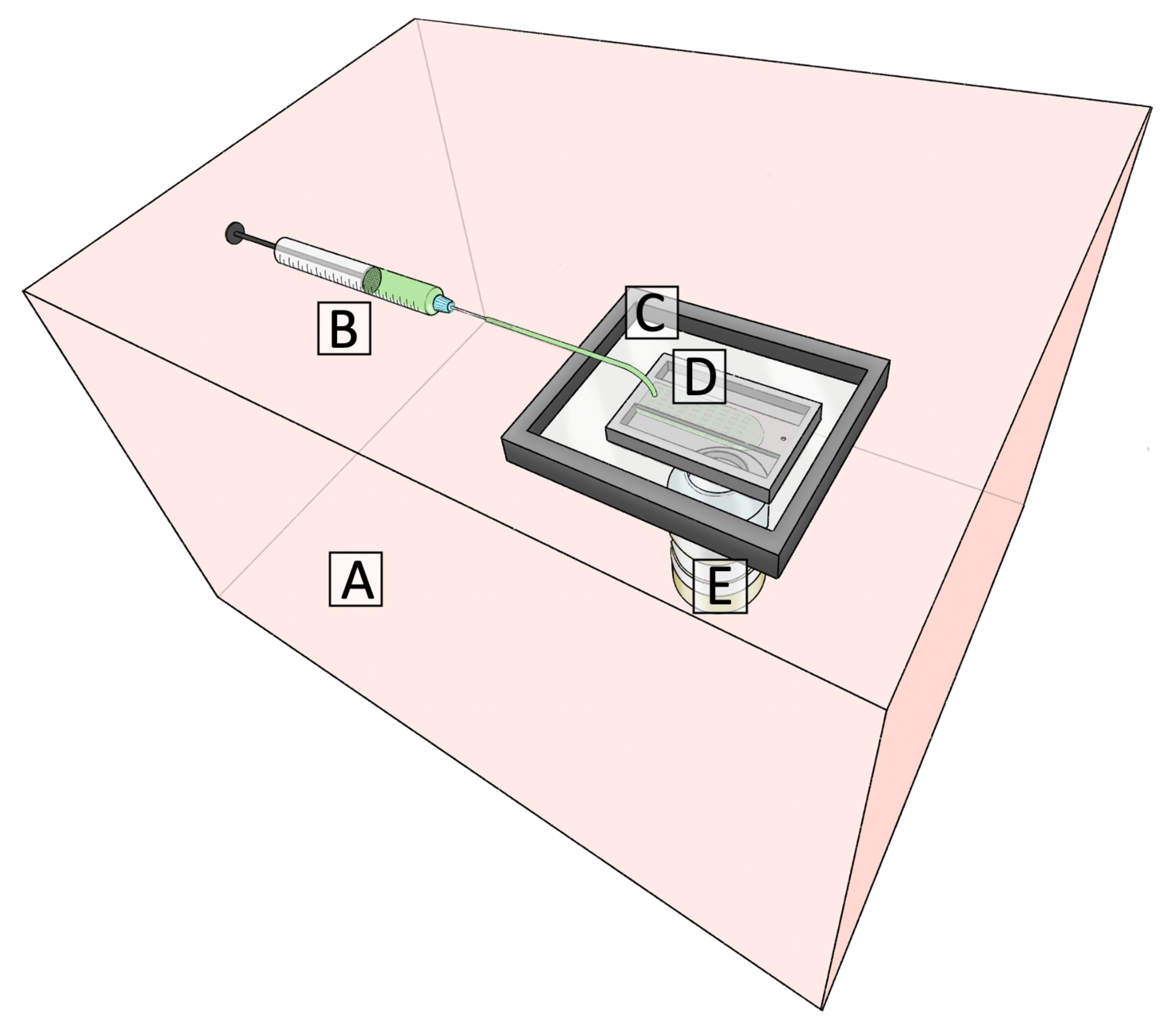
Figure 1: Schematic of the platform for sequential capillarity-assisted assembly in a microfluidic channel. <...
Toegang beperkt. Log in of start een proefperiode om deze inhoud te bekijken.
Materialen
| Naam | Bedrijf | Catalogusnummer | Opmerkingen |
|---|---|---|---|
| BD 10 mL Syringe (Luer-Lock) | BD | 300912 | Used to flush fresh Lysogeny broth into the microfluidic channel |
| Box Incubator | Life Imaging Services | Used to ensure a uniform and constant temperature in the channel | |
| Centrifuge | Eppendorf | 5424R | Used to replace the overnight media with fresh minimal media |
| Centrifuge vial | Eppendorf | 30120086 | 1.5 mL |
| CETONI Base 120 | CETONI GmbH | Syringe pump | |
| H401-T-CONTROLLER | Okolab | Controller of the heated glass plate | |
| H601-NIKON-TS2R-GLASS | Okolab | Heated glass plate | |
| Insulin syringes, U 100, with luer | Codan Medical ApS | CODA621640 | 1 mL syringe used to withdraw the liquid suspension during the patterning process |
| LB Broth, Miller (Luria-Bertani) | Fisher Scientific | 244610 | Lysogeny broth flushed into the microfluidic channel |
| Masterflex transfer tubing | Masterflex | HV-06419-05 | 0.020'' ID, 0.06'' OD |
| MOPS (10x) | Teknova | M2101 | Diluted tenfold with milliQ water and used to replace the overnight medium |
| Nikon Eclipse Ti2 | Nikon Instruments | Microscope | |
| Potassium phosphate dibasic | Sigma Aldrich | P3786 | Added to MOPS 1x |
| Tween 20 | Sigma Aldrich | P1379 | Used to ensure an optimal receding contact angle during the patterning process |
