This video describes a procedure for performing natural transformation in filamentous cyanobacteria using plasmid DNA to achieve stable GFP expression and antibiotic resistance through homologous recombination.
Een abonnement op JoVE is vereist om deze inhoud te bekijken. Log in of start vandaag met uw gratis proefperiode.
Methodenartikel
Natural Transformation and GFP Expression in the Filamentous Cyanobacterium Phormidium lacuna
555 weergaven
⸱
30 oktober 2025
In dit artikel
Samenvatting
Protocol
1. Natural transformation and GFP expression
NOTE: Transformation is based on a plasmid vector propagated in Escherichia coli (E. coli); pGEM-T or pUC19 may be used as backbone vectors. Examples for vectors for superfolder green fluorescent protein (sfGFP) expression are described in the representative results section.
- Perform all steps using sterile material under sterile laboratory conditions (clean bench, sterile glassware).
- Inoculate 2 x 50 mL of f/2 liquid medium in two 250 mL flasks with 2 x 1 mL of Phormidium lacuna (P. lacuna) filaments from a running culture. Cultivate in white light (50 µmol m-2 s-1) under agitation (horizontal rotation, 50 rpm) for ~5 days at 25 °C.
- Prepare ~200 µg of the transformation vector DNA using a midi prep kit according to the manufacturer's instructions.
- Homogenize 100 mL of P. lacuna cell suspension (see the Table of Materials) at 10,000 rpm for 3 min. Measure OD at 750 nm (desired value = 0.35).
- Centrifuge the cell suspension for 15 min at 6,000 × g. Remove the supernatant and suspend the pellet in 800 µL (total volume including residual liquid and filaments) of the remaining liquid and additional f/2+ medium.
- Take eight f/2+ bacto-agar plates (10 cm diameter) containing 120 µg/mL kanamycin. Pipette 10 µg of DNA into the middle of each agar plate. Immediately pipette 100 µL of cell suspension into the middle of each agar plate (on top of the DNA).
- Keep the agar plate without a lid on the clean bench to allow the excess liquid to evaporate. Close the plate and cultivate it in white light at 25 °C for 2 days.
- Distribute the filaments of each agar plate with an inoculation loop onto several fresh f/2+ bacto-agar plates containing 120 µg/mL kanamycin. Cultivate the plates in white light at 25 °C and check the cultures regularly under a microscope.
- Identify living, transformed filaments after 7-28 days under the microscope. Look for healthy, green filaments (Figure 1) that are different from other filaments. If these green filaments can be identified, continue with the next step; otherwise, keep the plate for another 7 days.
- Use forceps to transfer these identified living filaments into 50 mL of liquid f/2+ medium with 250 µg/mL kanamycin. Cultivate in white light at 25 °C on a shaker (horizontal rotation, 50 rpm). Observe growth for up to four weeks.
- Transfer the filaments back to agar medium containing 250 µg/mL kanamycin and wait for the filaments to grow. After several days, transfer single filaments to a fresh agar plate with a higher concentration of kanamycin, e.g., 500 µg/mL. Keep the original plate.
- Ensure that the filaments are propagated in a high concentration of kanamycin in liquid culture or on agar. Increase the kanamycin concentration again to speed up segregation.NOTE: Transformed P. lacuna grows in up to 10,000 µg/mL kanamycin. Other species might not tolerate such high concentrations.
- For GFP expression: observe single filaments with a fluorescence microscope at a magnification of the objective set at 40x or 63x. Capture a brightfield transmission image and a fluorescence image. Use the following settings for GFP: 470 nm bandpass for excitation, 525 nm bandpass for emission, and a 495 nm beam splitter, initial exposure time of 500 ms.
- Adjust the exposure time for clear fluorescence signals, avoiding saturating intensities. Try to use the same setting for all samples.
- As the wild-type filaments will also display fluorescence, capture images with the same settings as above for this background fluorescence.NOTE: The strain expressing GFP must have a higher signal; otherwise, it is not expressing GFP.
- Based on exposure times and the pixel intensities of the fluorescence images, calculate and compare the GFP content of the different filaments.
Toegang beperkt. Log in of start een proefperiode om deze inhoud te bekijken.
Resultaten
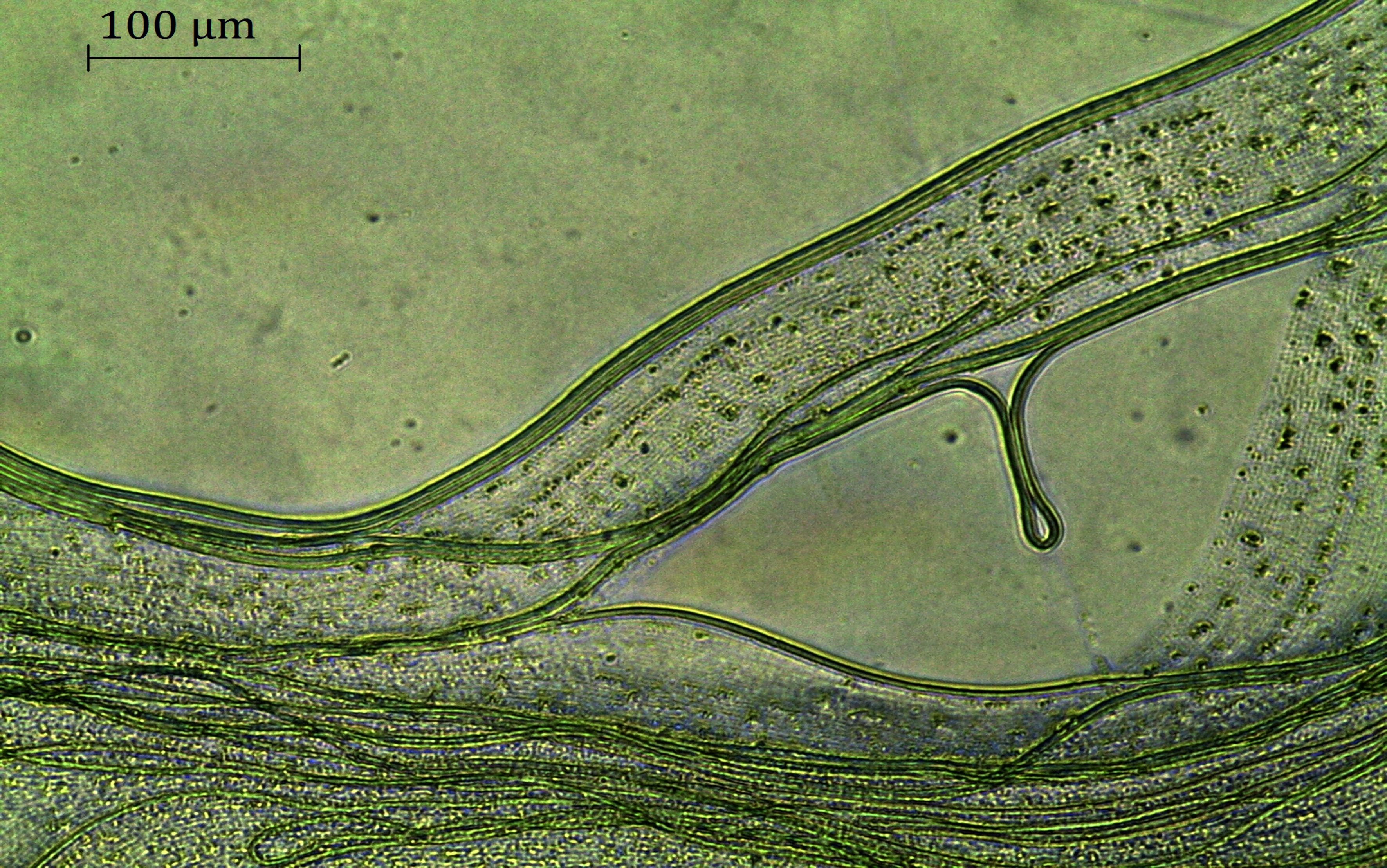
Figure 1. Phormidium lacuna filaments 5 weeks aft...
Toegang beperkt. Log in of start een proefperiode om deze inhoud te bekijken.
Materialen
| Naam | Bedrijf | Catalogusnummer | Opmerkingen |
|---|---|---|---|
| Bacto Agar | OttoNorwald | 214010 | |
| Fluorescence microscope ApoTome | Zeiss | ||
| Fluorescence microscope Axio Imager 2 | Zeiss | ||
| Kanamycin | Sigma-Aldrich | 25389-94-0 | |
| Microscope DM750 | Zeiss | ||
| Midi prep plasmid extraction kit NucleoBond Xtra Midi kit | Macherey-NAGEL GmbH & Co. KG | REF740410.50 | |
| Light source, fluorescent tube L18W/954 daylight | OSRAM | Cultivation of cyanobacteria | |
| Light source, LED panel XL 6500K 140 W | Bloom Star | N/A | Cultivation of cyanobacteria |
| Petri dishes polystyrole, 100 mm x 20 mm | Merck | P5606-400EA | |
| Petri dishes polystyrole, 60 mm x 15 mm | Merck | P5481-500EA | |
| Pipetman 100-1,000 µL | Gilson | SKU: FA10006M | |
| Pipetman 10-100 µL | Gilson | SKU: FA10004M | |
| Plastic pipettes 10 mL, sterile | Greiner | 607107 | |
| Shaker Unimax 2010 | Heidolph Instruments | For cultivation | |
| Plastic tube, sterile, 50 mL | Greiner | 227261 | |
| Ultraturrax Silent Crusher M | Heidolph Instruments | Homogenizer | |
| F/2+ liquid medium | f/2-medium, with 10 times increased NaNO₃ and NaH₂PO₄ (0.88 mM NaNO₃, 36 µM NaH₂PO₄ | ||
| F/2+-agar | 3 % (w/v) bacto agar, artificial seawater, 0.1 % (v/v) trace element solution, 0.05 % (v/v) vitamin solution ,8.8 mM NaNO₃, 0.36 mM NaH₂PO₄ |
