This video demonstrates the preparation and imaging of Myxococcus xanthus, a slow-growing soil bacterium. The culture is grown in a medium, applied to a coverslip with fluorescent microspheres, and overlaid with a pre-warmed agarose pad to support hydration and nutrient supply. This setup enables long-term, time-lapse live imaging of cellular processes during bacterial cell division.
Een abonnement op JoVE is vereist om deze inhoud te bekijken. Log in of start vandaag met uw gratis proefperiode.
Methodenartikel
Preparation of Myxococcus xanthus Samples for Live-Cell Imaging
424 weergaven
⸱
28 november 2025
In dit artikel
Samenvatting
Protocol
1. Preparation of Microscopy Samples
Note: Cells to be viewed by microscopy are placed on a microscope coverslip and then covered by an agarose pad containing nutrients. The coverslip is glued to a plastic or metal frame to provide mechanical support. In preparation for the microscopy, a large pad of 1% agarose/ TPM/0.2% CTT should be prepared in advance as described in steps 2.1 - 2.3. Please also refer to the Table of Materials for specific products used here.
1. Prepare 500 mL of TPM buffer (10 mM Tris-HCl pH 7.6, 1 mM KH2PO4 pH 7.6, 8 mM MgSO4) and autoclave or filter sterilize using a bottle top filter.
NOTE: The sterile buffer can be stored for several months at room temperature.
2. Prepare 1% agarose microscopy solution containing 0.2% CTT (mix 1 g of agarose with 80 mL of TPM buffer and 20 mL of 1% CTT medium). Heat in a microwave oven until the agarose is molten.
NOTE: The 0.2% CTT is sufficient to allow cells to grow and prevent starvation. Higher concentrations of CTT in the microscopy medium will result in high background fluorescence.
3. Fill a Petri dish with the molten agarose to a thickness of 0.5 cm (for an 11.5 cm x 11.5 cm square Petri dish, approximately 60 mL of molten agarose is required) and let it cool down to room temperature.
NOTE: The agarose pad can be stored at 4 °C in a humid environment for up to 2 days.
1. Pre-warm the 1% agarose/TPM/0.2% CTT pad at 32 °C for at least 15 min prior to use.
NOTE: To prepare the cells for microscopy, follow steps 2.4 - 2.8.
4. Place a sterile glass coverslip (60 mm x 22 mm, thickness: 0.7 mm) on a plastic or metal frame that has a hole in the middle (Figure 1A); this frame serves as a mechanical support for the thin coverslip and helps to reduce drift during microscopy. Fix the coverslip to the frame with tape.
1. To prepare the frame, cut out a 75 mm × 25 mm frame from a 1 mm thick metal plate, then cut out an appropriately sized hole (20 mm × 30 mm in this experiment) in the middle.
5. Add 10 - 20 µL of exponentially grown M. xanthus cells on the coverslip.
6. Add fluorescent 0.5 µm microspheres as fiducial markers to the cells to simplify tracking of cells or proteins in time-lapse recordings.
1. Dilute the microspheres 1:100 in TPM buffer and store at 4 °C for up to several months. Shake thoroughly before use and add 5 - 10 µL of the diluted microspheres to the cells.
NOTE: Here, microspheres that are fluorescent in all common blue, green, yellow, and red fluorescent channels were used.
7. Cut out a small pad approximately the size of the coverslip of the large pre-warmed 1% agarose/TPM/0.2% CTT pad and place it on top of the cells (Figure 1B). Place a coverslip on top of the 1% agarose/TPM/0.2% CTT agarose pad to prevent evaporation and to maintain cells in a humid environment.
NOTE: The coverslip alone will prevent significant evaporation for at least 2 h. For longer time-lapse recordings, the 1% agarose/TPM/0.2% CTT pad and coverslip sandwich should be sealed with paraffin film to prevent evaporation.
8. Incubate the microscopy sample at 32 °C for 15 - 20 min to let the cells attach to the bottom of the agarose pad. Then start the time-lapse microscopy recordings.
Toegang beperkt. Log in of start een proefperiode om deze inhoud te bekijken.
Resultaten
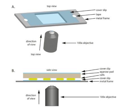
Figure 1: Schematic of the experimental set-up used throughout this study. (A) A...
Toegang beperkt. Log in of start een proefperiode om deze inhoud te bekijken.
Materialen
| Naam | Bedrijf | Catalogusnummer | Opmerkingen |
|---|---|---|---|
| DMI6000B with AFC | Leica microsystems | 11888945 | Automated inverted widefield fluorescence microscope with adaptive focus control |
| Universal mounting frame | Leica microsystems | 11532338 | Stage holder for different sample sizes |
| HCX PL FLUOTAR 100x/1.30 oil PH3 | Leica microsystems | 11506197 | Phase contrast objective |
| Orca Flash 4.0 camera | Hamamatsu | 11532952 | 4.0 megapixel sCMOS camera for picture aquisition |
| Filter set TXR ET, k | Leica microsystems | 11504170 | Fluorescence filter set, Ex: 560/40 Em: 645/75 |
| Filter set L5 ET, k | Leica microsystems | 11504166 | Fluorescence filter set, Ex: 480/40 Em: 527/30 |
| Filter set YFP ET, k | Leica microsystems | 11504165 | Fluorescence filter set, Ex: 500/20 Em: 535/30 |
| ProScan III | Prior | H117N1, V31XYZEF, PS3J100 | Microscope automation controller with interactive control center |
| EL 6000 light source | Leica microsystems | 11504115 | External fluorescence light source |
| Incubator BLX Black | Pecon | 11532830 | Black incubation chamber surrounding the microscope |
| Tempcontrol 37-2 digital | Leica microsystems | 11521719 | Automated temperature control for incubation chamber |
| Gentmycin sulphate | Carl Roth | 233.4 | Gentamycin |
| Oxytetracylin dihydrate | Sigma Aldrich | 201-212-8 | Oxytetracyclin |
| Kanamycin sulphate | Carl Roth | T832.3 | Kanamycin |
| Filtropur BT25 0.2 bottle top filter | Sarstedt | 831,822,101 | Bottle top filter for sterilization of buffers |
| Deckgläser | VWR | 630-1592 | Glass cover slip (60 x 22 mm, thickness: 0.7 mm) |
| Seakem LE agarose | Lonza | 50004 | Agarose for microscopy slides |
| Leica Metamorph AF | Leica microsystems | 11640901 | Microscope control software and software for picture analysis |
| Tetraspeck Microsperes, 0.5 µm | ThermoFisher | T7281 | Fluorescent microspheres |
| petri dish | Greiner Bio-one | 688102 | 120 mm x 120 mm x 17 mm squared petri dish for agarose pads |
| BD Bacto Casitone | Becton Dickinson | 225930 | Casitone |
| Parafilm M | VWR | 291-1213 | Parafilm |
| Tris(hydroxymethyl)-aminomethane | Carl Roth | AE15.2 | Tris |
| Magnesium sulphate heptahydrate | Carl Roth | P027.2 | Magnesium sulphate |
| Potassium dihydrogen phosphate p.a. | Carl Roth | 3904.1 | Potassium dihydrogen phosphate |
| 1% CTT medium: 1 % (w/v) BD Bacto™ casitone, 10 mM Tris-HCl ph 8.0, 1 mM potassium phosphate buffer pH 7.6, 8 mM MgSO₄ | Cultivation medium for M.xanthus | ||
| TPM buffer: 10 mM Tris-HCl ph 8.0, 1 mM potassium phosphate buffer pH 7.6, 8 mM MgSO₄ | Buffer for preparation of microscopy slides for M.xanthus |
