This video demonstrates a protocol for isolating and quantifying Mycobacterium abscessus from an infected zebrafish embryo. The procedure involves embryo euthanasia, tissue lysis, and homogenization to release the bacteria, followed by centrifugation and resuspension in a surfactant buffer. Finally, the bacterial suspension is serially diluted, plated on antibiotic-supplemented agar, and incubated to enable colony formation and quantification.
Een abonnement op JoVE is vereist om deze inhoud te bekijken. Log in of start vandaag met uw gratis proefperiode.
Methodenartikel
Isolation and Cultivation of Bacteria from Zebrafish Embryos to Assess Infection Load
358 weergaven
⸱
30 januari 2026
In dit artikel
Samenvatting
Protocol
- Preparation and Storage of the M. abscessus Inoculum
- M. abscessus growth conditions
- Plate out M. abscessus from a -80°C glycerol stock onto a Middlebrook 7H10 agar containing 10% of OADC and 0.5% glycerol (7H10ᴼᴬᴰᶜ) and supplemented with the appropriate antibiotic depending on the resistance marker carried by the vector encoding the fluorescent protein. Incubate the plates at 30 °C for 4-5 days.
- Pick up a fluorescence-positive mycobacterial colony and inoculate 1 ml Middlebrook 7H9 broth supplemented with OADC, 0.2% glycerol, 0.05% Tween 80 (7H9ᴼᴬᴰᶜ/ᵀ) with the required antibiotic in a 15 ml sterile plastic tube. Incubate at 30 °C for 1 week without shaking.
NOTE: Tween 80 restricts clumping and bacterial aggregation. - Resuspend the pre-culture obtained in 2.1.2 in 50 ml Middlebrook 7H9ᴼᴬᴰᶜ/ᵀ in 150 cm2 tissue culture flasks to a final 0.1 OD600 (corresponding to around 5.107 bacteria/ml) and incubate further at 30 °C for 4 days to obtain an exponentially-growing culture (OD600 = 0.6 to 0.8).
NOTE: To minimize clumping, especially of the rough variant, incubation should not exceed 5 days. Both smooth and rough variants display a similar growth rate.
- M. abscessus growth conditions
- Preparation of the M. abscessus inocula
NOTE: Due to the high propensity of rough M. abscessus to form large aggregates and to produce cords, a specific treatment is necessary to generate homogeneous and quantitatively controlled inocula prior to microinjections in the embryos. This treatment is also applied to smooth bacteria that produce aggregates, albeit to a lesser extent than the rough strain.- Harvest exponentially-growing cultures from 150 cm2 tissue culture flasks by centrifugation at 4,000 x g for 15 min at RT in a 50 ml sterile plastic tube and resuspend the bacterial pellet in 1 ml 7H9ᴼᴬᴰᶜ/ᵀ. Aliquot 200 µl of bacterial suspensions into 1.5 ml microcentrifuge tubes.
NOTE: Working with small volumes in 1 ml syringes is necessary to obtain highly homogeneous suspensions. - Homogenize the bacterial suspensions with a 26-G needle (15 up-and-down sequences), sonicate three times for 10 sec (with 10 sec breaks between each sonication sequence) using a waterbath sonicator. Add 1 ml of 7H9ᴼᴬᴰᶜ/ᵀ and vortex briefly. Centrifuge 3 min at 100 x g.
- Carefully collect the mycobacteria-containing supernatants to avoid the clumps and pool the homogenous suspensions in a 50 ml sterile plastic tube.
- Check visually for the presence of eventual remaining bacterial aggregates.
NOTE: If aggregates are still present, proceed to an additional centrifugation step at 100 x g for 2 min, and collect the supernatant. - Centrifuge the suspension at 4,000 x g for 5 min, resuspend the pellet in 200 µl 7H9ᴼᴬᴰᶜ/ᵀ and proceed to the injection.
- Assess the final bacterial concentration by plating serial dilutions (in 1x PBST) on 7H10ᴼᴬᴰᶜ agar and counting colony-forming units (CFU) after 4 days of incubation at 30 °C. CFUs should be determined for each new batch.
- Prepare frozen inocula stocks by storing 5 µl aliquots at -80 °C.
NOTE: The CFU assessment of the -80°C frozen aliquots is a prerequisite to determine the exact number of viable bacteria, as freeze/thawing may affect bacterial viability of the inoculum. These frozen inocula are ready to be used for subsequent injections. Since all aliquots contain the same number of CFU, they allow for injecting bacteria in a reproducible manner from one experiment to another.
- Harvest exponentially-growing cultures from 150 cm2 tissue culture flasks by centrifugation at 4,000 x g for 15 min at RT in a 50 ml sterile plastic tube and resuspend the bacterial pellet in 1 ml 7H9ᴼᴬᴰᶜ/ᵀ. Aliquot 200 µl of bacterial suspensions into 1.5 ml microcentrifuge tubes.
- Inoculum quality control
NOTE: Ziehl-Neelsen staining (specific to mycobacteria) can be used to compare the quality of the bacterial suspensions prior to and after the homogenization procedures.- Spot and spread out one droplet of the homogenized bacterial suspension on a glass slide and allow it to dry completely. Fix the smear for at least 30 min on a hot plate set to 65-70 oC and stain using a Ziehl-Neelsen staining protocol.
- Observe the bacterial smear with a microscope using a 100X objective and compare with a smear of a non-processed bacterial suspension (Figure 1).
2. Preparing Zebrafish Embryos
- Spawning and collecting zebrafish eggs
- The day before spawning, breed adult zebrafish by placing 2 males and 1 female (usually a 2:1 ratio allows optimal fertilization rate) into a breeding chamber, consisting of a separate fish tank with an egg collection basket.
NOTE: Egg collection baskets are used to protect the eggs from being eaten by the parents and facilitate egg harvesting. - At 1 hpf, harvest eggs and only properly developed embryos in a 100 mm Petri dish filled with 25 ml blue fish water (maximum 100 embryos per dish) and keep them at 28.5 °C. Non-fertilized eggs are discarded.
- The day before spawning, breed adult zebrafish by placing 2 males and 1 female (usually a 2:1 ratio allows optimal fertilization rate) into a breeding chamber, consisting of a separate fish tank with an egg collection basket.
- Preparation of zebrafish embryos for injections
- At about 24 hr post-fertilization (hpf), dechorionate the embryos with fine tweezers in a 100 mm Petri dish and keep them at 28.5 °C.
- Collect the 30-48 hpf embryos using a pipette and transfer them to a “V”-shaped positioning chamber filled with 25 ml of fish water containing 270 mg/L tricaine. Lay the embryos properly in the channels using a homemade microloader tip tool (a homemade clipped microloader tip) optimized for easier micro-manipulation.
NOTE: Orient the dorsal side downwards for intravenous injections in the caudal vein or the dorsal side upwards for tail muscle injections.
3. Micro-injection Procedure
NOTE:. To assess the chronology of M. abscessus infection (survival, bacterial loads, kinetics, and characteristics of infection), injections in the caudal vein of 30 hpf embryos are preferred. To visualize the recruitment of immune cells, injections are done in localized sites, such as in the tail muscles of a 48 hpf embryo.
- Dilute the mycobacterial inoculum in 1X PBST (depending on the number of CFU to be administered), containing 10% red phenol to check proper injection.
- Load a microcapillary injection needle with 5-10 µl of the bacterial inoculum using a microloader tip, connect the microinjection needle into the holder of the three-dimensional micromanipulator connected to the injector, and break off the top of the needle with fine tweezers to obtain an opening diameter of 5-10 µm.
- Calibrate the injection volume by adjusting the microinjection pressure (20-50 hPa) and time (0.2 sec).
NOTE: The required injection volume is calibrated by visual determination of the diameter of a droplet expelled into the yolk of an embryo. - For caudal vein injection, position the embryo with the ventral side facing the needle and place the needle tip close to the urogenital opening, targeting the caudal vein, and gently push the tip of the needle into the embryo until it just pierces the caudal vein region. Deliver the desired volume of bacterial preparation, usually 1-3 nl containing around 100 CFU/nl.
- For intramuscular injection, position the embryo with the dorsal side facing the needle, place the needle over one somite, and inject a small volume (1 nl) of inoculum.
NOTE: As for caudal vein injections, intramuscular injections are also challenging to perform. Care must be taken to avoid overloading, which may injure the surrounding tissues. - At the end of the injection procedure, control the size of the inoculum by injecting the same volume of bacterial suspension in a drop of sterile PBST, followed by plating on 7H10ᴼᴬᴰᶜ and counting the CFU. Around 100 embryos can be injected with a needle.
NOTE: Because rough M. abscessus may continue to form aggregates in the needle, injections need to be done immediately after preparation of the inoculum. - Transfer the infected fish individually to 24-well plates containing 2 ml/well of fish water and incubate at 28.5 °C. Embryos injected with 1-3 nl 1X PBS can be used as controls.
- Proper infection of fluorescent bacteria in embryos is monitored using a fluorescence microscope.
4. Bacterial Burden Quantification
- Determination by CFU enumeration
- At the desired time point, collect groups of infected embryos (5 per condition) in 1.5 ml microcentrifuge tubes (1 embryo/tube), cryo-anesthetize the embryos by incubation on ice for 10 min, and euthanize using an overdose of tricaine (300-500 mg/L)
- Wash the embryos with sterile water twice and dispense them in a new tube. Lyse each embryo with 2% Triton X-100 diluted in 1X PBST using a 26-G needle and homogenize the suspension until complete lysis. Centrifuge the suspension and resuspend the pellet in 1X PBST with 0.05% Tween 80. Serial dilutions of the homogenates are plated on Middlebrook 7H10ᴼᴬᴰᶜ and supplemented with BBL MGIT PANTA, as recommended by the supplier.
NOTE: M. abscessus is more susceptible to NaOH treatment than M. marinum. Decontamination of fish lysates without affecting M. abscessus growth can be successfully achieved using the BBL MGIT PANTA antibiotic cocktail. - Incubate the plates for 4 days at 30 °C and count colonies.
Toegang beperkt. Log in of start een proefperiode om deze inhoud te bekijken.
Resultaten
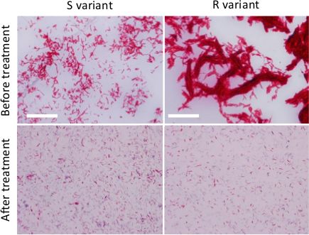
Figure 1. Preparation of dispersed M. abscessus inocula. Ziehl-Neelsen staining of R or S variants grown on broth medium prior to any treatment (upper panels) or after treatment (lower panels) to reduce the size ...
Toegang beperkt. Log in of start een proefperiode om deze inhoud te bekijken.
Materialen
| Naam | Bedrijf | Catalogusnummer | Opmerkingen |
|---|---|---|---|
| BBL MGIT PANTA | BD Biosciences | 245114 | |
| Bovine Serum Albumin | Euromedex | 04-100-811-E | |
| Catalase from Bovine Liver | Sigma-Aldrich | C40 | |
| Difco Middlebrook 7H10 Agar | BD Biosciences | 262710 | |
| Difco Middlebrook 7H9 Broth | BD Biosciences | 271310 | |
| Ethyl 3-aminobenzoate methanesulfonate salt (Tricaine) | Sigma-Aldrich | A5040 | |
| Oleic Acid | Sigma-Aldrich | O1008 | |
| Paraformaldehyde | Delta Microscopie | 15710 | |
| Phenol Red | Sigma-Aldrich | 319244 | |
| Tween 80 | Sigma-Aldrich | P4780 | |
| Agar | Gibco Life Technologie | 30391-023 | |
| Low melting agarose | Sigma-Aldrich | ||
| Instant Ocean Sea Salts | Aquarium Systems Inc | ||
| Borosilicate glass capillaries | Sutter instrument Inc | BF100-78-10 | 1 mm O.D. X 0.78 mm I.D. |
| Micropipette puller device | Sutter Instrument Inc | Flamming/Brown Micropipette Puller p-87 | |
| Microinjector | Tritech Research | Digital microINJECTOR, MINJ-D | |
| Tweezers | Sciences Tools inc | Dumont # M5S | |
| Microloader Tips | Eppendorf |
