This video demonstrates the procedure for establishing a bacterial infection in Drosophila larvae by placing them into a bacterial droplet and pricking them with a tungsten needle. The larvae are then incubated on a grooved, nutrient-rich agar plate to study host-pathogen interactions.
Een abonnement op JoVE is vereist om deze inhoud te bekijken. Log in of start vandaag met uw gratis proefperiode.
Methodenartikel
Establishing Bacterial Infection in Drosophila Larvae
353 weergaven
⸱
31 maart 2026
In dit artikel
Samenvatting
Protocol
1. In vivo infection
- Warm the Drosophila fruit juice agar plate at room temperature for 15 min.
- Add 30 g of agar to 700 mL of water and autoclave it for 40 min.
- Dissolve 0.5 g of methyl paraben in 10 mL of absolute ethanol.
- Add the methyl paraben solution to 300 mL of fruit juice concentrate.
- Quickly mix the juice concentrate into the autoclaved agar solution and dispense 5 mL into 10 × 35 mm Petri dishes.
- After the plates have cooled for 15 min, store them at 4 °C.
- Prepare 3rd instar larvae.
- Place the yeast paste on an agar plate. Make a fine cut in the agar plate where larvae can migrate to avoid drying (Figure 1A). Assemble a 0.001 mm pointed tungsten needle with holding forceps using paraffin film (Figure 1B, C).
- Pipette 50 µL of high-titer mCherry expressing-Coxiella burnetii (5.95×10⁹ GE/mL) onto the paraffin film under the stereo microscope and place the larvae into the pool of bacteria.
- Place the larvae in the pool of bacteria, prick them with a tungsten needle (Figure 3D), and transfer them onto an agar plate (Figure 1E).
- Transfer the remaining pathogen medium onto the agar plate and seal the plate with paraffin film.
- Keep the larvae on the plate in moist air until the desired time post-infection (Figure 1F). In this experiment, C. burnetii-infected larvae are on the plate for 24 h.
Toegang beperkt. Log in of start een proefperiode om deze inhoud te bekijken.
Resultaten
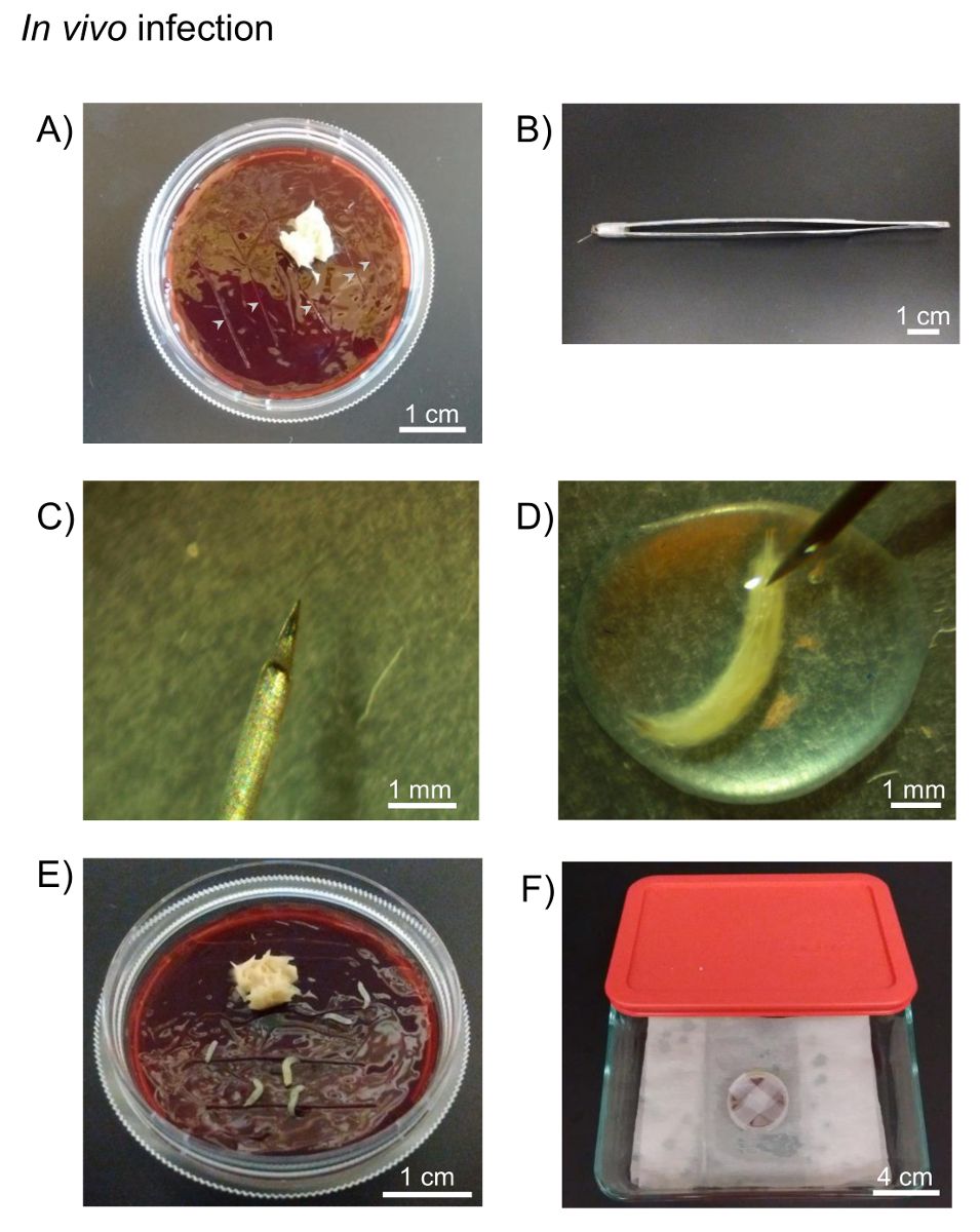
Figure 1: In vivo infection. A) Drosophila fruit juice agar plates and yeast paste...
Toegang beperkt. Log in of start een proefperiode om deze inhoud te bekijken.
Materialen
| Naam | Bedrijf | Catalogusnummer | Opmerkingen |
|---|---|---|---|
| Stereo microscope | Amscope | SM-1BSZ-L6W | |
| Forceps | VWR | 82027-402 | |
| Coxiella burnetii - mCherry | Dr. Heinzen, R. | ||
| Drosophila fruit juice plates | Cold Spring Harbor Protocols | http://cshprotocols.cshlp.org/content/2007/9/pdb.rec11113.full | |
| Agar | Fisher Bioreagents | BP1423-500 | |
| Methyl paraben | Amresco | 0572-500G | |
| Absolute ethanol | Fisher Bioreagents | BP2818-500 | |
| Welch's 100% Grape juice frozen concentrate, 340 mL | Amazon | B0025UJVGM | |
| Petri dishes, 10 x 35 mm | Fisher Scientific | 08-757-100A | |
| Yeast, Bakers Dried Active | MP Biomedicals | 210140001 | Add 2 parts of water to 1 part of yeast (v/v) |
| Tungsten needle | Fine Science Tools | 10130-20 | |
| Holding forceps | VWR | HS8313 | |
| Listeria monocytogenes | ATCC | strain: 10403S | Listeria monocytogenes strain 10403S (Bishop and Hinrichs, 1987) was grown in Difco Brain-heart infusion (BHI) broth (BD Biosciences) containing 50 µg/ml streptomycin at 30 °C. |
