Methodenartikel

Functional Chemoresistance Characterization: A Method to Evaluate Drug Resistance in Cancer Cells Using Clonogenic Assay

30 april 2023

In dit artikel

Samenvatting

Loading...
$$\rightleftharpoonup{xx}$$ $$\longleftharp{xx}$$, $$\longrightharp{xx}$$,

Source: Mohr, L. et al. Generation of Prostate Cancer Cell Models of Resistance to the Anti-mitotic Agent Docetaxel. J. Vis. Exp. (2017)

This video describes a method for evaluating chemotherapeutic drug resistance in prostate cancer cells using colony formation assay. This assay can be used as a functional tool to identify novel molecular and cellular mechanisms of chemoresistance in cancer cells.

Protocol

Loading...
$$\rightleftharpoonup{xx}$$ $$\longleftharp{xx}$$, $$\longrightharp{xx}$$,

1. Functional Characterization of Acquired Docetaxel Resistance by Colony Formation Assay

NOTE: In this protocol, chemoresistance has been evaluated using colony formation assays. Alternative methods used to evaluate cell viability (i.e., MTS assays) can be used based on investigators' preferences.

  1. Plate 2,000 cells (DU145 or 22Rv1, both parental and Docetaxel-resistant) per well in 6-well plates, using 2 mL of media per well.
  2. After 24 h, add increasing concentrations of Docetaxel (parental cells: 0.25, 0.5, 1, 2.5 and 5 nM; DR cells: 50, 125, 250, 500 and 1,000 nM). For both DU145 and 22Rv1 cell lines. Add DMSO only to one well as a control at the same volume used for the highest Docetaxel dose.
  3. After 72 h, aspirate the drug-containing media and add fresh Docetaxel-free media.
  4. Incubate plates for 1-2 weeks until colonies are visible under the microscope.
  5. To stain the colonies, wash them gently with 2-3 mL PBS and incubate with 2-3 mL crystal violet solution (0.1% w/v in 10% formalin) for 20 min inside the tissue culture hood or a fume hood.
  6. Remove staining solution, wash plates with 2-3 mL of H2O, remove H2O and air-dry plates.
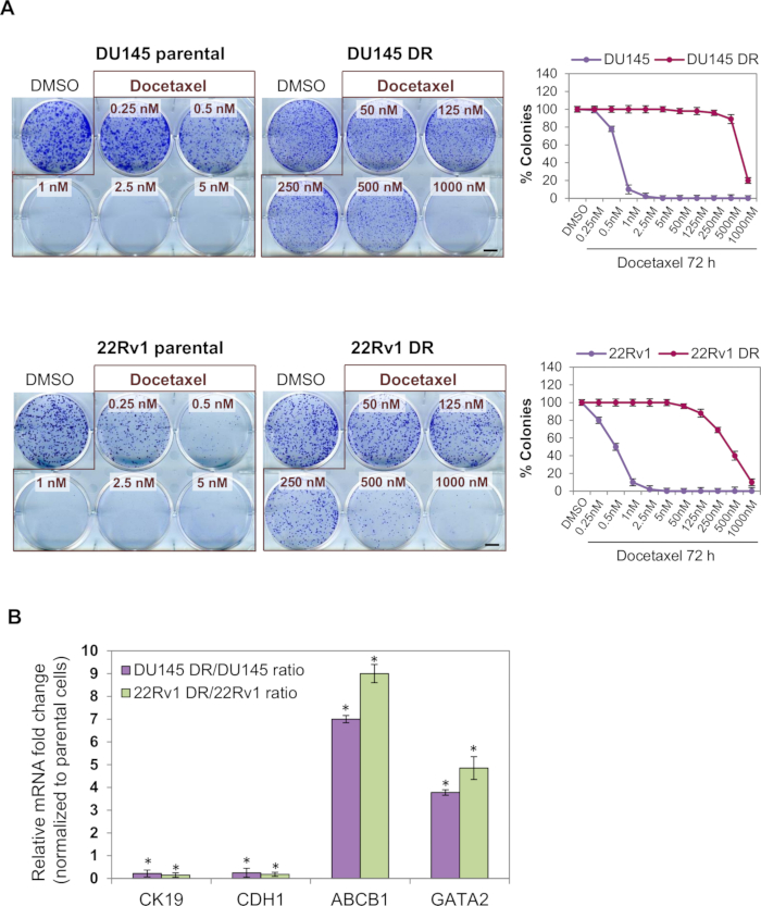
  7. Analyze the result by visualizing the wells and manually counting colonies with the help of a marker pen (to avoid counting the same colonies twice) and represent the percentage of cell viability in a graph. Take digital images of the plates for figure representation (Figure 1A).
Toegang beperkt. Log in of start een proefperiode om deze inhoud te bekijken.

Resultaten

Loading...
$$\rightleftharpoonup{xx}$$ $$\longleftharp{xx}$$, $$\longrightharp{xx}$$,

Cell viability assay, docetaxel effect; result charts, colony formation analysis, IC50 determination.
Figure 1: Functional and phenotypic characterization of the Docetaxel-resistant cell models. (A) Representative colony formation assays of parental and Docetaxel-resistant cells treated with the indicated Docetaxel concentrations for 7...

Toegang beperkt. Log in of start een proefperiode om deze inhoud te bekijken.

Openbaarmakingen

Loading...
$$\rightleftharpoonup{xx}$$ $$\longleftharp{xx}$$, $$\longrightharp{xx}$$,
No conflicts of interest declared.

Materialen

Lijst van materialen gebruikt in dit artikel
NaamBedrijfCatalogusnummerOpmerkingen
DU145 cells ATCC HTB-81
22Rv1 cells  ATCCCRL-2505
Docetaxel Selleck Chemicals S1148 in DMSO (10mM)
Tissue culture flask 150 sq cm   Falcon355001
6-well tissue culture plates  Falcon353046
RPMI 1640 Medium  Gibco11875093Supplemented with 10% FBS and 1% Penicillin/Streptomycin
Foundation B Fetal Bovine Serum  Gemini900208
1X Phosphate-Buffered Saline (PBS) Corning 21-040-CM
0.05%Trypsin-EDTA Gibco 25300-062
Penicillin-Streptomycin  Gibco15140122
Crystal Violet  Sigma-AldrichC0775
10% Formalin Solution  Sigma-AldrichHT501128

Herprints en machtigingen

Toestemming aanvragen om de tekst of afbeeldingen van dit JoVE-artikel te hergebruiken

Toestemming aanvragen

Trefwoorden

Evaluatie van geneesmiddelresistentiekolonievormingsassayprostaatkankercellendocetaxelbehandelingcellevensvatbaarheidsanalysekristalvioletkleuringmethode voor koloni tellingfunctionele resistentie assay

Gerelateerde artikelen